Review
. 2014 Oct;11(5):549-60.
doi: 10.1586/14789450.2014.939635. Epub 2014 Jul 25.

The necessity of and strategies for improving confidence in the accuracy of western blots

Affiliations
Review

The necessity of and strategies for improving confidence in the accuracy of western blots

Rajeshwary Ghosh et al. Expert Rev Proteomics. 2014 Oct.

Abstract

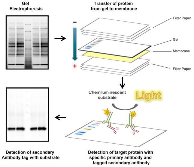
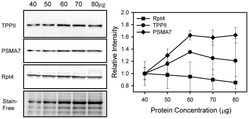
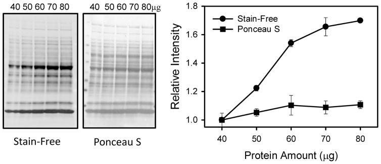
Western blotting is one of the most commonly used laboratory techniques for identifying proteins and semi-quantifying protein amounts; however, several recent findings suggest that western blots may not be as reliable as previously assumed. This is not surprising since many labs are unaware of the limitations of western blotting. In this manuscript, we review essential strategies for improving confidence in the accuracy of western blots. These strategies include selecting the best normalization standard, proper sample preparation, determining the linear range for antibodies and protein stains relevant to the sample of interest, confirming the quality of the primary antibody, preventing signal saturation and accurately quantifying the signal intensity of the target protein. Although western blotting is a powerful and indispensable scientific technique that can be used to accurately quantify relative protein levels, it is necessary that proper experimental techniques and strategies are employed.

Keywords: housekeeping protein; immunoblot; loading control; quantification; stain-free gel; total protein normalization; western blotting; western blotting accuracy; western blotting strategy.


Figures

Figure 1
Figure 1. Schematic diagram of a typical western blot
The typical western blot requires gel electrophoresis to separate proteins based upon molecular mass with subsequent transfer of the proteins from the gel to a protein binding membrane. The membrane is then incubated with the primary antibody to the target protein and the primary antibody is detected by a tagged secondary antibody. The tagged secondary antibody catalyzes an enzymatic reaction with the substrate which can be detected by film or a digital imager.
Figure 2
Figure 2. The linear dynamic range for each target protein may be different
Western blotting of heart homogenates (40 to 80μg) using anti-TPPII (Santa Cruz), anti-PSMA7 (Abcam) and anti-Rpt4 (Enzo). A Stain-Free image of a representative region of the blot is shown. The figure shows the relative intensity of the different target proteins without any normalization. Both anti-TPPII and anti-PSMA7 were linear within the range of linearity of the total protein stain (see Figure 4) but the anti-Rpt4 antibody was not linear within the protein range utilized. Use of the anti-Rpt4 antibody in samples in which 50μg of total protein was loaded would give incorrect results.
Figure 3
Figure 3. Schematic diagram of human glyceraldehyde-3-phosphate dehydrogenase (GAPDH) showing known post-translational modifications identified by different methods including mass spectrometry
The diagram is drawn to scale to show the location of the post-translational modifications (PTMs) on the 355 residue long GAPDH. The residue number for each PTM is shown. Ub-K, ubiquitinated lysine; Ac-K, acetylated lysine; pT, phosphorylated threonine; pS, phosphorylated serine; pY, phosphorylated tyrosine. PTMs on GAPH were obtained from PhosphoSitePlus (http://www.phosphosite.org/).
Figure 4
Figure 4. The linear dynamic range for total protein detection varies with the staining reagent utilized
Both Stain-Free and Ponceau S staining of total proteins (40 to 80μg) in heart homogenates show that both stains were linear between 40 to 60 μg of total protein. Images shown are nitrocellulose membranes post-transfer. Due to the larger relative changes in intensity using the Stain-Free method when compared to the Ponceau S stain, it is likely that smaller differences in target protein changes could be distinguished using the Stain-Free method.

References

    1. Gomes AV, Young GW, Wang Y, et al. Contrasting proteome biology and functional heterogeneity of the 20 S proteasome complexes in mammalian tissues. Molecular & cellular proteomics : MCP. 2009;8(2):302–315. - PMC - PubMed
    1. Gomes AV, Zong C, Edmondson RD, et al. Mapping the murine cardiac 26S proteasome complexes. Circulation research. 2006;99(4):362–371. - PubMed
    1. Burnette WN. “Western blotting”: electrophoretic transfer of proteins from sodium dodecyl sulfate--polyacrylamide gels to unmodified nitrocellulose and radiographic detection with antibody and radioiodinated protein A. Analytical biochemistry. 1981;112(2):195–203. This paper was the first to introduce the term “western blotting.”. - PubMed
    1. Towbin H, Staehelin T, Gordon J. Electrophoretic transfer of proteins from polyacrylamide gels to nitrocellulose sheets: procedure and some applications. Proceedings of the National Academy of Sciences of the United States of America. 1979;76(9):4350–4354. This is the original publication which introduced the western blotting methodology. - PMC - PubMed
    1. Kurien BT, Scofield RH. Western blotting. Methods. 2006;38(4):283–293. - PubMed

Publication types