. 2014 Jul 25:14:207.
doi: 10.1186/s12866-014-0207-5.

Differential responses of osteoblasts and macrophages upon Staphylococcus aureus infection

Affiliations

Differential responses of osteoblasts and macrophages upon Staphylococcus aureus infection

Therwa Hamza et al. BMC Microbiol. .

Abstract

Background: Staphylococcus aureus (S. aureus) is one of the primary causes of bone infections which are often chronic and difficult to eradicate. Bacteria like S. aureus may survive upon internalization in cells and may be responsible for chronic and recurrent infections. In this study, we compared the responses of a phagocytic cell (i.e. macrophage) to a non-phagocytic cell (i.e. osteoblast) upon S. aureus internalization.

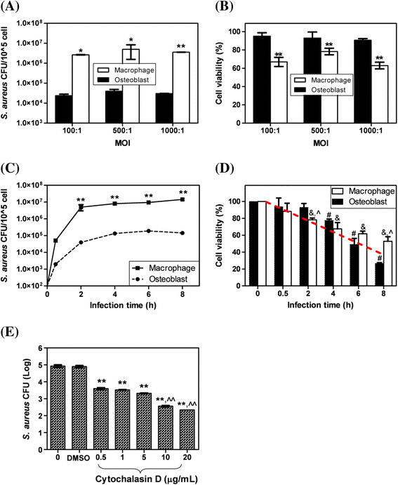
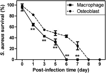
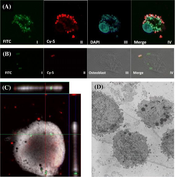
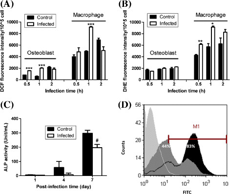
Results: We found that upon internalization, S. aureus could survive for up to 5 and 7 days within macrophages and osteoblasts, respectively. Significantly more S. aureus was internalized in macrophages compared to osteoblasts and a significantly higher (100 fold) level of live intracellular S. aureus was detected in macrophages compared to osteoblasts. However, the percentage of S. aureus survival after infection was significantly lower in macrophages compared to osteoblasts at post-infection days 1-6. Interestingly, macrophages had relatively lower viability in shorter infection time periods (i.e. 0.5-4 h; significant at 2 h) but higher viability in longer infection time periods (i.e. 6-8 h; significant at 8 h) compared to osteoblasts. In addition, S. aureus infection led to significant changes in reactive oxygen species production in both macrophages and osteoblasts. Moreover, infected osteoblasts had significantly lower alkaline phosphatase activity at post-infection day 7 and infected macrophages had higher phagocytosis activity compared to non-infected cells.

Conclusions: S. aureus was found to internalize and survive within osteoblasts and macrophages and led to differential responses between osteoblasts and macrophages. These findings may assist in evaluation of the pathogenesis of chronic and recurrent infections which may be related to the intracellular persistence of bacteria within host cells.


Figures

Figure 1
Figure 1
S. aureus infection of osteoblasts and macrophages.(A) Live intracellular S. aureus and (B) viability of osteoblasts and macrophages at different MOIs (100:1, 500:1, and 1000:1) for 2 h. *p < 0.05 and **p < 0.001 compared to osteoblasts at the same MOI. (C) Live intracellular S. aureus and (D) viability of osteoblasts and macrophages at an MOI of 500:1 for various infection times. **p < 0.001 compared to osteoblasts at the same infection time, &p < 0.01 compared to macrophages at infection times 0 and 0.5 h, ^p < 0.01 compared to osteoblasts at the same infection times, and #p < 0.05 compared to osteoblasts at infection times 0 and 0.5 h. (E) Effect of cytochalasin D on S. aureus internalization in osteoblasts. **p < 0.001 compared to the controls and ^^p < 0.001 compared to 0.5, 1, and 5 μg/mL.
Figure 2
Figure 2
Survival of intracellular S. aureus within osteoblasts and macrophages after infection at an MOI of 500:1 for 2 h.**p < 0.001 compared to osteoblasts at the same post-infection time.
Figure 3
Figure 3
Visualization of intracellular S. aureus within (A) macrophages and (B-D) osteoblasts. Osteoblasts and macrophages were infected with S. aureus at an MOI of 500:1 for 2 h. (A and B) S. aureus was stained with FITC before infection. Infected osteoblasts and macrophages were fixed, blocked, stained first with primary antibody to S. aureus surface protein A, and then secondary antibody conjugated to Cy-5. The nuclei of the macrophages were additionally stained with DAPI. Visualized at (I) 488 nm, (II) 633 nm, and (III) 405 nm. (IV) Merged images of (I), (II), and (III). As a result, intracellular S. aureus is shown in green (FITC) and extracellular S. aureus is co-localized with both red (Cy-5) and green (FITC). (C) Z-stack sections were used to confirm that all live S. aureus was inside osteoblasts as determined by the X-Y planes. Live S. aureus are green (Syto 9) and dead S. aureus are red (PI). Osteoblasts were infected with Syto 9-labeled S. aureus, then extracellular bacteria were killed with gentamicin and washed. Osteoblasts were detached from the wells and stained with PI. Approximately 20 cells (randomly selected) were examined. (D) Confirmation of intracellular S. aureus within osteoblasts using TEM.
Figure 4
Figure 4
Cellular and molecular responses of osteoblasts and macrophages infected with S. aureus at an MOI of 500:1 for 2 h. (A) DCF fluorescence intensity as an indicator of H2O2 production by non-infected controls and S. aureus-infected osteoblasts and macrophages. (B) DHE fluorescence intensity as an indicator of O.2 production by non-infected controls and S. aureus-infected osteoblasts and macrophages. (C) Osteoblast ALP activity measured at post-infection days 1, 4, and 7. (D) Macrophage phagocytosis activity (ingestion). Light gray area: control macrophages without incubation with S. aureus; dark gray area: non-infected macrophages; black area: infected macrophages. *p < 0.01, **p < 0.001, ***p < 0.0001, and #p < 0.05 compared to control.

Similar articles

Cited by

References

    1. Darouiche RO. Treatment of infections associated with surgical implants. N Engl J Med. 2004;350(14):1422–1429. doi: 10.1056/NEJMra035415. - DOI - PubMed
    1. von Eiff C, Becker K, Metze D, Lubritz G, Hockmann J, Schwarz T, Peters G. Intracellular persistence of Staphylococcus aureus small-colony variants within keratinocytes: A cause for antibiotic treatment failure in a patient with Darier’s disease. Clin Infect Dis. 2001;32(11):1643–1647. doi: 10.1086/320519. - DOI - PubMed
    1. Lentino JR. Prosthetic joint infections: Bane of orthopedists, challenge for infectious disease specialists. Clin Infect Dis. 2003;36(9):1157–1161. doi: 10.1086/374554. - DOI - PubMed
    1. Berendt T, Byren I. Bone and joint infection. Clin Med. 2004;4(6):510–518. doi: 10.7861/clinmedicine.4-6-510. - DOI - PMC - PubMed
    1. Lew DP, Waldvogel FA. Osteomyelitis. Lancet. 2004;364(9431):369–379. doi: 10.1016/S0140-6736(04)16727-5. - DOI - PubMed

Publication types

Substances