. 2014 Sep 12;289(37):25460-7.
doi: 10.1074/jbc.R114.583906. Epub 2014 Jul 24.

Harnessing the unique structural properties of isolated α-helices

Affiliations

Harnessing the unique structural properties of isolated α-helices

Carter J Swanson et al. J Biol Chem. .

Abstract

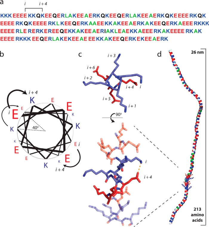
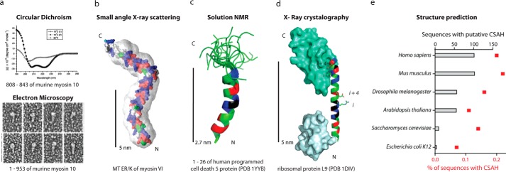
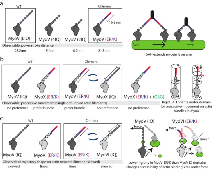
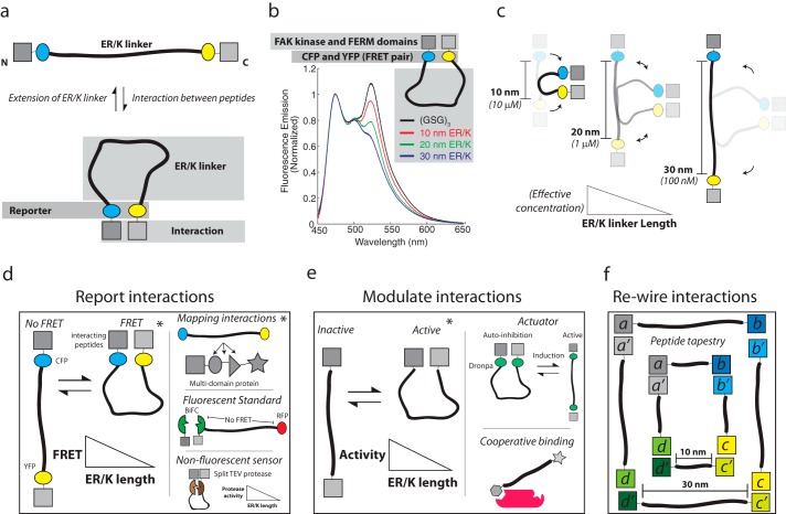
The α-helix is a ubiquitous secondary structural element that is almost exclusively observed in proteins when stabilized by tertiary or quaternary interactions. However, beginning with the unexpected observations of α-helix formation in the isolated C-peptide in ribonuclease A, there is growing evidence that a significant percentage (0.2%) of all proteins contain isolated stable single α-helical domains (SAH). These SAH domains provide unique structural features essential for normal protein function. A subset of SAH domains contain a characteristic ER/K motif, composed of a repeating sequence of ∼4 consecutive glutamic acids followed by ∼4 consecutive basic arginine or lysine (R/K) residues. The ER/K α-helix, also termed the ER/K linker, has been extensively characterized in the context of the myosin family of molecular motors and is emerging as a versatile structural element for protein and cellular engineering applications. Here, we review the structure and function of SAH domains, as well as the tools to identify them in natural proteins. We conclude with a discussion of recent studies that have successfully used the modular ER/K linker for engineering chimeric myosin proteins with altered mechanical properties, as well as synthetic polypeptides that can be used to monitor and systematically modulate protein interactions within cells.

Keywords: Biosensor; Coiled-coil; ER/K Alpha-Helix; ER/K Linker; Fluorescence Resonance Energy Transfer (FRET); Myosin; Protein Structure; Protein-Protein Interaction; Single Alpha-helical Domain (SAH).


Figures

FIGURE 1.
FIGURE 1.
Glu and Arg/Lys side chain interactions stabilize a monomeric α-helix in solution. a, the primary amino acid sequence of the ER/K motif in Kelch motif family protein from Trichomonas vaginalis. Positively charged residues (Arg and Lys) are shown in blue, negatively charged Glu is depicted in red, polar residues are in black, and hydrophobic residues are in green. Note the recurrence of Glu and R/K spaced at (i, i + 4) intervals. b, a pinwheel diagram representing the spacing of residues along an α-helix. The (i, i + 4) spacing in the ER/K motif positions amino acids 40° apart, with a distance of 0.6 nm along the helical backbone. (Pinwheel adapted from Ref. .) c, ionic interactions and H-bonded salt bridges occur between Glu and R/K side chains with (i, i + 4) spacing. Top panel, top view down the backbone from the N terminus of one heptad of (E4K4)2 peptide from an MD simulation (18). Bottom panel, a 90° rotation visualizing the entire 16-amino acid peptide with a E-K interaction highlighted. d, a representative snap shot from a Monte Carlo simulation of the Kelch motif family protein ER/K α-helix (as in a (29)) highlighting the extended α-helical conformation in a large polypeptide (∼30 kDa).
FIGURE 2.
FIGURE 2.
SAH domains have been observed in natural peptides or full-length proteins with multiple structural techniques. a, from Ref. , the putative coiled-coil region of myosin 10 forms an SAH domain. Top, CD of a peptide from murine myosin 10 has a canonical α-helical spectrum at 10 °C with a negative ellipticity at 222 nm (before and after heating to 80 °C) and a random coil spectra (with loss of ellipticity at 222 nm) at 80 °C. Bottom, rotary shadowed transmission electron microscopy of recombinant murine myosin 10 fragments. b, from Ref. , the predicted structure of the medial tail (MT) ER/K region (residues 916–981) of myosin VI docked into its SAXS envelope. c, from Ref. , an alignment of 20 solution states, determined by NMR, for the programed cell death 5 protein residues 1–26 following 1H-15N HSQC. PDB, Protein Data Bank. d, from Ref. , the x-ray crystal structure of ribosomal protein L9. e, from Ref. , the number and percentage of total sequences of putative CSAH domains identified from the primary amino acid sequences of the indicated organisms listed in Swiss-Prot and TrEMBL databanks utilizing the CSAH server. b–d, residues in SAH domains are colored as in Fig. 1.
FIGURE 3.
FIGURE 3.
The ER/K α-helix is a modular genetic motif that can be used to create myosin chimeras with altered mechanical properties. a, Baboolal et al. (33) created a myosin V (MyoV) chimera containing the putative ER/K α-helix from Dictyostelium myosin M. Left, the power stroke distances of WT myosin V, myosin V with truncation of two or four calmodulin stabilized IQ domains, and a chimera of myosin V with two native IQ domains and a 16.8-nm ER/K α-helix. Right, an extended and rigid ER/K α-helix can propagate force generated in the myosin catalytic domain to facilitate long processive steps on actin filaments. b, Nagy and Rock (34) generated multiple chimeras between myosin V and myosin X (MyoX) to assess structural elements that allow myosin X to preferably move on fascin-actin bundles. Left, representative chimeras that were used to identify that in myosin X, the ER/K α-helix and not the motor domain or step size dictates processive movement on fascin-actin bundles. Right, insertion of unstructured Gly-Ser-Gly residues between the SAH and IQ domains of myosin X disrupts preferential processivity on fascin-actin bundles. The ER/K α-helix alters the orientation of the motor domain, allowing it to favorably bind actin sites uniquely presented in fascin-actin bundles. c, Hariadi et al. (35) generated chimeras swapping the ER/K α-helix from myosin VI (MyoVI) with the IQ domains from myosin V while investigating the collective movement of multiple myosins tethered together. Left, multiple myosin V proteins, but not myosin VI, display meandering trajectories while traversing actin meshworks. Swapping regions of the lever arm containing the ER/K α-helix can reverse this phenomenon. Right, the IQ domains are likely more rigid than the ER/K α-helix, such that the inter-myosin force can selectively alter the accessibility of actin binding sites for the less rigid myosin VI.
FIGURE 4.
FIGURE 4.
The ER/K α-helix dictates the effective concentration of peptides attached to its distal ends and can be used for protein/cellular engineering applications. a, top, The ER/K α-helix adopts an extended conformation in the absence of an interaction between polypeptides fused to its ends. Bottom, interaction between peptides stabilizes the closed conformation of the ER/K α-helix, which is detected by a reporter system (e.g. FRET between CFP and YFP). b, from Ref. , an example of the schematic depicted in a, in which the interacting FAK, FERM, and kinase domains, as well as the fluorescent protein FRET pair CFP and YFP, are separated by a disordered GSG linker or by ER/K linkers with extended lengths of 10–20 and 30 nm. Fluorescence emission of these polypeptides was monitored at concentrations significantly lower than the bimolecular dissociation constant for the kinase-FERM domain interaction; FRET is assessed by the characteristic increase in fluorescence at 525 nm. c, Sivaramakrishnan and Spudich (36) found that the effective concentration of the intramolecular interaction was dependent on the ER/K α-helix length. Longer ER/K α-helix length leads to a lower effective concentration. d–f, sample applications, some experimentally demonstrated (*) and others conceptual, of a modular ER/K linker in protein engineering. d, left, reporting protein-protein interactions using fluorescent protein FRET reporters between conditionally interacting protein/peptide pairs, as demonstrated by Malik et al. (39) investigating G protein-coupled receptor-G protein interactions. Top right, ER/K linker with flanking FRET reporters inserted between domains of a multidomain protein as reported by Swanson et al. (41) investigating protein kinase C. Middle right, tethering a bimolecular fluorescence complementation (BiFC) (42) pair to fluorescent protein with an ER/K α-helix, places the fluorescent protein beyond FRET distance to allow for quantification of expression levels of the sensor. Bottom right, a non-fluorescent readout of a conditional protein-protein interaction, for example, enzymatic activity of a split tobacco etch virus (TEV) protease, or deriving antibiotic resistance from a split β-lactamase (43). These approaches allow for increased stringency of detection by increasing the ER/K α-helix length, while controlling for stoichiometry of interacting proteins. e, left, ER/K α-helix length modulates protein-protein interactions to control the activity resulting from the interaction. For instance, an activity that is dependent on two proteins interacting can occur more or less frequently depending on the length of the ER/K α-helix as demonstrated by Ritt et al. (40) investigating the intramolecular interaction between FERM and kinase domains of FAK. Top right, the ER/K α-helix can be used to generate single polypeptide actuators, using inducible protein interaction pairs. For instance, the optogenetically controlled dimeric dronpa fluorescent proteins (44) can be used to modulate autoinhibition of a catalytic domain. Bottom right, the ER/K α-helix can be used to control co-recruitment of peptides to an intermolecular complex. Although the initial interaction will be dependent on polypeptide concentration, recruitment of the second peptide tethered by the ER/K linker will be dependent on the linker length. f, ER/K α-helices can be used to engineer structural scaffolds from polypeptides. A schematic of one such design is depicted in which the size of the structure can be adjusted by the length of the ER/K α-helix.

References

    1. Knight P. J., Thirumurugan K., Xu Y., Wang F., Kalverda A. P., Stafford W. F., 3rd, Sellers J. R., Peckham M. (2005) The predicted coiled-coil domain of myosin 10 forms a novel elongated domain that lengthens the head. J. Biol. Chem. 280, 34702–34708 - PubMed
    1. Woolfson D. N. (2005) The design of coiled-coil structures and assemblies. Adv. Protein Chem. 70, 79–112 - PubMed
    1. Sunitha M. S., Nair A. G., Charya A., Jadhav K., Mukhopadhyay S., Sowdhamini R. (2012) Structural attributes for the recognition of weak and anomalous regions in coiled-coils of myosins and other motor proteins. BMC Res. Notes 5, 530. - PMC - PubMed
    1. Kreuzer S. M., Elber R. (2013) Coiled-coil response to mechanical force: global stability and local cracking. Biophys. J. 105, 951–961 - PMC - PubMed
    1. Scholtz J. M., Baldwin R. L. (1992) The mechanism of α-helix formation by peptides. Annu. Rev. Biophys. Biomol. Struct. 21, 95–118 - PubMed

Publication types

LinkOut - more resources