Review
. 2015 Feb;26(2):258-69.
doi: 10.1681/ASN.2014030278. Epub 2014 Jul 24.

A potential role for mechanical forces in the detachment of podocytes and the progression of CKD

Affiliations
Review

A potential role for mechanical forces in the detachment of podocytes and the progression of CKD

Wilhelm Kriz et al. J Am Soc Nephrol. 2015 Feb.

Abstract

Loss of podocytes underlies progression of CKD. Detachment of podocytes from the glomerular basement membrane (GBM) rather than apoptosis or necrosis seems to be the major mechanism of podocyte loss. Such detachment of viable podocytes may be caused by increased mechanical distending and shear forces and/or impaired adhesion to the GBM. This review considers the mechanical challenges that may lead to podocyte loss by detachment from the GBM under physiologic and pathophysiologic conditions, including glomerular hypertension, hyperfiltration, hypertrophy, and outflow of filtrate from subpodocyte spaces. Furthermore, we detail the cellular mechanisms by which podocytes respond to these challenges, discuss the protective effects of angiotensin blockade, and note the questions that must be addressed to better understand the relationship between podocyte detachment and progression of CKD.

Keywords: foot process effacement; mechanical forces; podocyte detachment; progression of chronic renal failure.


Figures

Figure 1.
Figure 1.
Foot process (FP) pattern under various pressures in the isolated perfused kidney (IPK) showing intact FPs even under conditions of high perfusion pressure and GBM expansion. FP pattern in the IPK with increasing perfusion pressures of (A) 65, (B) 105, and (C) 125 mmHg. (Top and middle panels) Outer surfaces of glomerular capillaries showing that, correlated with an expansion of capillaries from A–C, FPs become longer and change into a more regular pattern, in which FPs are strictly arranged in parallel (arrows in B and C). In A, FPs show a less regular pattern. (Lower panels) Cross-sections through the filtration barrier showing intact slit membranes. As confirmed by measurements (Table 1), there is no difference in slit membrane width between A and B. In C, the slit membrane appears to be wider, possibly on the basis of a different processing of the tissue; measurements were not done. Note that neither surface structure nor structure nor cross-section even exposure to 125 mmHg (C) created any defects. Isolated rat kidneys perfused for 100 minutes. Upper and middle panels, scanning electron microscopy; lower panels, transmission electron microscopy. Scale bars, 5 μm in top panel; 2 μm in middle panel; 1 μm in lower panel. Unpublished pictures are reprinted from refs. and , with permission.
Figure 2.
Figure 2.
Podocyte detachment from the GBM occurs in stages, including formation of pseudocysts. (A) The GBM is highlighted in yellow, and capillary lumens are in green. Three podocytes are seen undergoing focal detachment. The podocyte shown in light violet covers most of the surface of two capillary profiles. At several sites, detachments from the GBM are seen (arrows), which likely resulted from retraction of foot processes from a former neighboring podocyte; above them, small pseudocysts have developed. At other sites (notably with foot process effacement), the same podocyte is closely attached to the GBM (arrowheads). A second podocyte (shown in dark violet) exhibits large pseudocysts that open toward bare GBM (double arrows); note the normal chromatin pattern of the cell nucleus. A third podocyte (pastel) spans over the second podocyte, emphasizing the chaotic process of podocyte detachments. Note that the detaching podocytes contact each other at several sites. (B) Scanning electron microscopy of a detaching podocyte showing broadened, likely retracting foot processes and an area of bare GBM in between them. (A) Rat, uninephrectomy in young rats. Unpublished transmission electron microscopy is reprinted from ref. , with permission. (B) Rat, puromycin aminonucleoside nephrosis. Scanning electron microscopy is reprinted from ref. , with permission. Scale bars, 2 μm.
Figure 3.
Figure 3.
Prolapse of podocytes into the urinary orifice and shedding into the tubule occurs under the influence of elevated shear forces at the orifice. A cluster of at least seven podocytes (1–7) has come to lie within the urinary orifice. They are frequently found to be bottle-shaped (1, 3, 4, 6, and 7), which is likely a result of high shear stress from the high-flow velocities of filtrate at this site. As verified in serial sections, they all seem to have some remnant contact to the GBM, and in addition, they have contacts among each other. (B) The cluster of detaching podocytes in a subsequent section. Rat, growth stimulation with fibroblast growth factor 2 (FGF-2) for 13 weeks. Light micrographs from two sections of a complete section series. Unpublished light micrographs are from ref. . Scale bars, 10 μm.
Figure 4.
Figure 4.
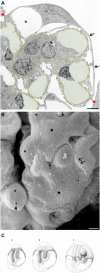
Stretching, attenuation, and pseudocyst formation of podocytes result from imbalanced glomerular growth and podocyte hypertrophy. (A) The GBM is shown in yellow, and capillary lumens are in green. Mismatch between the growth of the tuft and the hypertrophy of podocytes leads to stretching and attenuation of podocyte cell bodies of podocytes (arrows). Note that the podocyte spans a distance of four capillary loops (in between the two red arrowheads). *The attenuated cytoplasm bulges to a pseudocyst. (B) A surface view shows the great extent of cell body stretching (stars) and pseudocyst formation (asterisks). Clefts that allow an escape for the filtrate out of subpodocyte spaces are rare and frequently narrow (arrows). Gaps in the roof of pseudocysts (arrowhead) may provide more direct escape routes. Also, primary processes may undergo considerable stretching (double arrows). (C) Schematic to show the mechanism of pseudocyst formation. The tuft is depicted as a globe that grows from a to c. (b and c) Podocytes that cannot adequately adapt by cell hypertrophy undergo stretching and cell body attenuation. (c) Resistance to free exit of filtrate from subpodocyte spaces leads to a bulging of the attenuated cytoplasm called pseudocysts. (A) Rat, desoxycorticosterone-salt hypertension. Transmission electron microscopy reprinted from ref. , with permission. (B) uninephrectomy in young rats. Scanning electron microscopy reprinted from ref. , with permission. Scale bars, 5 μm in A; 2 μm in B. (C) Reprinted from ref. , with permission.
Figure 5.
Figure 5.
A chambered system of pseudocysts develops and favors podocyte detachment. The GBM is shown in yellow, capillary lumens are in green, and pseudocysts are in yellowish orange. Podocytes frequently detach in groups associated with the development of a communicating system of pseudocysts. (A) Tuft area with a group of at least five podocytes (1–5) undergoing detachment. Only podocytes 1 and 2 (possibly also podocyte 3) still have contact with the GBM, whereas podocytes 4 and 5 are attached to the podocytes beneath. Red arrows indicate communications between pseudocysts. (B) Two podocytes (1 and 2) are in the process of detachment. Podocyte 2 is attached by its apical cell portion to the parietal basement membrane (brown). Note, in A and B, that the communicating system of pseudocysts starts above bare areas of GBM (red dots); red arrows show that communications between pseudocysts captured in this section. (C) Schematic to show the suggested filtrate flow through the system of pseudocysts, finally emptying into Bowman’s space. (A) Rat, growth stimulation with fibroblast growth factor 2 (FGF-2) for 8 weeks. Transmission electron microscopy. Scale bar, 5 µm. Reprinted from ref. , with permission. (B) Rat, desoxycorticosterone-salt hypertension. Transmission electron microscopy. Scale bar, 10 µm. Reprinted from ref. , with permission.
Figure 6.
Figure 6.
Closing the slits and foot process effacement are measures to seal the filtration barrier and limit shear stresses on the FPs. In A and B, the GBM is shown in yellow, and capillary lumens are in green. (A) Capillary profile showing different degrees of sealing the barrier. At most sites, slit membranes are replaced by occluding junctions (arrows). Retraction of foot processes has led to broadened processes and stretches of complete foot process effacement (arrowheads). No sites of detachment are seen. (B) Filtration barrier undergoing foot process effacement. Just a single seemingly normal slit membrane is seen (arrowhead); at all other sites, the slit membranes are replaced by occluding junctions (arrows) between broadened processes. (C) Replacement of slit membranes by occluding junctions. Note that the slit membranes (sd) are still seen displaced above the newly formed occluding-type junctions (arrows). (A) Rat, desoxycorticosterone-salt hypertension. Scale bar, 2 µm. Unpublished transmission electron microscopy reprinted from ref. , with permission. (B) uninephrectomy in young rats. Scale bar, 40 µm. Unpublished transmission electron microscopy reprinted from ref. , with permission. (C) Rat, puromycin aminonucleoside nephrosis. Transmission electron microscopy reprinted from ref. , with permission.

References

    1. Pagtalunan ME, Miller PL, Jumping-Eagle S, Nelson RG, Myers BD, Rennke HG, Coplon NS, Sun L, Meyer TW: Podocyte loss and progressive glomerular injury in type II diabetes. J Clin Invest 99: 342–348, 1997 - PMC - PubMed
    1. Wharram BL, Goyal M, Wiggins JE, Sanden SK, Hussain S, Filipiak WE, Saunders TL, Dysko RC, Kohno K, Holzman LB, Wiggins RC: Podocyte depletion causes glomerulosclerosis: Diphtheria toxin-induced podocyte depletion in rats expressing human diphtheria toxin receptor transgene. J Am Soc Nephrol 16: 2941–2952, 2005 - PubMed
    1. Vogelmann SU, Nelson WJ, Myers BD, Lemley KV: Urinary excretion of viable podocytes in health and renal disease. Am J Physiol Renal Physiol 285: F40–F48, 2003 - PMC - PubMed
    1. Kriz W, Shirato I, Nagata M, LeHir M, Lemley KV: The podocyte’s response to stress: The enigma of foot process effacement. Am J Physiol Renal Physiol 304: F333–F347, 2013 - PubMed
    1. Craici IM, Wagner SJ, Bailey KR, Fitz-Gibbon PD, Wood-Wentz CM, Turner ST, Hayman SR, White WM, Brost BC, Rose CH, Grande JP, Garovic VD: Podocyturia predates proteinuria and clinical features of preeclampsia: Longitudinal prospective study. Hypertension 61: 1289–1296, 2013 - PMC - PubMed

Publication types

MeSH terms