. 2014 Sep 15;53(38):10020-31.
doi: 10.1002/anie.201403689. Epub 2014 Jul 24.

Multiscale modeling of biological functions: from enzymes to molecular machines (Nobel Lecture)

Affiliations

Multiscale modeling of biological functions: from enzymes to molecular machines (Nobel Lecture)

Arieh Warshel. Angew Chem Int Ed Engl. .

Abstract

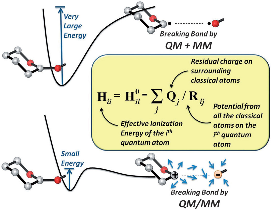
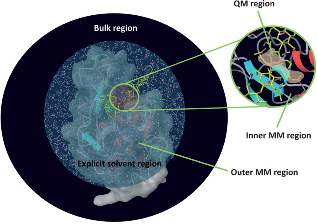
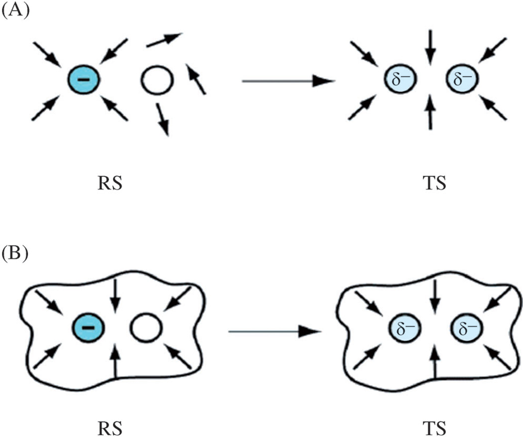
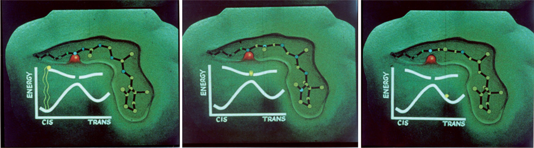
A detailed understanding of the action of biological molecules is a pre-requisite for rational advances in health sciences and related fields. Here, the challenge is to move from available structural information to a clear understanding of the underlying function of the system. In light of the complexity of macromolecular complexes, it is essential to use computer simulations to describe how the molecular forces are related to a given function. However, using a full and reliable quantum mechanical representation of large molecular systems has been practically impossible. The solution to this (and related) problems has emerged from the realization that large systems can be spatially divided into a region where the quantum mechanical description is essential (e.g. a region where bonds are being broken), with the remainder of the system being represented on a simpler level by empirical force fields. This idea has been particularly effective in the development of the combined quantum mechanics/molecular mechanics (QM/MM) models. Here, the coupling between the electrostatic effects of the quantum and classical subsystems has been a key to the advances in describing the functions of enzymes and other biological molecules. The same idea of representing complex systems in different resolutions in both time and length scales has been found to be very useful in modeling the action of complex systems. In such cases, starting with coarse grained (CG) representations that were originally found to be very useful in simulating protein folding, and augmenting them with a focus on electrostatic energies, has led to models that are particularly effective in probing the action of molecular machines. The same multiscale idea is likely to play a major role in modeling of even more complex systems, including cells and collections of cells.

Keywords: biomolecules; computational chemistry; free energy calculations; molecular modeling; multiscale models.


Figures

Figure 1
Figure 1
Showing the energetics of breaking a C–O bond in an uncoupled QM + MM (upper diagram) and when the electrostatic and steric effects of the environment are included in a coupled QM/MM (lower diagram). The dipoles designate the effect of the surrounding residual charges. As seen from the Figure it is very hard to break the bond without including the coupling between the QM and MM regions.
Figure 2
Figure 2
A QM/MM model of the lysozyme active site. The enzyme is divided to a small reactive QM region, and to the rest of the system, which is described by a classical MM model.
Figure 3
Figure 3
Schematic demonstration of the reorganization of the environment dipoles in an SN2 reaction, where the charges change from being on one atom in the reactant state (RS), to being delocalized in the transition state (TS) in A) water and B) an enzyme active site.[26]
Figure 4
Figure 4
Snapshots from the simulated MD trajectory of the primary event in the vision process. The trajectory starts with 11-cis retinal in the ground state, and, upon absorption of light, the system moves to the excited state where the 11–12 torsional angle rotates without a barrier to 90°, and the trajectory crosses to the ground state in the trans direction. The motion involves only a small change in the overall structure, since the other torsional angles move in the opposite direction to the 11–12 torsional angle. The snapshots are taken from a movie that used the original trajectory presented in Ref. [42].
Figure 5
Figure 5
Exploring the coupling between the rotation of the γ-stalk to ATP hydrolysis in F1-ATPase. The relevant system (namely F1-ATPase) is shown from the membrane side (A), and along the vertical direction parallel to the central γ-stalk (B). The α catalytic subunits are shown in deep blue, deep green and orange, while the β units are shown in cyan, light green and yellow. The γ-stalk is shown in magenta. The nucleotide occupancies of the β subunits are depicted as T (ATP bound), D (ADP bound) or E (empty) states. (C) The CG electrostatic free energy surface of the rotation of the γ-stalk coupled to the α/β conformational changes. This landscape reflects the stepwise 80°/40° features discussed in the main text. The combination of the diagram of (C) with the energetic of the chemical steps (which is given in Ref. [62]) provides a structure-based description of the action of F1-ATPase. This Figure is taken from Ref. [62].
Figure 6
Figure 6
Simulations of the coupling between the ribosome and the translocon (TR). The simulation addresses the effect of the TR on stalled peptides, where for some lengths of the linker, L, the coupling to the TR helps to release the stalled peptide. The time dependence of xistall and x1 for a peptide chain with 40 and 36 units is shown here, which corresponds to L = 31 (blue) and 27 (red), respectively. The x coordinate designates the insertion coordinate and is defined in Ref. [72]. The barriers used for the LD simulations were obtained by scaling down the energy terms by 0.43. This allowed for the simulation of the insertion process in a relatively short timescale, and then estimating the relevant time for the actual barriers by using the corresponding Boltzmann probability. The snapshots on the top and bottom of the plot show the configuration of the nascent peptide chain for L = 31 and L = 27, respectively. The ribosome and TR are shown schematically, the starting configuration of the nascent chain is in cyan, the leading particle (x1) is in red, and all other particles added to the growing chain are shown in magenta. The interpolated times (that should be obtained without scaling) for L = 31 and L = 27 are 6 min and 36 min, respectively. This Figure is taken from Ref. [72], which also gives a complete description of the problem and the simulations performed.

References

    1. Quantum Chemical Models (Nobel Lecture): Pople JA. Angew. Chem. 1999;111:2014–2023. Angew. Chem. Int. Ed. 1999;38:1894–1902.

    1. Theoretical Studies of Enzymic Reactions – Dielectric, Electrostatic and Steric Stabilization of Carbonium-Ion in Reaction of Lysozyme: Warshel A, Levitt M. J. Mol. Biol. 1976;103:227–249.

    1. Fersht A. Structure and Mechanism in Protein Science: A Guide to Enzyme Catalysis and Protein Folding. New York: Freeman; 1999. p. 631.
    1. Warshel A. Computer Modeling of Chemical Reactions in Enzymes and Solutions. Hoboken: Wiley-Interscience; 1997. p. 256.
    1. Jencks WP. Catalysis in Chemistry and Enzymology. New York: McGraw-Hill; 1969. p. 644.