. 2014 Sep 15;74(18):4996-5007.
doi: 10.1158/0008-5472.CAN-13-1807. Epub 2014 Jul 24.

Neutralizing murine TGFβR2 promotes a differentiated tumor cell phenotype and inhibits pancreatic cancer metastasis

Affiliations

Neutralizing murine TGFβR2 promotes a differentiated tumor cell phenotype and inhibits pancreatic cancer metastasis

Katherine T Ostapoff et al. Cancer Res. .

Abstract

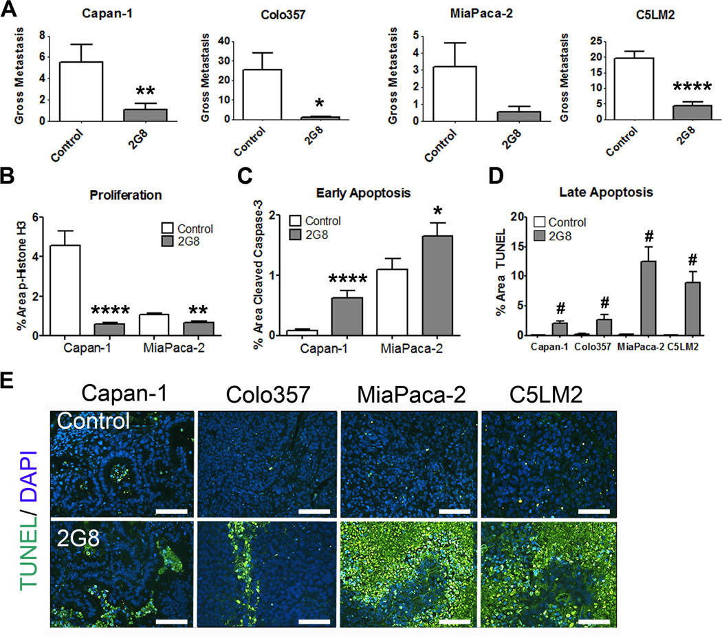
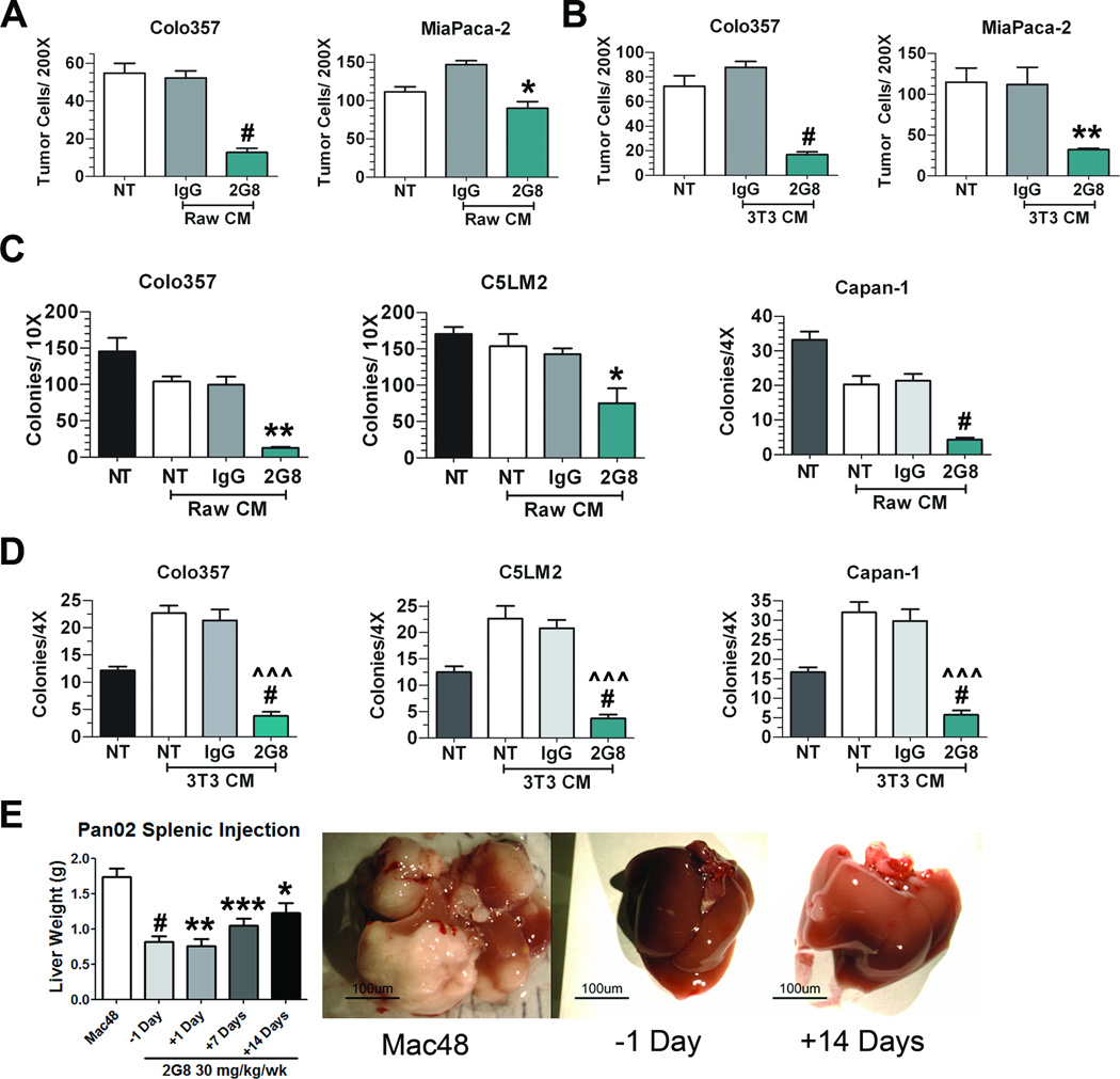
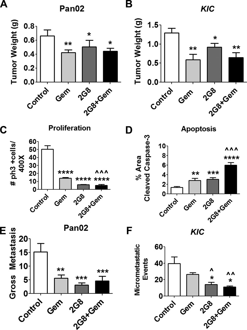
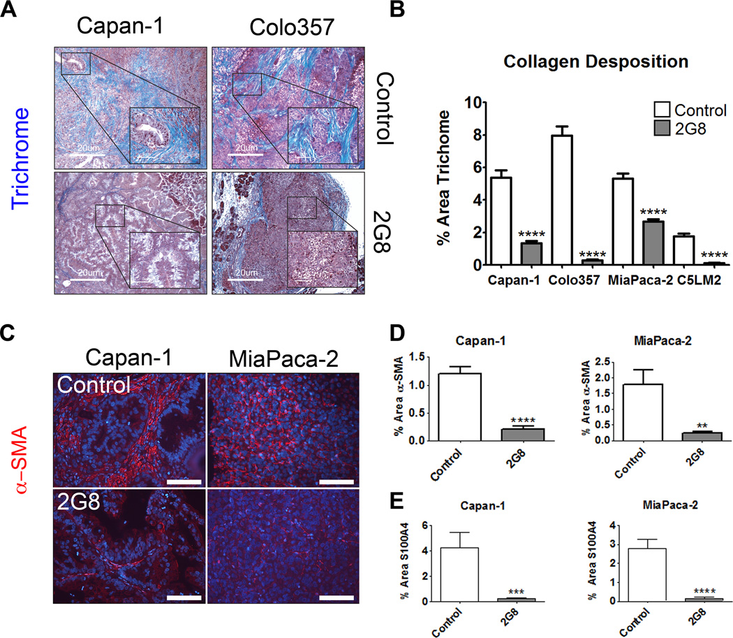
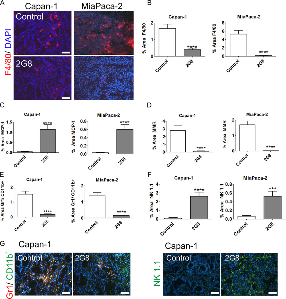
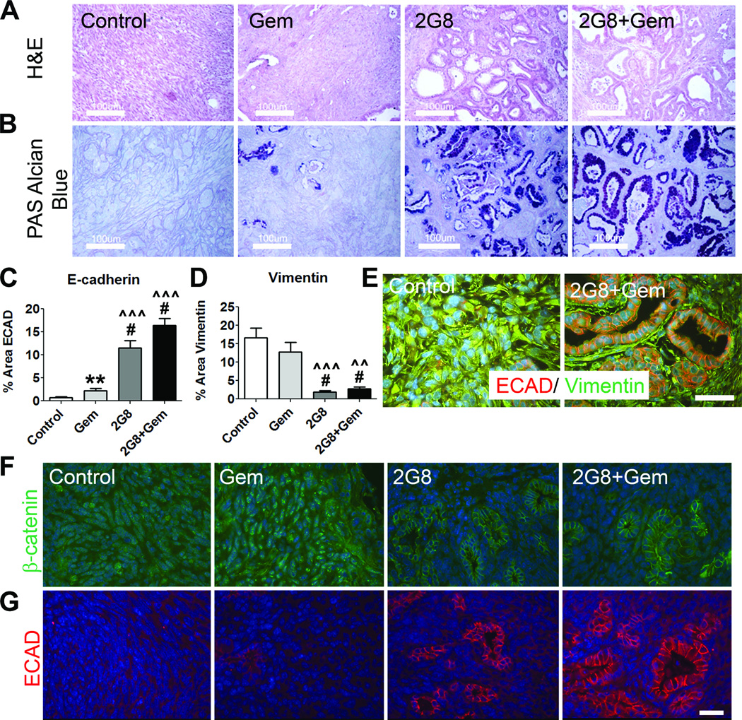
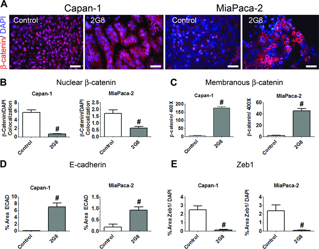
Elevated levels of TGFβ are a negative prognostic indicator for patients diagnosed with pancreatic cancer; as a result, the TGFβ pathway is an attractive target for therapy. However, clinical application of pharmacologic inhibition of TGFβ remains challenging because TGFβ has tumor suppressor functions in many epithelial malignancies, including pancreatic cancer. In fact, direct neutralization of TGFβ promotes tumor progression of genetic murine models of pancreatic cancer. Here, we report that neutralizing the activity of murine TGFβ receptor 2 using a monoclonal antibody (2G8) has potent antimetastatic activity in orthotopic human tumor xenografts, syngeneic tumors, and a genetic model of pancreatic cancer. 2G8 reduced activated fibroblasts, collagen deposition, microvessel density, and vascular function. These stromal-specific changes resulted in tumor cell epithelial differentiation and a potent reduction in metastases. We conclude that TGFβ signaling within stromal cells participates directly in tumor cell phenotype and pancreatic cancer progression. Thus, strategies that inhibit TGFβ-dependent effector functions of stromal cells could be efficacious for the therapy of pancreatic tumors. Cancer Res; 74(18); 4996-5007. ©2014 AACR.


Conflict of interest statement

Conflict of Interest Statement Xiaohong Xu, Desiree Nugent, and Kyla D. Carroll are employees of Imclone Systems, a wholly-owned subsidiary of Eli Lilly and Company. This work was supported in part by a sponsored research agreement from Imclone Systems to Rolf A. Brekken.

Figures

Figure 1
Figure 1. Inhibition of stromal Tgfβr2 reduces metastasis in vivo
Human pancreatic cancer cell lines Capan-1, Colo357, MiaPaCa-2 and C5LM2 were orthotopically implanted into NOD-SCID mice. After tumor establishment, mice were randomized to receive saline (control) or 2G8 (30 mg/kg/week, n= 7–12/group). Total gross metastases were determined by evaluation of liver, diaphragm, GI lymph nodes, and lung at the time of sacrifice (A). B and C, Cell proliferation (B) and apoptosis (C) in control and 2G8 treated Capan-1 and MiaPaCa-2 tumors were assessed by immunohistochemistry for phospho-histone H3 (B) or cleaved caspase 3 (C), respectively. D and E, Control and 2G8 treated tumors from each model were evaluated for apoptosis using TUNEL. (TUNEL, green; scale bar, 100 µm). 5 animals/group were analyzed with 5 representative pictures taken and analyzed per animal. Results are expressed as mean+/−SEM. *, p<0.05; **, p<0.01; ***, p<0.001; ****, p<0.0001; #, p<0.0001 vs. control.
Figure 2
Figure 2. Inhibition of Tgfβr2 on stromal cells limits tumor cell migration and colony formation in vitro and in vivo
A and B, Transwell migration of tumor cells (Colo357 and MiaPaCa-2) towards RAW 264.7 (A) or NIH 3T3 (B) cells treated with serum free media (NT), control Rat IgG (IgG) or 2G8 for 24 hours. After removing treatment conditions, Colo357 and MiaPaCa-2 cells were plated in transwell chambers and allowed to migrate overnight towards previously treated stromal cells. Bar graphs represent number of cells/200× field. The assays were performed in triplicate in two independent experiments. *, p<0.05; **, p<0.01; #, p<0.0001 vs. Rat IgG. C and D, Anchorage independent growth of tumor cells (Colo357, C5LM2, and Capan-1) in the presence of growth media with 10% serum (NT), conditioned media from RAW 264.7 (C) or NIH 3T3 (D) cells grown in media with 10% serum with or without control Rat IgG (IgG) or 2G8. Colony formation was quantified by number of colonies per well at 4× or 10× magnification. Bar graph represents mean +/− SEM of a single experiment performed in triplicate with similar results found upon 2 independent experiments. *, p<0.05; **, p<0.01; **, p<0.01; #, p<0.0001 vs. NT; ^^^, p<0.0001 vs Rat IgG. E, Pan02 cells were injected intrasplenically and liver weight used as a surrogate for tumor burden in the liver. Mice were treated with a control rat IgG (Mac48, n=14) or 2G8 initiated at -1 (n=10), +1 (n=3), +7 (n=8) or +14 (n=7) days post splenic injection. Representative images of livers from mice are shown. Results are expressed as mean+/−SEM. *, p<0.05; **, p<0.01; ***, p<0.001; #, p<0.0001 vs. control
Figure 3
Figure 3. Inhibition of tumor and stromal Tgfβr2 results in reduced primary pancreatic tumor growth and metastasis in murine models
A, Orthotopic Pan02 tumors were established and mice randomized and treated for 4 weeks with vehicle (control), gemcitabine (Gem, 25 mg/kg/week), 2G8 (60 mg/kg/week) or 2G8+Gem. 2G8 alone and in combination with gemcitabine reduced primary tumor growth (n=7–10/group). B, LSL-KrasG12D; Cdkn2alox/lox; p48Cre (KIC) mice (n = 8–12/group) were randomized when 4 weeks old and treated for 4 weeks as above with Gem (12.5 mg/kg 3×/week), 2G8 (120 mg/kg/week) or the combination. At sacrifice, mice treated with 2G8 and 2G8+Gem had reduced tumor growth. C and D, Pan02 tumors were harvested and sections were evaluated by immunohistochemistry for proliferation (phospho-histone H3 (ph3+), C) and apoptosis (cleaved caspase-3, D). 2G8 reduced the number of ph3+ cells and increased the number of cells positive for cleaved caspase 3 compared to control. E and F, Total gross metastases in Pan02 bearing animals were determined by evaluation of liver, diaphragm, GI lymph nodes, and lung at the time of sacrifice. Metastatic burden in the KIC model was determined by histologic evaluation of H&E stained liver tissue. 2–3 sections of the anterior lobe of the liver (n=at least 5/group) were scored for lesions. 2G8 alone suppressed metastasis in each tumor model. Results are expressed as mean+/−SEM. *, p<0.05; **, p<0.01; ***, p<0.001; ****, p<0.0001 vs. control; ^, p<0.05; ^^, p<0.01; ^^^, p<0.0001 vs. Gem.
Figure 4
Figure 4. Inhibition of mouse Tgfβr2 blunts collagen deposition within xenografts
A and B, The level of fibrillar collagen deposited in human tumor xenografts from mice treated with saline (Control) or 2G8 was determined by trichrome histology (Trichrome, blue; scale bar, 20 µm, insets 5 µm A). B, The intensity of trichrome staining was quantified and shows that 2G8 significantly reduced collagen deposition within each tumor (5 animals/group, 5 pictures/200× field). C-E, To determine the level of fibroblast investment, xenografts from control and 2G8 treated animals were stained for α-smooth muscle actin (C and D) and S100A4 (E). Results are expressed as mean+/−SEM. **, p<0.01; ***, p<0.001; #, p<0.0001 vs. control.
Figure 5
Figure 5. Inhibition of mouse Tgfβr2 promotes a pro-inflammatory immune cell phenotype
A-D, The level of F4/80 (A and B), MCP-1 (C), and MMR (D) expressing macrophages in Capan-1 and MiaPaCa-2 xenografts was determined by immunohistochemistry. The effect of 2G8 on the level of MDSC (E) and NK cells (F) in Capan-1 and MiaPaCa-2 xenografts was also evaluated by immunohistochemistry with example images shown in panel G. Graphs represent 5 animals/group with 5/pictures per animal. Results are expressed as mean+/−SEM. ***, p<0.001; #, p<0.0001 vs. control.
Figure 6
Figure 6. 2G8 promotes an epithelial phenotype in murine tumors in vivo
A-E, LSL-KrasG12D; Cdkn2alox/lox; p48Cre (KIC) mice establish tumors and precursor PanIN lesions by 4 weeks old. Mice at this timepoint were randomized to receive saline (Control), gemcitabine (Gem), 2G8 or 2G8 + Gem for 4 weeks. A, Analysis of tumor architecture using Hemotoxylin and Eosin staining (scale bar, 100 µm). 2G8 treated tumors were noted to have significantly more PanIN and epithelial lesions than the mice treated with Gem or Control. B, This was confirmed with PAS-Alcian Blue staining that marks mucin-secreting PanIN lesions but not invasive lesions (PAS-Alcian Blue, purple; scale bar, 100 µm). Additionally, 2G8 tumors had significantly increased Ecad expression (C) and decreased vimentin expression (D) by immunohistochemistry. E, Representative images of these tumors demonstrate the predominant epithelial phenotype of 2G8-treated tumors (Ecad red, vimentin green; scale bar, 50 µm). F and G, 2G8 induces a similar epithelial phenotype in Pan02 tumors. Immunohistochemical analysis of β-catenin (F) and E-cadherin (Ecad, G) in Pan02 tumors treated with control, Gem, 2G8 or 2G8+GEM. Bar graphs represent mean+/−SEM. **, p<0.01; #, p<0.0001 vs. control; ^^, p<0.001; ^^^, p<0.0001 vs. Gem.
Figure 7
Figure 7. Inhibition of stromal Tgfβr2 promotes an epithelial phenotype in xenograft models of pancreatic cancer
A-C, The expression and localization of β-catenin in Capan-1 and MiaPaCa-2 xenografts treated with control or 2G8 was determined by immunohistochemistry (scale bar, 50 µm). Localization of β-catenin to the nucleus (B) or membrane (C) was quantified. D and E, The effect of 2G8 on the expression of E-cadherin (ECAD, D) and Zeb1 (E) was also determined by immunohistochemistry. Graphs represent 5–6 animals/group with 5/pictures per animal. Results are expressed as mean+/−SEM. #, p<0.0001 vs. control.

References

    1. Siegel R, Naishadham D, Jemal A. Cancer statistics 2012. CA Cancer J Clin. 2012;62(1):10–29. - PubMed
    1. Hanahan D, Coussens LM. Accessories to the crime: functions of cells recruited to the tumor microenvironment. Cancer Cell. 2012;21(3):309–322. - PubMed
    1. Corcoran RB, Contino G, Deshpande V, Tzatsos A, Conrad C, Benes CH, et al. STAT3 plays a critical role in KRAS-induced pancreatic tumorigenesis. Cancer Res. 2011;71(14):5020–5029. - PMC - PubMed
    1. Yang L, Pang Y, Moses HL. TGF-beta and immune cells: an important regulatory axis in the tumor microenvironment and progression. Trends Immunol. 2010;31(6):220–227. - PMC - PubMed
    1. Jones S, Zhang X, Parsons DW, Lin JC, Leary RJ, Angenendt P, et al. Core signaling pathways in human pancreatic cancers revealed by global genomic analyses. Science. 2008;321(5897):1801–1806. - PMC - PubMed

Publication types

MeSH terms

Substances