Meta-Analysis
. 2014 Dec 1;23(23):6407-18.
doi: 10.1093/hmg/ddu346. Epub 2014 Jul 24.

Salt-inducible kinase 3, SIK3, is a new gene associated with hearing

Affiliations
Meta-Analysis

Salt-inducible kinase 3, SIK3, is a new gene associated with hearing

Lisa E Wolber et al. Hum Mol Genet. .

Abstract

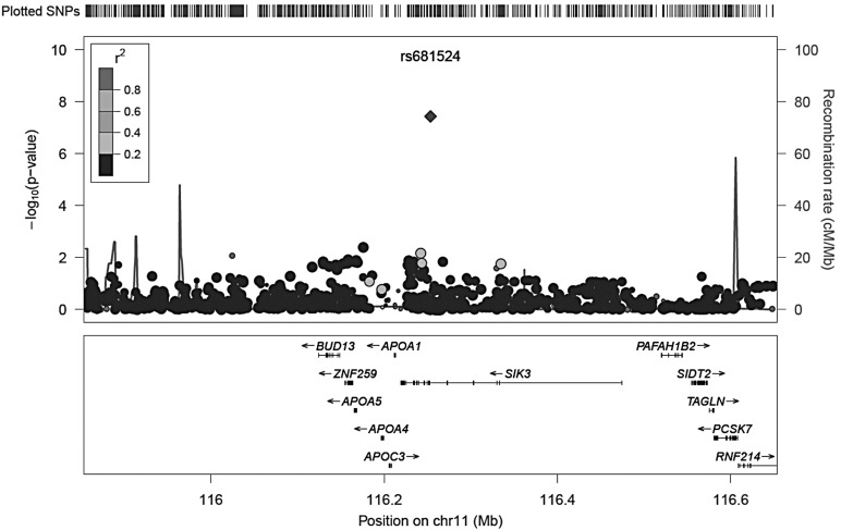
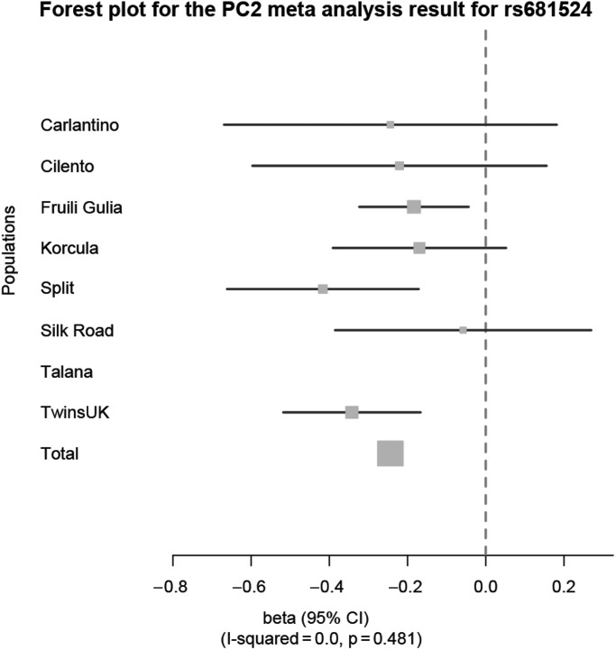
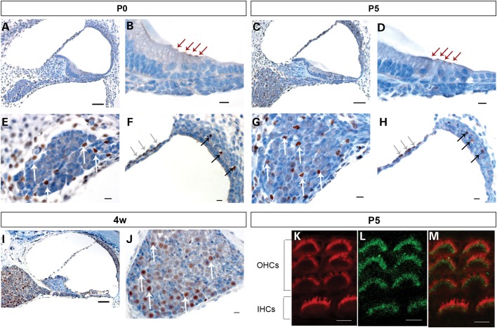
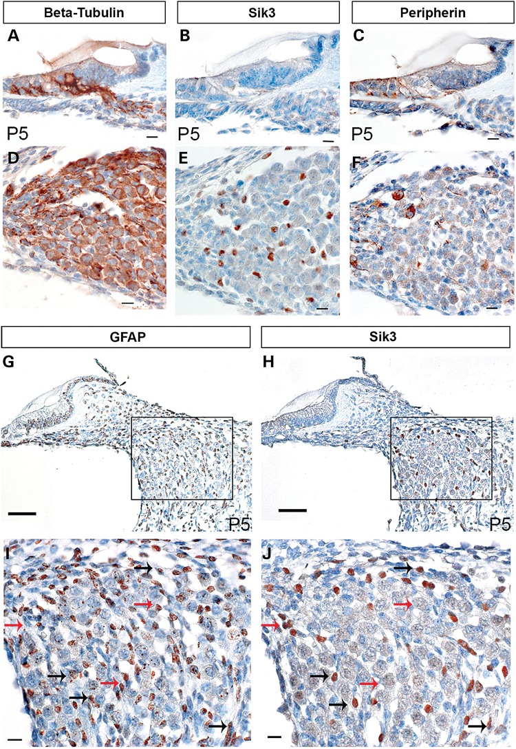
Hearing function is known to be heritable, but few significant and reproducible associations of genetic variants have been identified to date in the adult population. In this study, genome-wide association results of hearing function from the G-EAR consortium and TwinsUK were used for meta-analysis. Hearing ability in eight population samples of Northern and Southern European ancestry (n = 4591) and the Silk Road (n = 348) was measured using pure-tone audiometry and summarized using principal component (PC) analysis. Genome-wide association analyses for PC1-3 were conducted separately in each sample assuming an additive model adjusted for age, sex and relatedness of subjects. Meta-analysis was performed using 2.3 million single-nucleotide polymorphisms (SNPs) tested against each of the three PCs of hearing ability in 4939 individuals. A single SNP lying in intron 6 of the salt-inducible kinase 3 (SIK3) gene was found to be associated with hearing PC2 (P = 3.7×10(-8)) and further supported by whole-genome sequence in a subset. To determine the relevance of this gene in the ear, expression of the Sik3 protein was studied in mouse cochlea of different ages. Sik3 was expressed in murine hair cells during early development and in cells of the spiral ganglion during early development and adulthood. Our results suggest a developmental role of Sik3 in hearing and may be required for the maintenance of adult auditory function.


Figures

Figure 1.
Figure 1.
Locus zoom plot of the associated genetic markers on chromosome 11q23.3. The locus zoom depicts the location of genetic markers versus their significance of association with PC2. Significance of association is measured as the negative logarithm of the P-value. Genes located in the area 400 kb up-and downstream of rs681524 are displayed below the x-axis. The grey shading of each genetic marker indicates its correlation with the reference SNP rs681524. A legend for the correlation is shown in the upper left corner of the locus zoom. Recombination rate is highlighted as peaked lines.
Figure 2.
Figure 2.
Forest plot for the PC2 meta-analysis result for rs681524 in SIK3. The forest plot shows the effect sizes (beta with 95% CI) at rs681524 on PC2 for each population in comparison with a combined meta-analysis effect (total). A consistent negative effect for the minor C allele at rs681524 for all European populations could be determined. The percentage of variation owing to heterogeneity across populations was not significant (I2 = 0.0, P = 0.481). Corresponding data are listed in Table 5.
Figure 3.
Figure 3.
Sik3 is expressed in cochlear structures at different developmental stages. Mouse cochlear sections were stained with primary Sik3 antibody (brown) and counterstained with haematoxylin (blue). A, B, E and F: at the day of birth (P0), Sik3 expression was detected in the apex of inner and outer hair cells (B, red arrows), in the perilymph-facing layer of the Reissner's membrane (F, grey arrows), near blood vessels of the intermediate layer of the stria vascularis (F, black arrows), as well as in cells of the spiral ganglion (E, white arrows) and cells surrounding the ganglion. C, D, G and H: at 5 days postnatal (P5), Sik3 expression was found in the hair cells (D, red arrows) and small cells of the spiral ganglion (G, white arrows). I and J: at 4 weeks postnatal, Sik3 expression remained in the spiral ganglion (J, white arrows), Reissner's membrane and stria vascularis but could not be detected in the apex of the hair cells (I). K, L and M: confocal imaging of Sik3 expression in the stereocilia at 5 days postnatal (L, green), compared with Phalloidin expression (K, red) showed that Sik3 was expressed in the region around the base of the stereocilia (M, merged image of K and L). IHCs, inner hair cells; OHCs, outer hair cells. Scale bars: A, C and I: 50 µm; B, D, E, F, G, H and J: 10 µm; K, L and M: 5 µm.
Figure 4.
Figure 4.
Sik3 is expressed in small cells between the neurones of the spiral ganglion. To identify the cell type expressing Sik3 in the spiral ganglion, Sik3 expression was compared with beta-tubulin (A and D), Peripherin (C and F) and GFAP (G and I) expression in adjacent sections. Beta-tubulin is expressed in both Type 1 and 2 spiral ganglion neurones, whereas peripherin expression is limited to type 1 spiral ganglion neurones. Sik3 expression (B and E) did not coincide with either beta-tubulin (A and D) or peripherin expression (C and F) but seemed to be expressed in smaller cells interspersed between the neurones. To determine whether Sik3 could be expressed in glial cells, Sik3 expression (H and J) was compared with GFAP expression (G and I) in adjacent sections. Sik3 and GFAP expression overlapped partially. Sik3 expression coincided with GFAP expression in some cells (black arrows) but was absent in other glial cells (red arrows). All sections were prepared from mice at 5 days postnatal (P5). Expression of each antibody is indicated by a brown signal. Scale bars: A, B, C, D, E, F, I and J: 10 µm; G, H: 50 µm.

References

    1. Lenoir M., Shnerson A., Pujol R. Cochlear receptor development in the rat with emphasis on synaptogenesis. Anat. Embryol. (Berl) 1980;160:253–262. - PubMed
    1. Spoendlin H. The innervation of the outer hair cell system. Otol. Neurotol. 1982;3:274–278. - PubMed
    1. Viljanen A., Era P., Kaprio J., Pyykkö I., Koskenvuo M., Rantanen T. Genetic and environmental influences on hearing in older women. J. Gerontol. A Biol. Sci. Med. Sci. 2007;62:447–452. - PubMed
    1. Wolber L.E., Steves C.J., Spector T.D., Williams F.M.K. Hearing ability with age in Northern European women: a new web-based approach to genetic studies. PLoS One. 2012;7:e35500. - PMC - PubMed
    1. Karlsson K.K., Harris J.R., Svartengren M. Description and primary results from an audiometric study of male twins. Ear. Hear. 1997;18:114–120. - PubMed

Publication types