. 2014 Oct 16;124(16):2554-63.
doi: 10.1182/blood-2014-01-551820. Epub 2014 Jul 24.

A new form of macrothrombocytopenia induced by a germ-line mutation in the PRKACG gene

Affiliations

A new form of macrothrombocytopenia induced by a germ-line mutation in the PRKACG gene

Vladimir T Manchev et al. Blood. .

Abstract

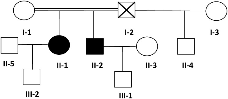
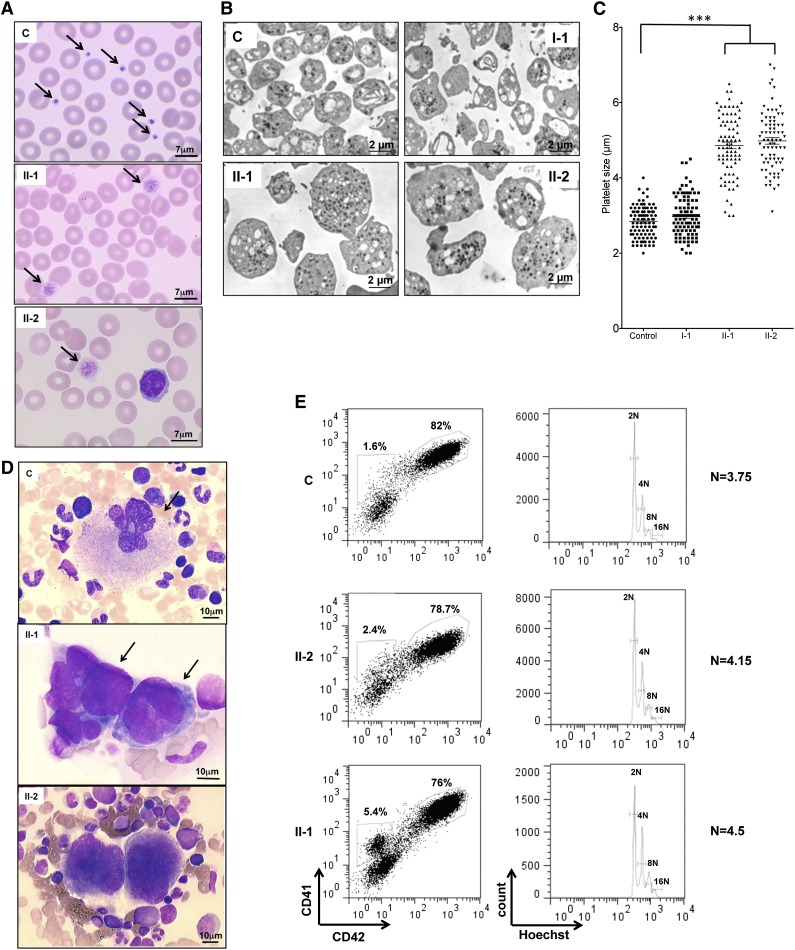
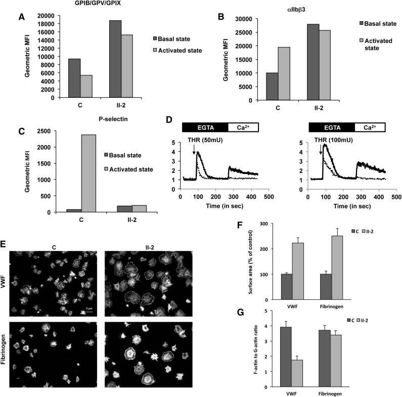
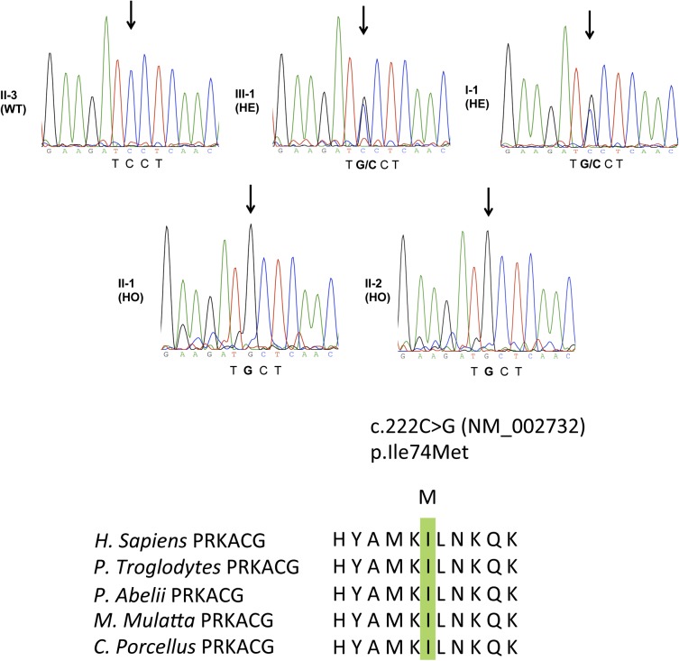
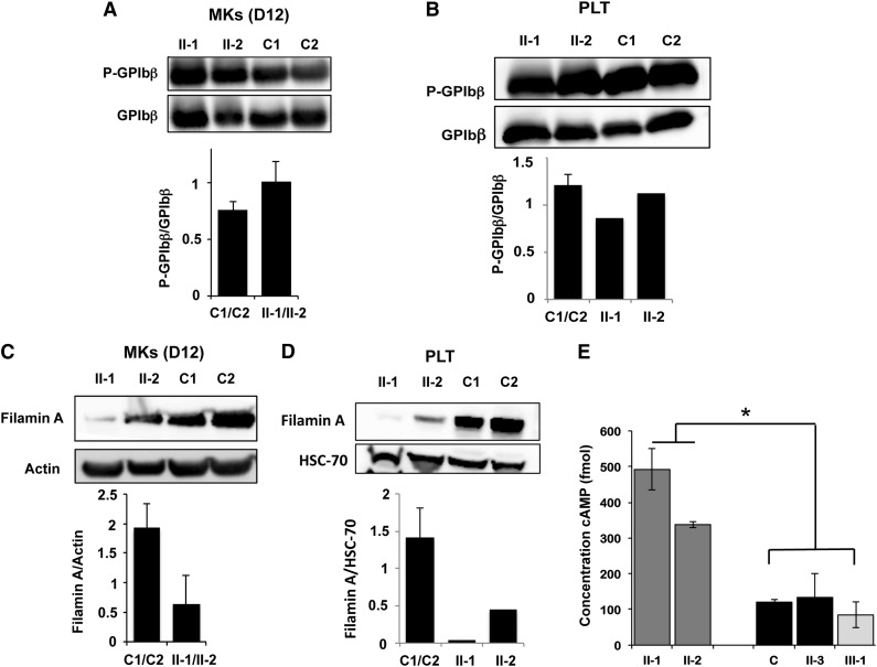
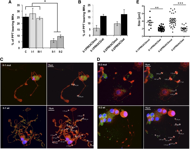
Macrothrombocytopenias are the most important subgroup of inherited thrombocytopenias. This subgroup is particularly heterogeneous because the affected genes are involved in various functions such as cell signaling, cytoskeleton organization, and gene expression. Herein we describe the clinical and hematological features of a consanguineous family with a severe autosomal recessive macrothrombocytopenia associated with a thrombocytopathy inducing a bleeding tendency in the homozygous mutated patients. Platelet activation and cytoskeleton reorganization were impaired in these homozygous patients. Exome sequencing identified a c.222C>G mutation (missense p.74Ile>Met) in PRKACG, a gene encoding the γ-catalytic subunit of the cyclic adenosine monophosphate-dependent protein kinase, the mutated allele cosegregating with the macrothrombocytopenia. We demonstrate that the p.74Ile>Met PRKACG mutation is associated with a marked defect in proplatelet formation and a low level in filamin A in megakaryocytes (MKs). The defect in proplatelet formation was rescued in vitro by lentiviral vector-mediated overexpression of wild-type PRKACG in patient MKs. We thus conclude that PRKACG is a new central actor in platelet biogenesis and a new gene involved in inherited thrombocytopenia with giant platelets associated with a thrombocytopathy.


Figures

Figure 1
Figure 1
Family tree. Circles, females; squares, males; black filled symbols, affected individuals; white symbols, nonaffected individuals; symbol with a diagonal line, deceased individual; double horizontal line, consanguinity.
Figure 2
Figure 2
Platelet and MK analysis. (A) Cytological investigation of blood platelets. The black arrows point to platelets in the blood smears. Note the much larger platelet size for patients (II-1 and II-2) compared with control (C). (B) Ultrastructural aspect of blood platelets. Large platelets were detected in blood of II-1 and II-2 patients, and platelets of normal size were detected in 1 control and in a I-1 family member with the heterozygous PRKACG mutation. (C) The size of 100 platelets for control, I-1, II-1, and II-2 individuals was measured. The results represent mean ± SEM. ***P < .0001, unpaired Student t test (2-tailed). (D) Cytological investigations of the bone marrow of II-1 and II-2 patients and control. (E) MK differentiation was induced from control or patient peripheral blood CD34+ cells and analyzed at day 10 of culture. Gates represent mature (CD41+CD42+) or immature (CD41+CD42) MKs (left). The ploidy level (N) was analyzed in the gate of CD41+CD42+ MKs and was based on the percentage of cells in 8N, 16N, and 32N gates.
Figure 3
Figure 3
Platelet functions. (A-C) Fluorescence-activated cell sorter flow analysis of the (A) GPIb-IX-V complex (anti-CD42a), (B) αIIbβ3 complex (anti-CD41a), and (C) P-selectin (anti-CD62P) on control (C) and patient (II-2) platelets before (basal state) and after activation by thrombin receptor agonist peptide (activated state). Histograms present geometric mean fluorescence intensity (geometric MFI). (D) Typical traces representative of normalized (to basal level) fluorescence intensity of Oregon green 488 BAPTA1-AM (representative of the cytosolic Ca2+ concentration) recorded using Accury flow cytometer. Platelets from the control (black line) and from the patient (dotted line) were treated, in the absence of calcium (EGTA, 100 µM), with thrombin (THR, 50 or 100 mU/mL) and Ca2+ (CaCl2, 300 µM). (E-G) Platelet spreading. (E) Platelets were adhered on fibrinogen or von Willebrand factor substrates and stained with both Alexa Fluor488-labeled phalloidin and Alexa Fluor594-labeled DNAse I. (F) Platelet surface area and (G) ratio of F-actin to G-actin were measured by ImageJ version 1.42k.
Figure 4
Figure 4
Germ-line PRKACG mutation. Electrophoregram of PRKACG gene (NM_00273), as sequenced by the Sanger method, revealed no mutation in individual II-3 designed as wild type (WT). Individuals III-1 and I-1 were found to be heterozygous (HE) for the c.222C>G mutation, without clinical features of macrothrombocytopenia. Patients II-1 and II-2 were found to be homozygous for the c.222C>G mutation and exhibited macrothrombocytopenia. The c.222C>G mutation caused substitution of the evolutionarily conserved Ile amino acid. Homologous sequences were aligned using the CLUSTALW Web site.
Figure 5
Figure 5
Analysis of PKA activity in patient platelets and megakaryocytes. (A-B) Western blot analysis and quantification of GPIbβ phosphorylated at Ser166 in MKs derived in vitro from (A) blood CD34+ progenitors and (B) in platelets of patients homozygous (II-1 and II-2) for the PRKACG p.74I>M mutation. Two external controls (C1 and C2) were analyzed. Total GPIbβ was used as a control of protein loading. MKs were investigated at (A) day 12 of culture. (C-D) Western blot analysis and quantification of filamin A in (C) MKs and (D) platelets of patients carrying the homozygous (II-1 and II-2) mutation. Two external controls (C1 and C2) were used. Actin or HSC70 was used as a control of protein loading. (E) Analysis of cAMP level in platelets isolated from one external (C1) and one internal control (II-3) and from 2 patients homozygous (II-1 and II-2) and 1 heterozygous for the PRKACG p.74I>M mutation (III-1). Error bars represent mean ± SD of triplicate. Experiments were performed 2 times with similar results. *P < .05, 2-tailed Mann-Whitney test.
Figure 6
Figure 6
Mutant PRKACG leads to defective PPT formation, which is rescued by wild-type PRKACG overexpression. (A-E) In vitro MK differentiation was induced from control or patient peripheral blood CD34+ progenitors in the presence of TPO and SCF. (B-E) CD34+ cells of patients II-1 and II-2 were transduced with a lentiviral vector harboring wild-type (wt) or mutant PRKACG cDNA (used as a control of experiment) at days 1 and 2 of culture. (A-B) The percentage of PPT-forming MKs was estimated by counting MKs exhibiting ≥1 cytoplasmic processes with areas of constriction at day 13 of culture. A total of 200 cells per well were counted. The histograms show 1 of 2 independent experiments with similar results. Each experiment was performed in triplicate. Data represent mean ± SD of triplicate. *P < .05, 2-tailed Mann-Whitney test. (C-D) Immunoconfocal analysis of platelet-like structures formed by PPTs generated from patients (C) II-1 and (D) II-2. MKs overexpressing wt or mutant PRKACG PPT-forming MKs were allowed to adhere on fibrinogen for 2 hours at day 13 of culture and stained with anti-tubulin (red) and rabbit anti-VWF (green) antibodies. Confocal imaging was performed on a Leica TCS SP8 inverted laser scanning confocal microscope (Leica Microsystems, Heidelberg, Germany), equipped with a 405-nm UV laser diode and visible optically pumped semiconductor lasers (488 and 552 nm). All images were acquired using an oil immersion 63× objective (1.4 numeric aperture). (E) At least 5 MKs for each condition were analyzed, and the size of platelet-like structures was measured by LAS AF version 2.4.1 software. Data are presented ± SEM. **P < .005 and ***P < .0001, unpaired Student t test with Welch’s correction.

Comment in

References

    1. Balduini CL, Savoia A, Seri M. Inherited thrombocytopenias frequently diagnosed in adults. J Thromb Haemost. 2013;11(6):1006–1019. - PubMed
    1. Stevenson WS, Morel-Kopp MC, Chen Q, et al. GFI1B mutation causes a bleeding disorder with abnormal platelet function. J Thromb Haemost. 2013;11(11):2039–2047. - PubMed
    1. Albers CA, Cvejic A, Favier R, et al. Exome sequencing identifies NBEAL2 as the causative gene for gray platelet syndrome. Nat Genet. 2011;43(8):735–737. - PMC - PubMed
    1. Kunishima S, Okuno Y, Yoshida K, et al. ACTN1 mutations cause congenital macrothrombocytopenia. Am J Hum Genet. 2013;92(3):431–438. - PMC - PubMed
    1. Gnirke A, Melnikov A, Maguire J, et al. Solution hybrid selection with ultra-long oligonucleotides for massively parallel targeted sequencing. Nat Biotechnol. 2009;27(2):182–189. - PMC - PubMed

Publication types

Substances