. 2014 Jul 1;37(7):1163-70.
doi: 10.5665/sleep.3830.

Sleep promotes cortical response potentiation following visual experience

Affiliations

Sleep promotes cortical response potentiation following visual experience

Sara J Aton et al. Sleep. .

Abstract

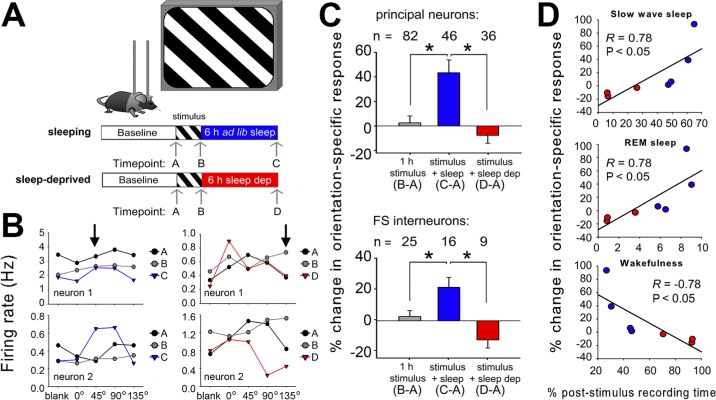
Study objectives: Sleep has been hypothesized to globally reduce synaptic strength. However, recent findings suggest that in the context of learning and memory consolidation, sleep may promote synaptic potentiation. We tested the requirement for sleep in a naturally occurring form of experience-dependent synaptic potentiation in the adult mouse visual cortex (V1), which is initiated by patterned visual experience.

Design: Visual responses were recorded in individual V1 neurons before and after presentation of an oriented grating stimulus, and after subsequent sleep or sleep deprivation.

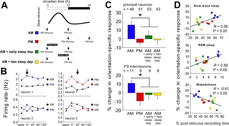
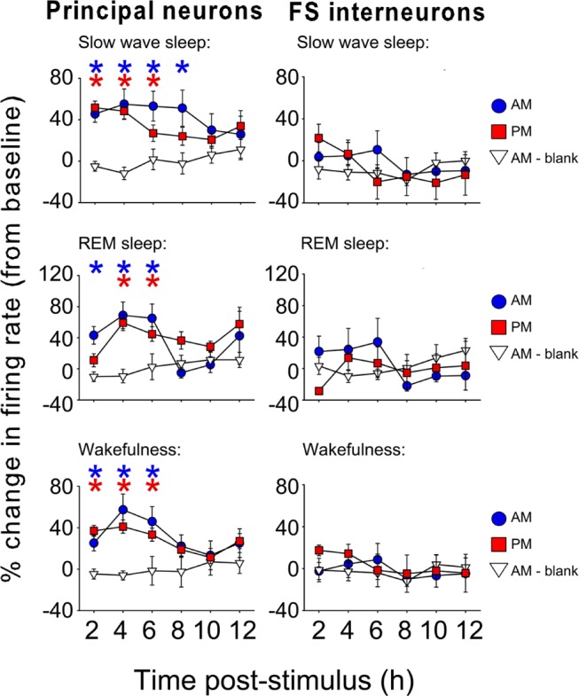
Measurements and results: We find that V1 response potentiation-associated with a shift in orientation preference in favor of the presented stimulus-occurs only after sleep and only during the entrained circadian sleep phase, and is blocked by sleep deprivation. Induction of plasticity following stimulus presentation is associated with an increase in principal neuron firing in V1, which is present in all behavioral states and occurs regardless of time of day. Sleep dependent potentiation is proportional to phase-locking of neuronal activity with thalamocortical spindle oscillations.

Conclusions: Our results suggest that sleep can promote cortical synaptic potentiation in vivo, and that this potentiation may be mediated by slow wave sleep spindles.

Citation: Aton SJ, Suresh A, Broussard C, Frank MG. Sleep promotes cortical response potentiation following visual experience.

Keywords: in vivo recording; sleep; synaptic plasticity; thalamocortical oscillations; visual system.


Figures

Figure 1
Figure 1
Spike data from continuously-recorded V1 neurons. Data are shown for two representative neurons recorded on the same stereotrode at baseline (0-2 h), following presentation of a stimulus to induce orientation-specific response potentiation (24-26 h) and following a 12-h poststimulus ad libitum sleep period (36-38 h). Spike waveform shape, relative spike amplitude on the two stereotrode wires, and clusters of spike data in three-dimensional principal component space are stable for the two neurons throughout the recording.
Figure 2
Figure 2
Orientation-specific response potentiation (OSRP) is consolidated during poststimulus sleep. (A) V1 neurons were recorded across a baseline period of ad libitum sleep and wakefulness, a 1-h period of oriented grating stimulus presentation in the awake mouse (starting at lights-on), and a 6-h period of either ad libitum sleep or sleep deprivation in the dark. Visual responses were recorded during presentation of a series of gratings (four orientations plus a blank screen) in the contralateral visual field at the intervals indicated (arrows): timepoint A, after baseline recording; timepoint B, after stimulus presentation; and timepoint C, after subsequent ad libitum sleep or timepoint D, sleep deprivation. (B) Representative visual response data for two V1 neurons recorded the same experiment, in mice from either sleeping (left) or sleep deprived (right) groups. In each graph, mean firing rate responses of a single neuron are shown for each of the four stimulus orientations and for blank screen presentation, at each of three intervals (timepoints A, B, and either C or D). The orientation of the stimulus presented to induce OSRP for both neurons recorded from each experiment is indicated in the upper graphs (arrows). Relative firing rate responses increased for the orientation of the presented stimulus in sleeping mice (left, data shown in blue) but not in sleep deprived mice (right, data shown in red). These orientation-specific response changes were quantified as a measure of OSRP (see Materials and Methods). (C) Orientation preference for the presented stimulus did not change after 1-h stimulus presentation, but was enhanced after subsequent sleep in both non-fast spiking (principal) neurons and in fast spiking (FS) interneurons. OSRP was blocked by sleep deprivation. *P < 0.05, Holm-Sidak post hoc test. (D) OSRP was proportional to sleep time, and negatively correlated with wakefulness.
Figure 3
Figure 3
Orientation-specific response potentiation (OSRP) occurs specifically during the entrained circadian sleep phase. (A) Grating stimuli were presented to induce OSRP at the time of either lights-on (AM) or lights-off (PM). Visual responses to oriented gratings were recorded just before stimulus presentation, and again 12 h later. In both cases, mice were entrained to a 12 h:12 h light:dark cycle prior to OSRP induction, but were kept in complete darkness over the next 12 h to eliminate effects of patterned vision on OSRP consolidation. OSRP consolidation was tested across the day/sleep phase (AM, blue) or across the night/active phase (PM, red). Two additional groups of mice had OSRP assessed across the sleep phase, with sleep deprivation during either the first (AM + early sleep dep, green) or second (AM + late sleep dep, yellow) half of the circadian day. (B) Firing rate response data for two V1 neurons recorded from representative mice in either AM or PM stimulus conditions. In each graph, mean firing rate responses of a single neuron are shown for each of the four stimulus orientations and for blank screen presentation, at two intervals: after baseline, and 12 h later (after ad libitum sleep). The orientation of the stimulus presented to induce OSRP for both neurons recorded from each experiment is indicated in the upper graphs (arrows). Relative firing rate responses increased for the orientation of the presented stimulus in AM mice (left, data shown in blue) but not in PM mice (right, data shown in red). (C) V1 neurons showed OSRP following AM stimulus presentation, but not PM stimulus presentation. Sleep deprivation during either half of the circadian day blocked OSRP. (D) Across the four experimental groups, OSRP was proportional to sleep time, and negatively correlated with wakefulness.
Figure 4
Figure 4
Principal neuron firing increases after orientation-specific response potentiation induction. For the first several hours after either lights-on or AM (blue circles) or lights-our or PM (red squares) stimulus presentation, firing rates among principal neurons were significantly increased (* P < 0.05 versus 24-h average across baseline recording, and versus control mice [white triangles] presented with a blank screen at lights on). There were no significant changes in fastspiking interneuron activity.
Figure 5
Figure 5
Potentiation of visual responsiveness during orientation-specific response potentiation (OSRP) is proportional to slow wave sleep (SWS) spindle spike-field coherence. (A) Visual responsiveness (expressed as the evoked responsiveness index [ERI]) was enhanced after poststimulus sleep and blocked by poststimulus sleep deprivation (top). ERI increases were present across lights-on or AM stimulus experiments, but not evening or lights out or PM or AM-blank screen experiments (bottom). Values indicate mean ± standard error of the mean for all neurons (fastspiking interneurons + principal neurons). (B) For all freely sleeping mice (in experiments shown in Figure 2A and Figure 3A), ERI changes were proportional to OSRP (% change from pre-stimulus baseline shown). (C) SWS spike-field correlations between firing in a representative V1 neuron and local field potential (LFP) spindle and delta oscillatory activity are shown at baseline (solid lines), and in the first 2 h following stimulus presentation (dashed lines). Although synchrony (coherence) of firing with delta oscillations was not significantly changed after OSRP induction, coherence with spindle oscillations increased significantly during OSRP consolidation. (D) Among freely sleeping mice, mean response changes at each recording site (i.e., stereotrode bundle) were proportional to mean SWS spindle coherence across the poststimulus recording period.

Comment in

  • Sleep. 37:1157.

References

    1. Aton SJ, Seibt J, Frank MG. Encyclopedia of life science. Chichester: John Wiley and Sons, Ltd.; 2009. Sleep and memory.
    1. Aton SJ. Set and setting: how behavioral state regulates sensory function and plasticity. Neurobiol Learn Mem. 2013;106:1–10. - PMC - PubMed
    1. Tononi G, Cirelli C. Sleep and synaptic homeostasis: a hypothesis. Brain Res Bull. 2003;62:143–50. - PubMed
    1. Cirelli C, Gutierrez CM, Tononi G. Extensive and divergent effects of sleep and wakefulness on brain gene expression. Neuron. 2004;41:35–43. - PubMed
    1. Vyazovskiy VV, Cirelli C, Pfister-Genskow M, Faraguna U, Tononi G. Molecular and electrophysiological evidence for net synaptic potentiation in wake and depression in sleep. 2009;63:865–78. - PubMed

Publication types

LinkOut - more resources