

Brain tissue hypoxia and oxidative stress induced by obstructive apneas is different in young and aged rats
- PMID: 25061253
- PMCID: PMC4098810
- DOI: 10.5665/sleep.3848
Brain tissue hypoxia and oxidative stress induced by obstructive apneas is different in young and aged rats
Abstract
Study objectives: To test the hypotheses that brain oxygen partial pressure (PtO2) in response to obstructive apneas changes with age and that it might lead to different levels of cerebral tissue oxidative stress.
Design: Prospective controlled animal study.
Setting: University laboratory.
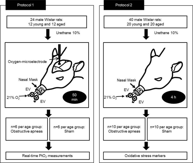
Participants: Sixty-four male Wistar rats: 32 young (3 mo old) and 32 aged (18 mo).
Interventions: Protocol 1: Twenty-four animals were subjected to obstructive apneas (50 apneas/h, lasting 15 sec each) or to sham procedure for 50 min. Protocol 2: Forty rats were subjected to obstructive apneas or sham procedure for 4 h.
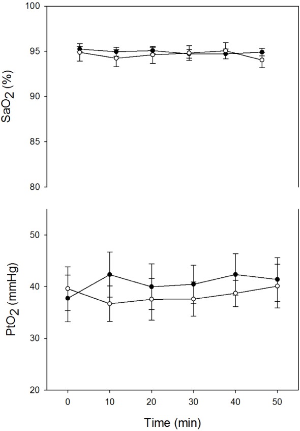
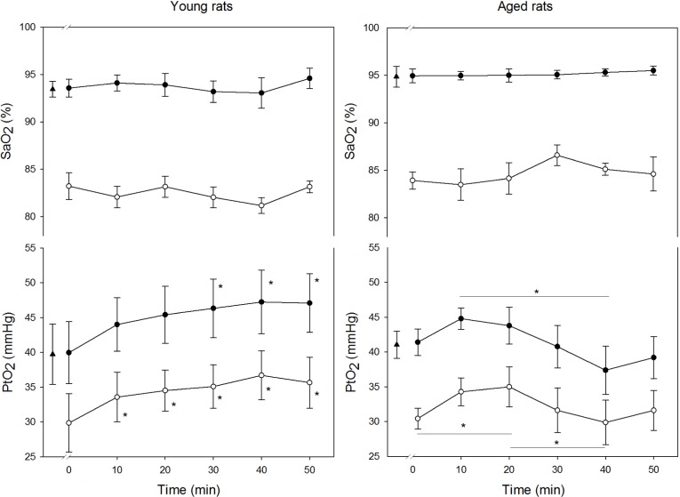
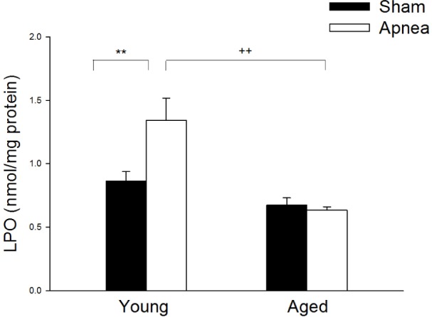
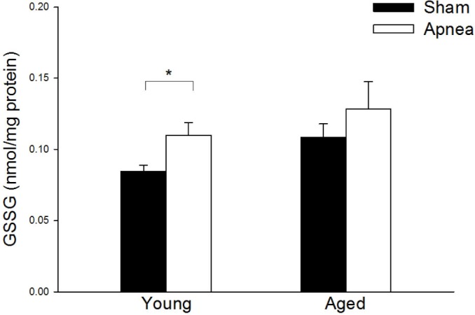
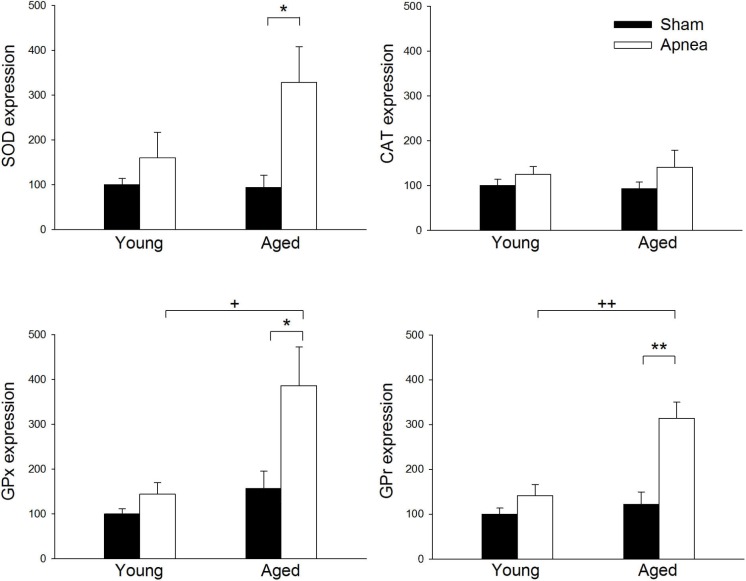
Measurements and results: Protocol 1: Real-time PtO2 measurements were performed using a fast-response oxygen microelectrode. During successive apneas cerebral cortex PtO2 presented a different pattern in the two age groups; there was a fast increase in young rats, whereas it remained without significant changes between the beginning and the end of the protocol in the aged group. Protocol 2: Brain oxidative stress assessed by lipid peroxidation increased after apneas in young rats (1.34 ± 0.17 nmol/mg of protein) compared to old ones (0.63 ± 0.03 nmol/mg), where a higher expression of antioxidant enzymes was observed.
Conclusions: The results suggest that brain oxidative stress in aged rats is lower than in young rats in response to recurrent apneas, mimicking obstructive sleep apnea. This could be due to the different PtO2 response observed between age groups and the increased antioxidant expression in aged rats.
Citation: Dalmases M, Torres M, Márquez-Kisinousky L, Almendros I, Planas AM, Embid C, Martínez-Garcia MA, Navajas D, Farré R, Montserrat JM. Brain tissue hypoxia and oxidative stress induced by obstructive apneas is different in young and aged rats.
Keywords: aging; animal model; obstructive apnea; oxidative stress; tissue oxygenation.
Figures
Comment in
- Sleep. 37:1161.
References
-
- Trzepizur W, Le Vaillant M, Meslier N, et al. Independent association between nocturnal intermittent hypoxemia and metabolic dyslipidemia. Chest. 2013;143:1584–9. - PubMed
-
- Bradley TD, Floras JS. Obstructive sleep apnoea and its cardiovascular consequences. Lancet. 2009;373:82–93. - PubMed
-
- Veasey S. Insights from animal models into the cognitive consequences of adult sleep-disordered breathing. ILAR J. 2009;50:307–11. - PubMed
Publication types
MeSH terms
Substances
LinkOut - more resources
Full Text Sources
Other Literature Sources
Medical
