

Acute pulmonary embolism caused by enlarged uterine leiomyoma: a rare presentation
- PMID: 25061497
- PMCID: PMC4108191
- DOI: 10.12659/AJCR.890607
Acute pulmonary embolism caused by enlarged uterine leiomyoma: a rare presentation
Abstract
Patient: Female, 42.
Final diagnosis: Acute pulmonary embolism.
Symptoms: Chest pain • dyspnea.
Medication: Streptokinase • Warfarin.
Clinical procedure: .-
Specialty: Cardiology and Neoplasm.
Objective: Management of emergency care.
Background: Deep venous thrombosis (DVT) and subsequent pulmonary embolism (PE) caused by pelvic vein compression are rare and life-threatening complications of leiomyoma of the uterus.
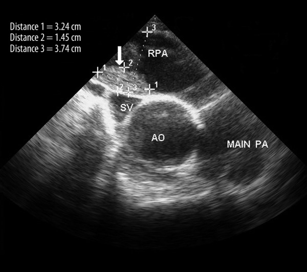
Case report: We report a 42-year-old virgin woman with a history of leiomyoma who presented to the emergency department with complaints of dyspnea and pleuritic chest pain with transient spotting. On physical examination, she had a non-tender abdomen with a 20-week size uterus. Imaging investigations revealed an acute DVT in her left leg and a huge uterine-derived mass compressing the common iliac veins. Transesophageal echocardiography (TEE) demonstrated an echogenic mass in her right pulmonary artery consistent with thrombosis. The patient was completely cured using thrombolytic therapy and myomectomy, and was well at 1 year after thrombolysis.
Conclusions: PE caused by pelvic vein compression is a rare complication of leiomyoma, which should be considered. Thrombolytic therapy associated with myomectomy can be implemented for treating such cases, and TEE can be used for diagnosing suspected high-risk PE.
Keywords: Echocardiography; Leiomyoma; Pulmonary Embolism; Thrombolytic Therapy.
Figures

Similar articles
-
Massive pulmonary embolism in a woman with leiomyomatous uterus causing pelvic deep venous thrombosis.Ann Ital Med Int. 2005 Apr-Jun;20(2):104-7. Ann Ital Med Int. 2005. PMID: 16052843
-
[A surgically treated case of acute pulmonary embolism owing to deep vein thrombosis of the leg mainly caused by uterine myoma].Kyobu Geka. 1992 Jul;45(7):631-4. Kyobu Geka. 1992. PMID: 1619829 Japanese.
-
Large uterine fibroid causing DVT and PE: Successful management with mechanical aspiration thrombectomy and hysterectomy: A case report and literature review.Medicine (Baltimore). 2024 Dec 6;103(49):e40862. doi: 10.1097/MD.0000000000040862. Medicine (Baltimore). 2024. PMID: 39654178 Free PMC article. Review.
-
Deep venous thrombosis and pulmonary thromboembolism associated with a huge uterine myoma--a case report.Angiology. 2000 Feb;51(2):161-6. doi: 10.1177/000331970005100210. Angiology. 2000. PMID: 10701725
-
Acute complications of fibroids.Best Pract Res Clin Obstet Gynaecol. 2009 Oct;23(5):609-17. doi: 10.1016/j.bpobgyn.2009.01.012. Epub 2009 Mar 4. Best Pract Res Clin Obstet Gynaecol. 2009. PMID: 19264555 Review.
Cited by
-
Vena cava balloon occlusion for pulmonary embolism prevention during resection of giant uterus fibroids.J Surg Case Rep. 2022 May 25;2022(5):rjac234. doi: 10.1093/jscr/rjac234. eCollection 2022 May. J Surg Case Rep. 2022. PMID: 35665394 Free PMC article.
-
Case series of seven women with uterine fibroids associated with venous thromboembolism and chronic thromboembolic disease.Pulm Circ. 2019 Jan-Mar;9(1):2045894018803873. doi: 10.1177/2045894018803873. Epub 2018 Sep 11. Pulm Circ. 2019. PMID: 30204062 Free PMC article.
-
Increased Risk of Venous Thromboembolism in Women with Uterine Leiomyoma: A Nationwide, Population-Based Case-Control Study.Acta Cardiol Sin. 2018 Jan;34(1):66-76. doi: 10.6515/ACS.201801_34(1).20170901B. Acta Cardiol Sin. 2018. PMID: 29375226 Free PMC article.
-
Uterine Fibroids and Their Association with Acute and Chronic Venous Thromboembolic Disease-An Expert Review of the Literature.J Clin Med. 2025 Jun 9;14(12):4065. doi: 10.3390/jcm14124065. J Clin Med. 2025. PMID: 40565811 Free PMC article. Review.
-
Cardiac Findings of Pulmonary Thromboembolism by Autopsy: A Review of 48 Cases.Med Sci Monit. 2016 Apr 27;22:1410-4. doi: 10.12659/msm.897695. Med Sci Monit. 2016. PMID: 27117720 Free PMC article.
References
-
- Konstantinides S. Clinical practice. Acute pulmonary embolism. N Engl J Med. 2008;359:2804–13. - PubMed
-
- Tapson VF. Acute pulmonary embolism. N Engl J Med. 2008;358:1037–52. - PubMed
-
- Gupta S, Manyonda IT. Acute complications of fibroids: Best Pract Res Clin Obstet Gynaecol. 2009;23:609–17. - PubMed
-
- Jaff MR, McMurtry MS, Archer SL, et al. Management of massive and sub-massive pulmonary embolism, iliofemoral deep vein thrombosis, and chronic thromboembolic pulmonary hypertension: a scientific statement from the American Heart Association. Circulation. 2011;123:1788–830. - PubMed
Publication types
MeSH terms
LinkOut - more resources
Full Text Sources
Other Literature Sources
Medical