. 2014 May 9;3(5):507-17.
doi: 10.1016/j.molmet.2014.04.009. eCollection 2014 Aug.

Inactivation of the cardiomyocyte glucagon-like peptide-1 receptor (GLP-1R) unmasks cardiomyocyte-independent GLP-1R-mediated cardioprotection

Affiliations

Inactivation of the cardiomyocyte glucagon-like peptide-1 receptor (GLP-1R) unmasks cardiomyocyte-independent GLP-1R-mediated cardioprotection

John R Ussher et al. Mol Metab. .

Abstract

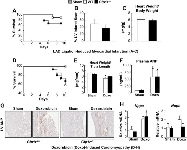
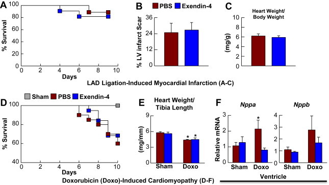
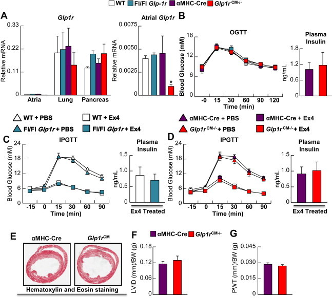
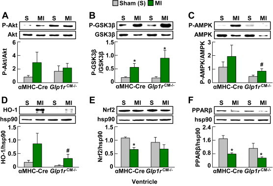
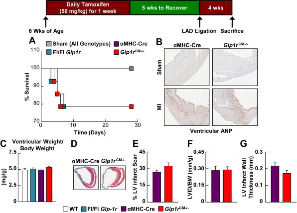
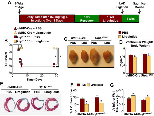
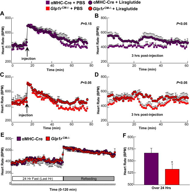
GLP-1R agonists improve outcomes in ischemic heart disease. Here we studied GLP-1R-dependent adaptive and cardioprotective responses to ventricular injury. Glp1r (-/-) hearts exhibited chamber-specific differences in gene expression, but normal mortality and left ventricular (LV) remodeling after myocardial infarction (MI) or experimental doxorubicin-induced cardiomyopathy. Selective disruption of the cardiomyocyte GLP-1R in Glp1r (CM-/-) mice produced no differences in survival or LV remodeling following LAD coronary artery occlusion. Unexpectedly, the GLP-1R agonist liraglutide still produced robust cardioprotection and increased survival in Glp1r (CM-/-) mice following LAD coronary artery occlusion. Although liraglutide increased heart rate (HR) in Glp1r (CM-/-) mice, basal HR was significantly lower in Glp1r (CM-/-) mice. Hence, endogenous cardiomyocyte GLP-1R activity is not required for adaptive responses to ischemic or cardiomyopathic injury, and is dispensable for GLP-1R agonist-induced cardioprotection or enhanced chronotropic activity. However the cardiomyocyte GLP-1R is essential for the control of HR in mice.

Keywords: Cardiomyopathy; GLP-1, glucagon-like peptide-1; GLP-1R, glucagon-like peptide-1 receptor; Glucagon-like peptide-1; Glucagon-like peptide-1 receptor; HR, heart rate; Heart failure; Incretin; Ischemia; LAD, left anterior descending; MI, myocardial infarction; Myocardial infarction; tGLP-1, truncated forms of GLP-1 such as GLP-1(9–36) or GLP-1(28–36).


Figures

Figure 1
Figure 1
Whole-body GLP-1R deficiency does not influence cardiovascular outcomes following LAD-ligation induced MI and Doxorubicin-induced cardiomyopathy. A: Survival in Glp1r−/− mice and their Glp1r+/+ littermates over 9 days following permanent LAD coronary artery occlusion (n = 9). B: % LV infarct scar formation from Masson's Trichrome fixed heart sections from Glp1r−/− and wild-type littermates at day 9 post-LAD ligation (n = 6). C: Cardiac hypertrophy at day 9 post-LAD ligation (n = 6–8). D: Survival in Glp1r−/− and Glp1r+/+ littermates following treatment with a single dose of doxorubicin (20 mg/kg i.p. injection) (n = 14–15). E: Heart weight/tibia length ratios at 10 days post-doxorubicin (n = 4–5). F: Plasma ANP levels 4 days post-doxorubicin treatment (Shams, n = 3, Doxorubicin, n = 5–7). G: Histology for ANP expression in LV from sham and doxorubicin-treated mice. H: Nppa and Nppb mRNA expression (normalized to Ppia) in ventricles (n = 3–5). Values represent mean ± SE. The significance of differences was determined by a two-way ANOVA followed by a Bonferroni post-hoc analysis. *significantly different from sham counterpart.
Figure 2
Figure 2
Systemic GLP-1R activation does not influence cardiovascular outcomes following LAD-ligation induced MI and Doxorubicin-induced cardiomyopathy. A: Survival in C57BL/6J mice treated with saline (PBS) or exendin-4 (5 nmol/kg BW i.p., twice daily) for 9 days following permanent LAD coronary artery occlusion (n = 9–11). B: % LV infarct scar formation in heart sections analyzed at day 9 post-LAD ligation (n = 6–7). C: Cardiac hypertrophy analyzed at day 9 post-LAD ligation (n = 8–9). D: Survival following treatment with a single dose of doxorubicin (20 mg/kg i.p. injection) (n = 19–20). Mice were injected for 7 days with saline (PBS) or exendin-4 (5 nmol/kg BW i.p. injection twice daily) with the first injection taking place concurrently with the doxorubicin treatment. E: Heart weight/tibia length ratios at 10 days post-doxorubicin (n = 4–6). F: Nppa and Nppb mRNA expression (normalized to Ppia) in ventricles from saline- and exendin-4-treated C57BL/6J mice at 10 days post-doxorubicin treatment (n = 4–5). Values represent mean ± SE. The significance of differences was determined by a two-way ANOVA followed by a Bonferroni post-hoc analysis. *significantly different from sham counterpart.
Figure 3
Figure 3
Characterization of cardiomyocyte-specific Glp1r knockout mice (Glp1rCM−/−). A: Glp1r mRNA (normalized to Ppia) in atria, lung and pancreas from Glp1rCM−/− mice 5 weeks following tamoxifen administration (n = 4). The right hand panel for atrial RNA depicts an expanded relative scale for assessment of levels of Glp1r mRNA transcripts. B–D: Glycemic control and plasma insulin levels in Glp1rCM−/− mice and control littermates during oral (B) glucose tolerance testing (OGTT, n = 6–8) and intraperitoneal (C–D; IPGTT, n = 5–7) glucose tolerance testing. During the IPGTT, mice were treated with exendin-4 (24 nmol/kg i.p.) (n = 5–7) or saline (PBS). E: Representative H&E heart cross sections depicting LV structure in αMHC-Cre littermate control vs. Glp1rCM−/− mice (n = 3), F: LV internal diameter (LVID) and G: LV posterior wall thickness (PWT). Values represent mean ± SE. Statistical significance was determined by a one-way ANOVA followed by a Bonferroni post-hoc analysis. *significantly different from all other groups.
Figure 4
Figure 4
Analysis of candidate cardioprotective proteins in hearts from Glp1rCM−/mice following MI. A: Akt phosphorylation, B: Glycogen synthase kinase 3β (GSK3β) phosphorylation, C: 5′ AMP activated protein kinase (AMPK) phosphorylation, D: Heme oxygenase-1 (HO-1) expression, E: Nuclear respiratory factor 2 (Nrf2) expression, and F: Peroxisome proliferator activated receptor β (PPARβ) expression in ventricles (viable myocardium) extracted from Glp1rCM−/− mice and their αMHC-Cre littermates 48 h post-LAD ligation (n = 3–4). Values represent mean ± SE. The significance of differences was determined by a two-way ANOVA followed by a Bonferroni post-hoc analysis. *significantly different from sham counterpart. #significantly different from corresponding αMHC-Cre littermates.
Figure 5
Figure 5
Cardiovascular outcomes following ischemic injury in Glp1rCM−/− mice. A: Survival in Glp1rCM−/− mice and control littermates following permanent LAD coronary artery occlusion (n = 14). B: Histology of LV cross sections for ANP expression in Glp1rCM−/− mice and their αMHC-Cre littermates at day 28 post-LAD coronary artery occlusion. C: Cardiac hypertrophy (ventricular weight/body weight) in Glp1rCM−/− mice and control littermates at day 28 post-LAD coronary artery occlusion (n = 4–9). D: Representative Masson's Trichrome stained heart sections depicting, E: % LV infarct scar formation, F: LV internal diameter (LVID), and G: LV infarct wall thickness in Glp1rCM−/− mice and their αMHC-Cre littermates at day 28 post-LAD coronary artery occlusion (n = 4–5). Values represent mean ± SE.
Figure 6
Figure 6
Liraglutide produces cardioprotection in Glp1rCM−/− mice. A: Experimental protocol for liraglutide treatment and LAD coronary artery ligation. B: Survival in PBS- and liraglutide-treated (30 μg/kg BW i.p. twice daily for 1 wk) Glp1rCM−/− mice and αMHC-Cre littermates following LAD coronary artery occlusion (n = 21–30). C–D: Cardiac hypertrophy in PBS and liraglutide-treated Glp1rCM−/− mice and control littermates at day 28 post-LAD ligation (n = 6–10). E: Representative Masson's Trichrome stained heart sections depicting, F: % LV infarct scar formation, and G: LV infarct wall thickness in PBS- and liraglutide-treated Glp1rCM−/− mice and αMHC-Cre littermates at day 28 post-LAD ligation (n = 4–6). Values represent mean ± SE. The significance of differences was determined by a Kaplan Meier survival analysis or a two-way ANOVA followed by a Bonferroni post-hoc analysis. *significantly different from corresponding PBS treated counterpart.
Figure 7
Figure 7
Heart rate in Glp1rCM−/− mice. A–B: HR in αMHC-Cre littermates following an acute liraglutide injection monitored for the first 60 min (A) or 3 h after injection (B) (30 μg/kg BW i.p., n = 3). C–D: HR in Glp1rCM−/− mice following an acute liraglutide injection for the first 60 min (C) or 3 h after injection (D) (30 μg/kg BW i.p., n = 4). Values represent mean ± SE. E: HR in Glp1rCM−/− mice and their αMHC-Cre littermates following a 24 h fast-refeeding protocol; data is presented for the final hr of the 24 h fast and the first hr immediately upon refeeding (n = 3–4). F: Baseline HR over 24 h in freely moving conscious Glp1rCM−/− mice and their αMHC-Cre littermates (n = 3–4). Statistical significance was determined by the use of an unpaired, two-tailed Student's t-test, or a repeated measures two-way ANOVA followed by a Bonferroni post-hoc analysis. *significantly different from αMHC-Cre littermate mice.

References

    1. Gerstein H.C., Miller M.E., Byington R.P., Goff D.C., Jr., Bigger J.T., Buse J.B. Effects of intensive glucose lowering in type 2 diabetes. New England Journal of Medicine. 2008;358(24):2545–2559. - PMC - PubMed
    1. Lindenfeld J., Masoudi F.A. Fluid retention with thiazolidinediones: does the mechanism influence the outcome? Journal of the American College of Cardiology. 2007;49(16):1705–1707. - PubMed
    1. Scirica B.M., Bhatt D.L., Braunwald E., Steg P.G., Davidson J., Hirshberg B. Saxagliptin and cardiovascular outcomes in patients with type 2 diabetes mellitus. New England Journal of Medicine. 2013;369(14):1317–1326. - PubMed
    1. Nikolaidis L.A., Mankad S., Sokos G.G., Miske G., Shah A., Elahi D. Effects of glucagon-like peptide-1 in patients with acute myocardial infarction and left ventricular dysfunction after successful reperfusion. Circulation. 2004;109(8):962–965. - PubMed
    1. Woo J.S., Kim W., Ha S.J., Kim J.B., Kim S.J., Kim W.S. Cardioprotective effects of exenatide in patients with ST-segment-elevation myocardial infarction undergoing primary percutaneous coronary intervention: results of exenatide myocardial protection in revascularization study. Arteriosclerosis, Thrombosis, and Vascular Biology. 2013;33(9):2252–2260. - PubMed

LinkOut - more resources