. 2014 Jul 25;9(7):e103383.
doi: 10.1371/journal.pone.0103383. eCollection 2014.

Silencer of death domains controls cell death through tumour necrosis factor-receptor 1 and caspase-10 in acute lymphoblastic leukemia

Affiliations

Silencer of death domains controls cell death through tumour necrosis factor-receptor 1 and caspase-10 in acute lymphoblastic leukemia

Adam Cisterne et al. PLoS One. .

Abstract

Resistance to apoptosis remains a significant problem in drug resistance and treatment failure in malignant disease. NO-aspirin is a novel drug that has efficacy against a number of solid tumours, and can inhibit Wnt signaling, and although we have shown Wnt signaling to be important for acute lymphoblastic leukemia (ALL) cell proliferation and survival inhibition of Wnt signaling does not appear to be involved in the induction of ALL cell death. Treatment of B lineage ALL cell lines and patient ALL cells with NO-aspirin induced rapid apoptotic cell death mediated via the extrinsic death pathway. Apoptosis was dependent on caspase-10 in association with the formation of the death-inducing signaling complex (DISC) incorporating pro-caspase-10 and tumor necrosis factor receptor 1 (TNF-R1). There was no measurable increase in TNF-R1 or TNF-α in response to NO-aspirin, suggesting that the process was ligand-independent. Consistent with this, expression of silencer of death domain (SODD) was reduced following NO-aspirin exposure and lentiviral mediated shRNA knockdown of SODD suppressed expansion of transduced cells confirming the importance of SODD for ALL cell survival. Considering that SODD and caspase-10 are frequently over-expressed in ALL, interfering with these proteins may provide a new strategy for the treatment of this and potentially other cancers.


Conflict of interest statement

Competing Interests: The corresponding author is an academic editor for PLOS ONE but this does not alter the authors' adherence to PLOS ONE editorial policies and criteria.

Figures

Figure 1
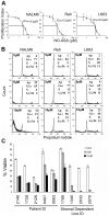
Figure 1. para-NO-ASA inhibit the proliferation and induces cell death in ALL cell lines and patient samples.
(A) The proliferation of the indicated ALL cell lines as determined by 3H-thymidine incorporation following 24 hours incubation with para-NO-ASA. Vertical bars represent standard deviation of quadruplicate determinations from one of 2 independent experiments. Curves were generated using the line of best fit in Excel software. IC50 values are indicated. (B) Cell cycle analysis of ALL cell lines treated with indicated concentrations of para-NO-ASA for 24 hours. Representative plots including the mean from duplicate assessments are shown. (C) The viability of pre-B ALL patient samples or patient derived stromal dependent cell lines was assessed after 24 hours of culture in the presence of DMSO (Cont), 5 or 10 µM para-NO-ASA by annexin V and 7AAD staining with dual negative cells being considered viable. Vertical bars represent standard deviation of quadruplicate determinations.
Figure 2
Figure 2. para-NO-ASA results in activation of executioner caspases and mitochondrial depolarization.
(A) Cleavage of caspase-3 in NALM6 cells by intracellular flow cytometry following 6 hour exposure to 10 µM para-NO-ASA. (B) Caspase-3 activation as measured by flow cytometry, in NALM6 cells untreated or treated with 5 µM or 10 µM para-NO-ASA for the given time. Bars indicate the mean and s.d. of 3 independent experiments. (C) Western blot analysis of PARP activation in NALM6 cells stimulated with the indicated concentrations of para-NO-ASA for 12 hours. The cleaved (89 kD) and uncleaved (116 kD) forms of PARP are indicated. Data is representative of 2 independent experiments. (D) NALM6 cells were treated with 5 µM para-NO-ASA or vehicle for 6 hours and assessed for ΔΨm using TMRM to label cells with polarized mitochondria, and apoptosis using annexin V and 7AAD. Representative plots are shown and the mean percentage of cells from 2 experiments in each quadrant indicated. (E) Representative histograms showing cytochrome c release following exposure of NALM6 cells to 5 µM para-NO-ASA for 6 hours. The thin line represents isotype control (Iso) staining and the heavy line cytochrome c (Cyto c) staining. (F) Quantitation of cytochrome c release at the indicated time points following addition of 5 µM para-NO-ASA. The mean and s.d. of replicates is shown. *p = 0.02. (G) NALM6 cells were pre-treated with 100 µM Z-VAD or 2.5 mM NAC for 1 hour prior to exposure to 5 µM para-NO-ASA for 6 h the mean ± s.e. is shown (n≥5). *p<0.05 compared to NO-ASA alone.
Figure 3
Figure 3. Multiple caspases are activated by para-NO-ASA.
(A) Western blot of cell lysates from cells treated with para-NO-ASA for the indicated times. Western Blot of caspases-8 (cleaved forms only), -9 (full length, p47 and cleaved forms), caspase-10 (full length only), Bid (full length and cleaved forms) and full length Noxa and Puma are shown. Note that the caspase-8 antibody (#9496) only recognised the cleaved forms and not the intact protein, while the caspase-10 antibody only recognised the full-length form of the protein. (B) NALM6 cells were treated with 5 µM NO-ASA for 6 h and cell lysates analysed for caspase-8 or caspase-9 activity. The mean±SD of 3 experiments is shown. *p<0.05 compared to control. (C) Representative histograms showing para-NO-ASA mediated activation of caspase 10 as determined by flow cytometry. Time of exposure to para-NO-ASA is indicated on the histograms. The mean and s.d. from one of two independent experiments is shown. (D) Gene expression was assessed using quantitative RT-PCR in NALM6 cells treated with 10 µM para-NO-ASA for 12 hours. *p<0.05.
Figure 4
Figure 4. Caspase-10 activation is required for para-NO-ASA induced cell death.
(A) The viability (top panel), as determined by 7AAD and annexin V staining, and mitochondrial depolarization (lower panel) of NALM6 cells treated with vehicle or 5 µM para-NO-ASA for the 30 min following pre-incubation with inhibitors of caspase-8, -9 or caspase-10. Mean and s.d. of 3 independent experiments is shown. *p<0.05. (B) Western blot showing the cleavage of caspase-8 and -9 following treatment with 10 µM para-NO-ASA with or without a 30 min pre-incubation with the caspase-10 inhibitor. (C) Activation of caspase-10 in patient samples exposed to 5 µM para-NO-ASA for 6 h as assessed by flow cytometry. The mean and s.d. of duplicate samples is shown. (D) Activation of caspase-10 in control treated NALM6 cells (left) or NALM6 cells treated with 10 µM para-NO-ASA (right) for 6 h. The mean and s.d. of duplicate assessments of caspase-10 activation is shown in the lower right panel.
Figure 5
Figure 5. para-NO-ASA induces formation of the death initiating signalling complex containing caspase-10 and suppression of SODD.
(A) NALM6 and LK63 cell lines were treated with 10 µM para-NO-ASA for 6 h and labelled for surface TNF-R1 (left panels). (B) NALM6 and REH cells were treated with para-NO-ASA for 6 h and assessed for TNF-α production by flow cytometry. PBMC treated with 50 ng/ml PMA and 1 µg/ml ionomycin are included as a positive control. The percentage of cells expressing TNF-α is shown on each plot. (C) NALM6 cells were treated with TNF-α (100 ng/ml), or para-NO-ASA (10 µM) as indicated and cell lysates prepared. TNF-R1 was immunoprecipitated and recovered complexes probed for caspase-10 or TNF-R1.
Figure 6
Figure 6. SODD is over-expressed in ALL cells and expression is required for ALL cell growth.
(A) SODD expression was determined by semi-quantitative RT-PCR in NALM6 and REH cells treated with 10 µM para-NO-ASA for 12 h. (B) SODD expression was determined by western blotting following treatment with 10 µM para-NO-ASA for the indicated times. (C) Western blot analysis of SODD in normal peripheral blood mononuclear cells (PBMC), indicated cell lines (upper blots) or patient samples (lower blots). Patient samples had been expanded in NOD/SCID mice to obtain sufficient cells for Western blotting. (D) NALM6 transduced with lentiviral constructs expressing GFP alone (Control) or containing one of two shRNA specific for SODD (SODD1 and SODD2). The level of SODD protein (D) was determined in LK63 cells after 44 days in culture while mRNA levels were assessed in NALM6 cells on day 6 of culture by qRT-PCR and was normalised to the levels of GAPDH (E). The mean and s.e.m. of three independent experiments is shown. (F) The total cell number (left panels), and the percentage (centre panels) and total number of GFP+ transduced cells (right panels) was monitored over time. The mean and s.d. of duplicate cultures is shown.
Figure 7
Figure 7. Schematic diagram illustrating the proposed mechanism of action of para-NO-ASA.
para-NO-ASA down regulates SODD allowing self-aggregation, or enhancing TNF-α-induced, activation of signalling through TNF-R1. TNF-R1 signalling triggers the extrinsic apoptosis cascade including cleavage of pro-caspase-10 and Bid, mitochondrial depolarization, outer membrane permeabilization and release of cytochrome c. This is followed by cleavage of pro-caspase-9 and the executioner caspases-3 and -7. While signalling through TNF-R1 can activate NF-κB this may be suppressed by caspase-mediated cleavage of TRAF. In contrast, TNF-R2 signal induces cell survival and proliferation pathways, predominantly through NF-κB but also MAPK signalling. In ALL cells where SODD is over expressed, exogenous TNF-α is likely to predominantly signal through TNF-R2 resulting in increased survival and proliferation. Activating TNF-R1 by down regulation of SODD provides a mechanism for inducing cell death without increasing proliferation and survival signals. FADD, Fas-associated death domain; IKK, inhibitor of κB kinase; I-κB, inhibitor of κB; TRADD, TNF-R1-associated death domain; TRAF, TNF receptor-associated factor.

Similar articles

Cited by

References

    1. Pui CH (2010) Recent research advances in childhood acute lymphoblastic leukemia. J Formos Med Assoc 109: 777–787. - PubMed
    1. Gokbuget N, Hoelzer D (2009) Treatment of adult acute lymphoblastic leukemia. Semin Hematol 46: 64–75. - PubMed
    1. Movassagh M, Foo R (2008) Simplified apoptotic cascades. Heart Fail Rev 13: 111–119. - PubMed
    1. Goldstein J, Waterhouse N, Juin P, Evan G, Green D (2000) The coordinate release of cytochrome c during apoptosis is rapid, complete and kinetically invariant. Nat Cell Biol 2(3):156–62 2(3): 156–162. - PubMed
    1. Li P, Nijhawan D, Budihardjo I, Srinivasula SM, Ahmad M, et al. (1997) Cytochrome c and dATP-dependent formation of Apaf-1/caspase-9 complex initiates an apoptotic protease cascade. Cell 91: 479–489. - PubMed

Publication types

MeSH terms

Substances

LinkOut - more resources