. 2014 Jul 25;9(7):e103272.
doi: 10.1371/journal.pone.0103272. eCollection 2014.

Characterization of the small exported Plasmodium falciparum membrane protein SEMP1

Affiliations

Characterization of the small exported Plasmodium falciparum membrane protein SEMP1

Olivier Dietz et al. PLoS One. .

Abstract

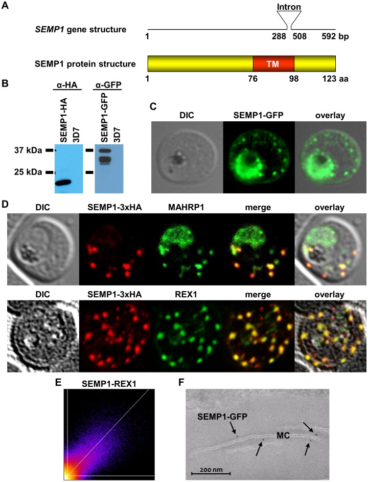
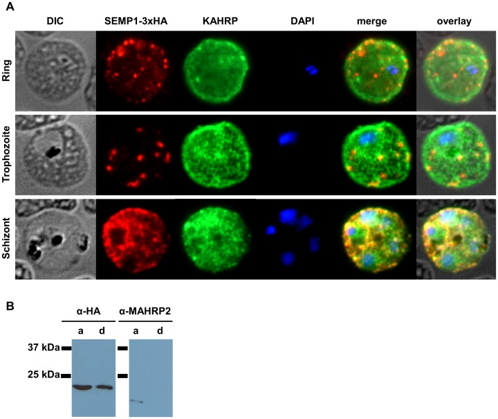
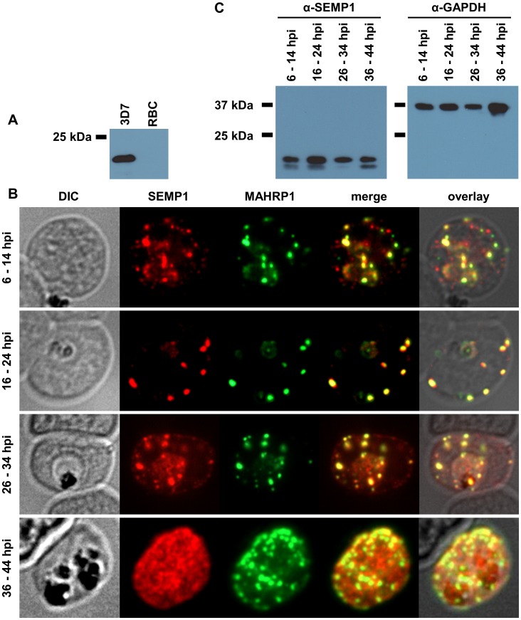
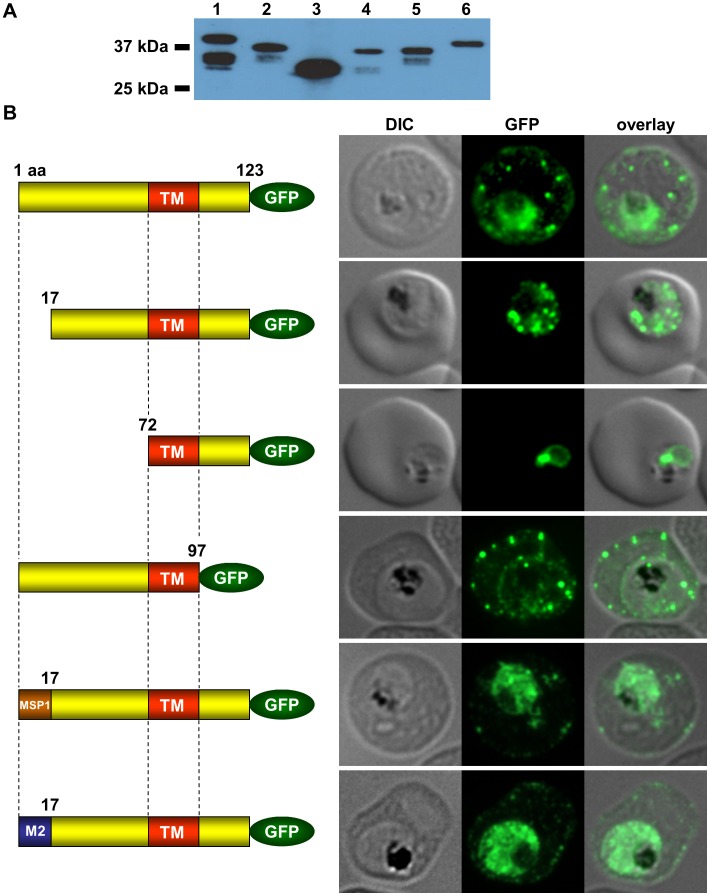
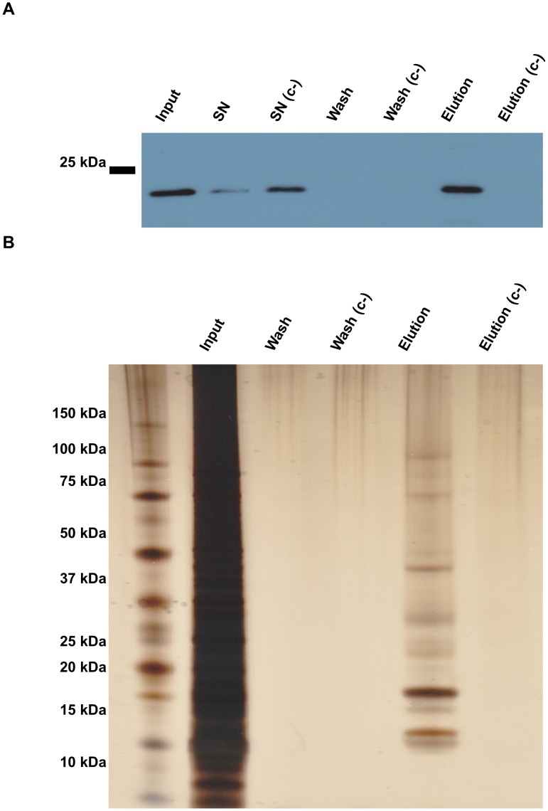
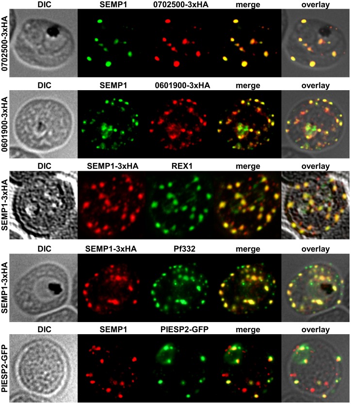
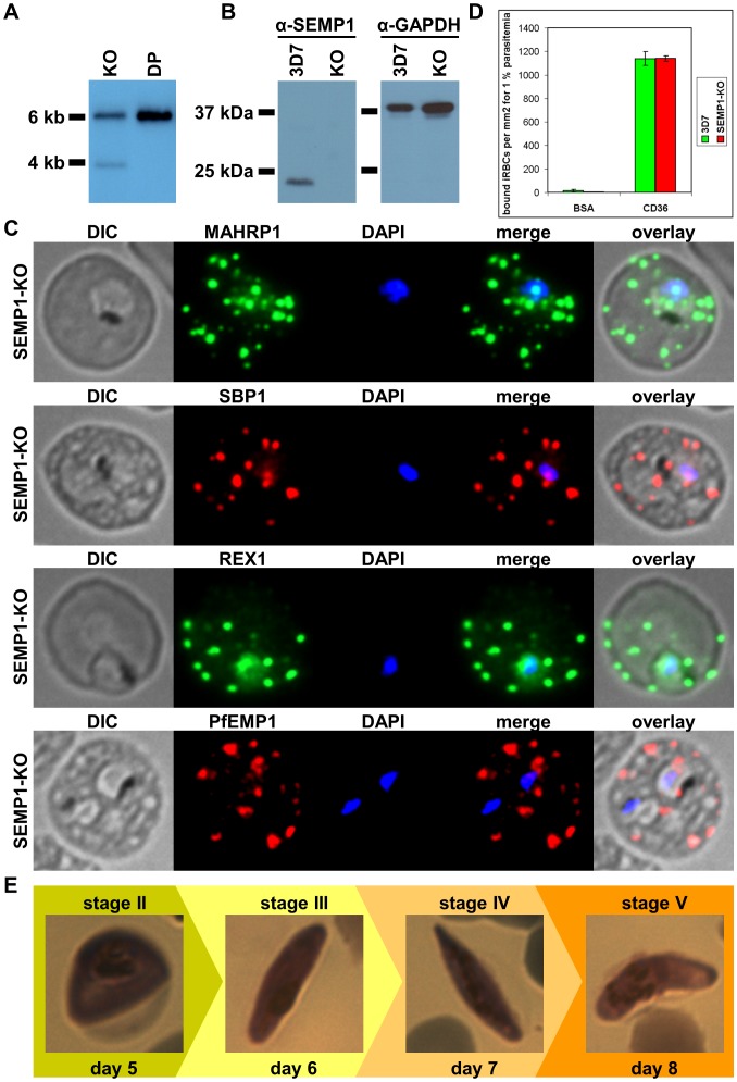
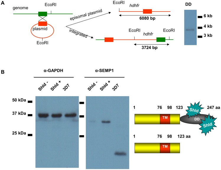
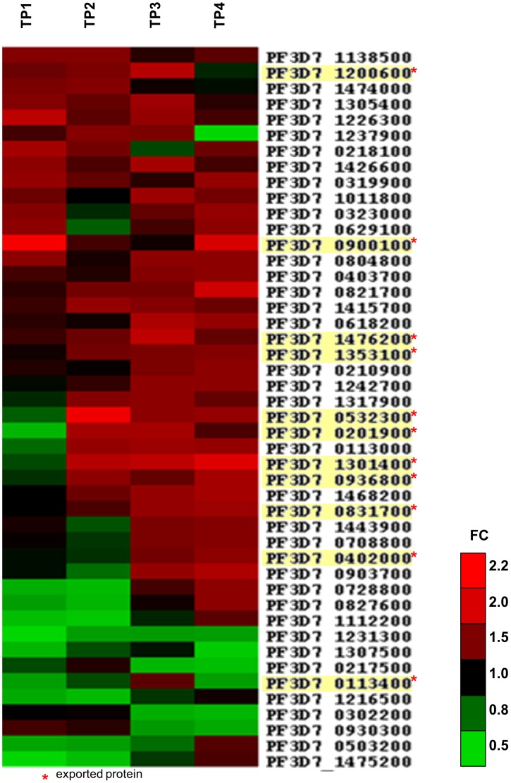
Survival and virulence of the human malaria parasite Plasmodium falciparum during the blood stage of infection critically depend on extensive host cell refurbishments mediated through export of numerous parasite proteins into the host cell. The parasite-derived membranous structures called Maurer's clefts (MC) play an important role in protein trafficking from the parasite to the red blood cell membrane. However, their specific function has yet to be determined. We identified and characterized a new MC membrane protein, termed small exported membrane protein 1 (SEMP1). Upon invasion it is exported into the RBC cytosol where it inserts into the MCs before it is partly translocated to the RBC membrane. Using conventional and conditional loss-of-function approaches we showed that SEMP1 is not essential for parasite survival, gametocytogenesis, or PfEMP1 export under culture conditions. Co-IP experiments identified several potential interaction partners, including REX1 and other membrane-associated proteins that were confirmed to co-localize with SEMP1 at MCs. Transcriptome analysis further showed that expression of a number of exported parasite proteins was up-regulated in SEMP1-depleted parasites. By using Co-IP and transcriptome analysis for functional characterization of an exported parasite protein we provide a new starting point for further detailed dissection and characterisation of MC-associated protein complexes.


Conflict of interest statement

Competing Interests: The authors have the following interest. Annette Gaida is employed by CSL Behring. There are no patents, products in development or marketed products to declare. This does not alter our adherence to all the PLOS ONE policies on sharing data and materials, as detailed online in the guide for authors.

Figures

Figure 1
Figure 1. SEMP1 expression and localization.
A: schematic representation of the semp1 gene (top) and SEMP1 protein structure (bottom). TM, transmembrane protein. B: Live cell imaging of 3D7 parasites expressing SEMP1 with C-terminal tagged GFP. C: Immunofluorescence assays of MeOH-fixed RBCs infected with 3D7 expressing SEMP1 with a C-terminal 3xHA tag, co-labelled with rat α-HA and either rabbit α-MAHRP1 or rabbit α-REX1 antibodies. D: Scatter plot of co-localization of SEMP1 and REX1 in SEMP1-3xHA parasites. E: Electron microscopy (EM) of RBCs infected with 3D7 expressing SEMP1 with a C-terminal GFP tag labelled with rabbit α-GFP antibodies and decorated with 5 nm gold conjugated Protein A.
Figure 2
Figure 2. SEMP1 expression during blood stage development.
A: Immunofluorescence assays of MeOH-fixed RBCs infected with 3D7 expressing SEMP1 with a C-terminal 3xHA tag, co-labelled with rat α-HA and mouse α-KAHRP antibodies. B: Parasite protein pellet of 3D7 expressing SEMP1-3xHA was fractionated using TritonX-114 into a soluble (aqueous phase) (a) and an insoluble (membrane) fraction (d). Proteins were visualized using mouse α-HA and rabbit α-MAHRP2 antibodies.
Figure 3
Figure 3. Localization and expression of endogenous SEMP1 in 3D7 wild-type parasites.
A: 3D7 wild-type parasite lysate (3D7) and uninfected red blood cells (RBC) were analysed by immunoblotting and probed with mouse serum raised against recombinant full-length SEMP1. B: IFAs of MeOH-fixed RBCs infected with 3D7 wild-type parasites synchronized at timepoints 6–14 hours post invasion (hpi), 16–24 hpi, 26–34 hpi and 36–44 hpi. Cells were co-labelled with mouse α-SEMP1 and rabbit α-MAHRP1 serum. C: 3D7 parasite pellets were generated at the respective timepoints by saponin lysis and analysed by immunoblot using mouse α-SEMP1 serum and mouse α-GAPDH (PF3D7_1462800) antibodies.
Figure 4
Figure 4. Requirements for SEMP1 export into the RBC.
A: Lysates of SEMP1-KO parasites expressing full-length and truncated or mutated forms of SEMP1 C-terminally fused to GFP were generated by saponin lysis and analysed by immunoblotting using mouse α-GFP antibodies. Lane 1: SEMP11–123-GFP (full-length), lane 2: SEMP117–123-GFP, lane3: SEMP172–123GFP, lane4: SEMP11–97-GFP, lane 5: MSP11–16SEMP117–123-GFP, lane 6: MAHRP21–16SEMP117–123-GFP. B: Immunofluorescence assays of MeOH-fixed RBCs infected with SEMP1-KO parasites expressing full-length and truncated or mutated forms of SEMP1 C-terminally fused to GFP. Expressed SEMP1 was labelled with rabbit α-GFP antibodies. The transmembrane domain is depicted in red (TM), a MSP1 signal peptide in brown (MSP1) and the MAHRP2 N-terminus in blue (M2).
Figure 5
Figure 5. Identification of potential SEMP1 interaction partners by Co-IP.
Co-IP was performed with 3D7 parasites expressing SEMP1 with a C-terminal 3xHA tag. Cultures were cross-linked with 1% formaldehyde and parasites were released by saponin treatment, lysed in 1%SDS followed by sonication. Lysate (Input) was incubated with α-HA affinity matrix. After centrifugation the matrix was washed three times with washing buffer (Wash) and proteins were eluted from by HA peptide elution. As a negative control, an excess of soluble HA peptides was added to the input to block the HA binding sites of the matrix (c-). Samples were analysed by Western blot with α-HA antibodies (A) and by silver staining (B). Proteins co-eluted with SEMP1-3xHA were identified by MS analysis of both TCA precipitated total elution (precipitation) and Coomassie-stained gel slices (gel extraction).
Figure 6
Figure 6. Localization of potential SEMP1 interacting proteins.
Immunofluorescence assays of MeOH-fixed RBCs infected with 3D7 expressing SEMP1/PF3D7_0702500/PF3D7_0601900 with a C-terminal 3xHA tag and 3D7 expressing PIESP2 with a C-terminal GFP-tag, co-labelled with mouse α-SEMP1 and rat α-HA (PF3D7_0702500-3xHA & PF3D7_0601900-3xHA) /α-GFP (PIESP2-GFP). For co-labelling of SEMP1-3xHA with REX1 and Pf332, rat α-HA and rabbit α-REX1 / mouse α-Pf332 antibodies were used.
Figure 7
Figure 7. Knockout of SEMP1 by gene disruption has no detectable phenotype.
A: ClaI- and XmaI-digested gDNA isolated from a SEMP1-KO clone (KO) pH-KO plasmid (DP) were analysed by Southern blot and probed with radioactively labelled hdhfr. In case of an integration of the KO plasmid into the parasite genome the expected fragment size is 3927 bp. B: lysates of 3D7 wild type and SEMP1-KO (KO) parasites were generated by saponin lysis and analysed by Western blot with α-SEMP1 and α-GAPDH (loading control) antibodies. C: Immunofluorescence assays of fixed RBCs infected with SEMP1-KO parasites, probed with α-MAHRP1, α-SBP1, α-REX1 and α-PfEMP1 antibodies. The parasite nuclei were stained with DAPI. D: Bar graph depicting differential binding to CD36 of RBCs infected with wild-type 3D7 and SEMP1-KO parasites. CD36 (or BSA as control) was immobilized on Petri dishes and bound infected RBCs (iRBCs) were calculated as iRBCs per mm2 for 1% parasitemia.
Figure 8
Figure 8. Conditional Knockdown of SEMP1.
A: EcoRI-digested gDNA isolated from SEMP1-DD parasites (DD) was analysed by Southern blot and probed with radioactively labelled hdhfr B: A culture of 3D7 parasites expressing SEMP1-DD was split and cultured for 2 weeks in presence (Shld +) or absence (Shld -) of the small molecule Shield-1. Whole parasite-lysates were generated after saponin lysis and analysed by Western blot using α-SEMP1. As a loading control the blot was additionally probed with antibodies against the housekeeping protein GAPDH.
Figure 9
Figure 9. Transcriptional changes in absence of SEMP1 identified by microarray analysis.
Summary of all significantly (p<0.05) up- and down-regulated parasite genes with a respective average fold change (RAFC) >1.5 or <0.6. The RAFC is thereby the average FC over the significantly up- / down-regulated time points (TPs) only. A graphic depiction of their FCs throughout the four time points (TPs) is shown in form of a heat map. Up-regulation (FC>1) is indicated in red, down-regulation (FC<1) in green. The time points were 6–14 hpi (TP1), 16–24 hpi (TP2), 26–34 hpi (TP3) and 36–44 hpi (TP4). Exported proteins are highlighted in yellow.

References

    1. World Health Organization (2012) WHO, World Health Report 2012.
    1. Miller LH, Baruch DI, Marsh K, Doumbo OK (2002) The pathogenic basis of malaria. Nature 415: 673–679. - PubMed
    1. Przyborski JM, Wickert H, Krohne G, Lanzer M (2003) Maurer's clefts—a novel secretory organelle? Mol. Biochem. Parasitol. 132: 17–26. - PubMed
    1. Bhattacharjee S, van Ooij C, Balu B, Adams JH, Haldar K (2008) Maurer's clefts of Plasmodium falciparum are secretory organelles that concentrate virulence protein reporters for delivery to the host erythrocyte. Blood 111: 2418–2426. - PMC - PubMed
    1. Hiller NL, Bhattacharjee S, van Ooij C, Liolios K, Harrison T, et al. (2004) A host-targeting signal in virulence proteins reveals a secretome in malarial infection. Science 306: 1934–1937. - PubMed

Publication types

Substances