. 2014 Jul 25;9(7):e103101.
doi: 10.1371/journal.pone.0103101. eCollection 2014.

Association between the intrinsically disordered protein PEX19 and PEX3

Affiliations

Association between the intrinsically disordered protein PEX19 and PEX3

Katarina Hattula et al. PLoS One. .

Abstract

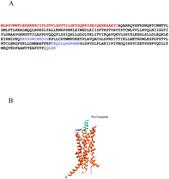
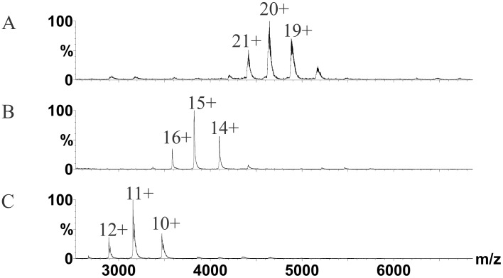
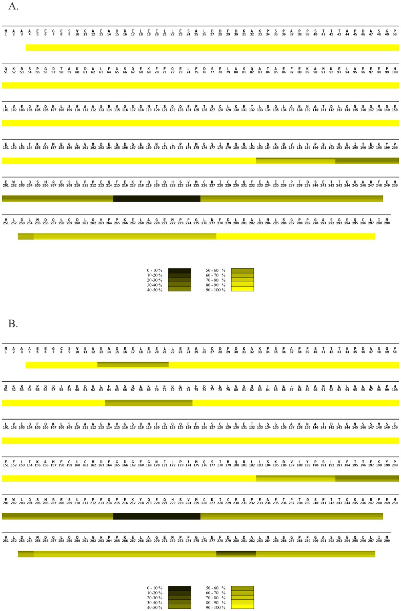
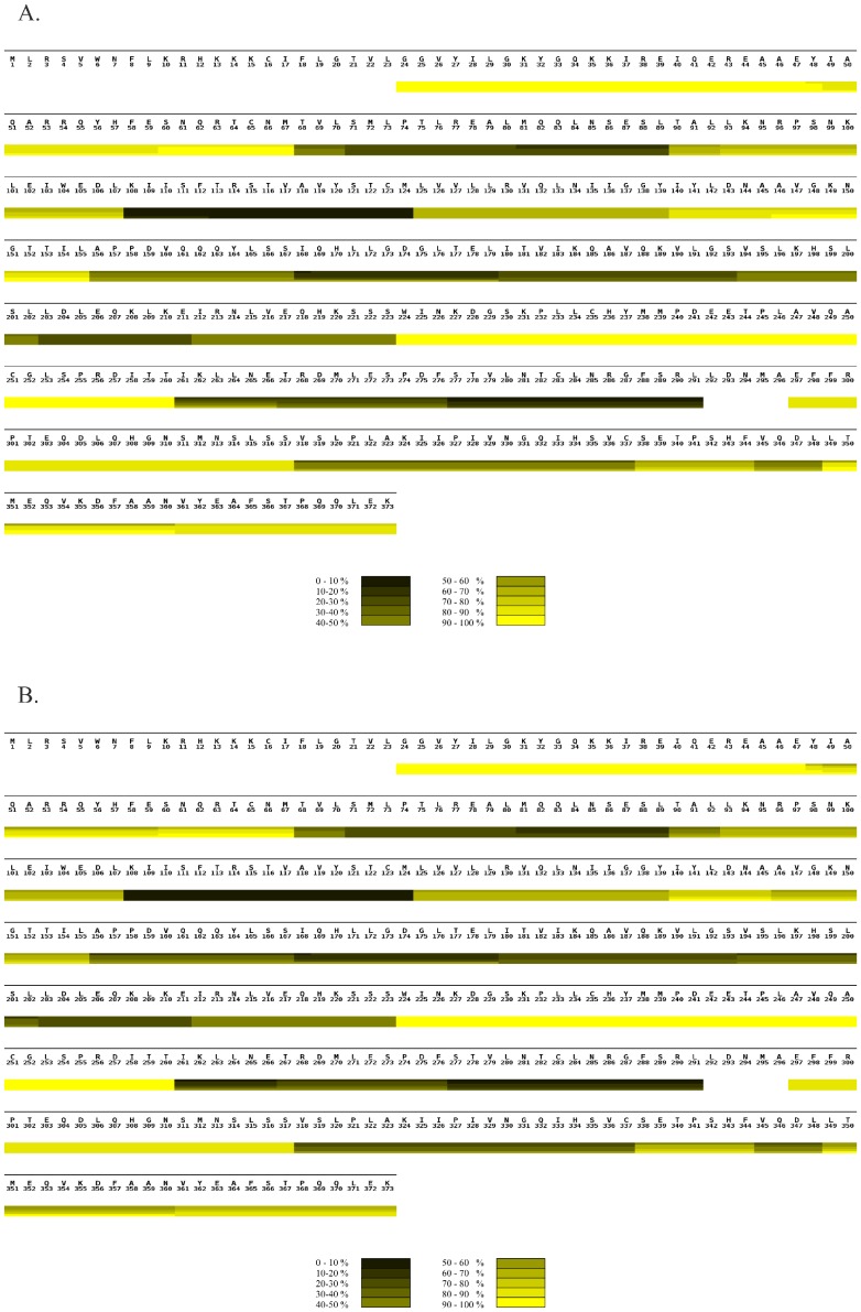
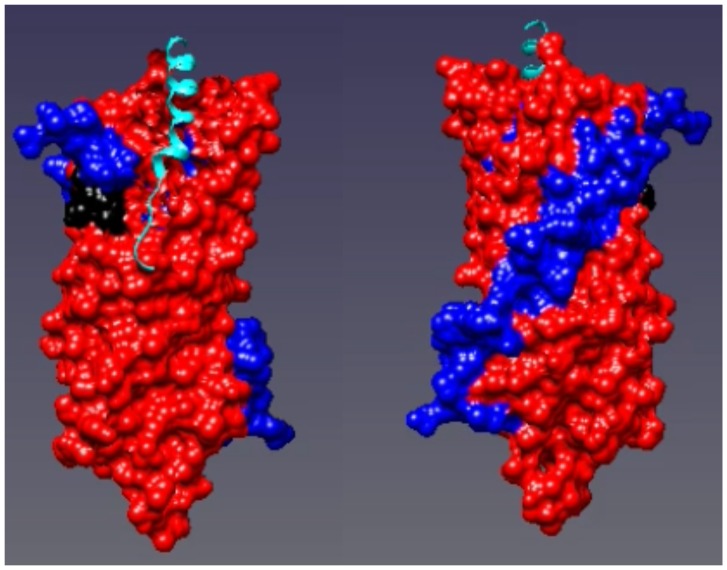
In peroxisomes, peroxins (PEXs) 3 and 19 are the principal protein components of the machinery required for early peroxisomal biogenesis. For further insight into the interaction of PEX3 and PEX19, we used hydrogen exchange mass spectrometry to monitor conformational changes during complex formation between PEX3 and PEX19 in vitro. Our data showed that PEX19 remained highly flexible during interaction with PEX3. However, we could detect three changes, one each in the N-and C-terminus along with a small stretch in the middle of PEX19 (F64-L74) which became shielded from hydrogen exchange when interacting with PEX3. PEX3 became more protected from hydrogen exchange in the binding groove for PEX19 with only small changes elsewhere. Most likely the N-terminus of PEX19 initiates the binding to PEX3, and then subtle conformational changes in PEX3 affect the surface of the PEX3 molecule. PEX19 in turn, is stabilized by folding of a short helix and its C-terminal folding core permitting PEX19 to bind to PEX3 with higher affinity than just the N-terminal interaction allows. Thus within the cell, PEX3 is stabilized by PEX19 preventing PEX3 aggregation.


Conflict of interest statement

Competing Interests: The authors have declared that no competing interests exist.

Figures

Figure 1
Figure 1. The full length human PEX3 protein sequence.
A) The red letters indicate amino acids which are not expressed in the recombinant protein. The black letters indicate amino acids modelled in the X-ray structure (PDB ID: 3AJB) . The blue letters indicate amino acids which are not modelled in the X-ray structure. B) X-ray structure of PEX3 with a PEX19 peptide bound (PDB ID: 3AJB). The secondary structure of PEX3 is colored as a) helix (orange), b) sheet (dark blue) and c) coil (pink). The PEX19 peptide is shown in turquoise.
Figure 2
Figure 2. Complex formation of PEX3 with PEX19 monitored by nano-electrospray MS.
The spectra were recorded under conditions that preserve non-covalent interactions showing A, MS on PEX3T-PEX19 heterodimer, B, PEX3 monomer and C, PEX19 monomer. Samples were analyzed at 10 µM concentrations for the complex and five-fold higher for the monomers. No homodimers were detected in the spectra. The numbers of the respective peaks represent the charge states. Relative intensities (%) are plotted against mass-over-charge (m/z).
Figure 3
Figure 3. Comparison of hydrogen exchange in PEX19 alone and in complex with PEX3 monitored by MS.
(A) HXMS heat map of PEX19 monomer summarizing the deuterium uptake over time. (B). HXMS heat map of PEX19 in the heterodimer with PEX3 summarizing the deuterium uptake over time. (A and B). The sequence of the protein is shown above the heat map. Although the recombinant protein includes an N-terminal tag only the full sequence of human PEX19 is shown with the numbering starting at the initial PEX19 methionine. The heat maps were assembled from individual peptic peptides using MSTools . The extent of the peptide is demarcated by vertical lines in each block, running through all four time point bars, when there is a difference in uptake from the preceding or following peptide. All peptic peptides are shown in Figure S1. The scale bar at the bottom of each heat map illustrates the color coding for deuterium uptake as a percentage. The horizontal bars from the top to the bottom in each block of the heat map indicate incubation times of 0, 3, 7, 40 and 60 minutes, respectively. White bars represent the residues for which no data were available.
Figure 4
Figure 4. Comparison of hydrogen exchange in PEX3 alone and in complex with PEX19 monitored by MS.
A) HXMS heat map of PEX3 monomer summarizing the deuterium uptake over time. B) HXMS heat map of PEX3 in the heterodimer with PEX19 summarizing the deuterium uptake over time. A) and B) The sequence of the PEX3 protein is shown above the heat map. The heat maps were assembled from individual peptic peptides using MSTools . The extent of the peptide is demarcated by vertical lines in each block, running through all four time point bars, when there is a difference in uptake from the preceding or following peptide. All peptic peptides are shown in Figure S2. The scale bar at the bottom of each heat map illustrates the color coding for deuterium uptake as a percentage. The horizontal bars from the top to the bottom in each block of the heat map indicate incubation times of 0, 3, 7, 40 and 60 minutes, respectively. White bars represent the residues for which no data were available.
Figure 5
Figure 5. The heat map of PEX3 plotted on the X-ray structure of PEX3 with a PEX19 peptide bound (PDB ID: 3AJB) .
Regions where the hydrogen/deuterium exchange decreased in the heterodimer compared to PEX3 alone are shown in red. Regions where the exchange remained the same are shown in blue. The left hand panel is rotated 180° along the Y axis compared to the right hand panel.

References

    1. DeDuve CABP (1966) Peroxisomes (microbodies and related particles). Physiol Rev 46: 323–357. - PubMed
    1. Distel B, Erdmann R, Gould SJ, Blobel G, Crane DI, et al. (1996) A unified nomenclature for peroxisome biogenesis factors. J Cell Biol 135: 1–3. - PMC - PubMed
    1. Sacksteder KA, Jones JM, South ST, Li X, Liu Y, et al. (2000) PEX19 binds multiple peroxisomal membrane proteins, is predominantly cytoplasmic, and is required for peroxisome membrane synthesis. J Cell Biol 148: 931–944. - PMC - PubMed
    1. Fagarasanu A, Fagarasanu M, Rachubinski RA (2007) Maintaining peroxisome populations: a story of division and inheritance. Annu Rev Cell Dev Biol 23: 321–344. - PubMed
    1. Geraghty MT, Bassett D, Morrell JC, Gatto GJ Jr, Bai J, et al. (1999) Detecting patterns of protein distribution and gene expression in silico. Proc Natl Acad Sci U S A 96: 2937–2942. - PMC - PubMed

Publication types