. 2014 Oct 1;549(1):134-40.
doi: 10.1016/j.gene.2014.07.056. Epub 2014 Jul 23.

A role for IL-10 in the transcriptional regulation of Roquin-1

Affiliations

A role for IL-10 in the transcriptional regulation of Roquin-1

Jeremy S Schaefer et al. Gene. .

Abstract

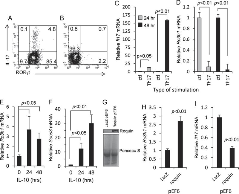
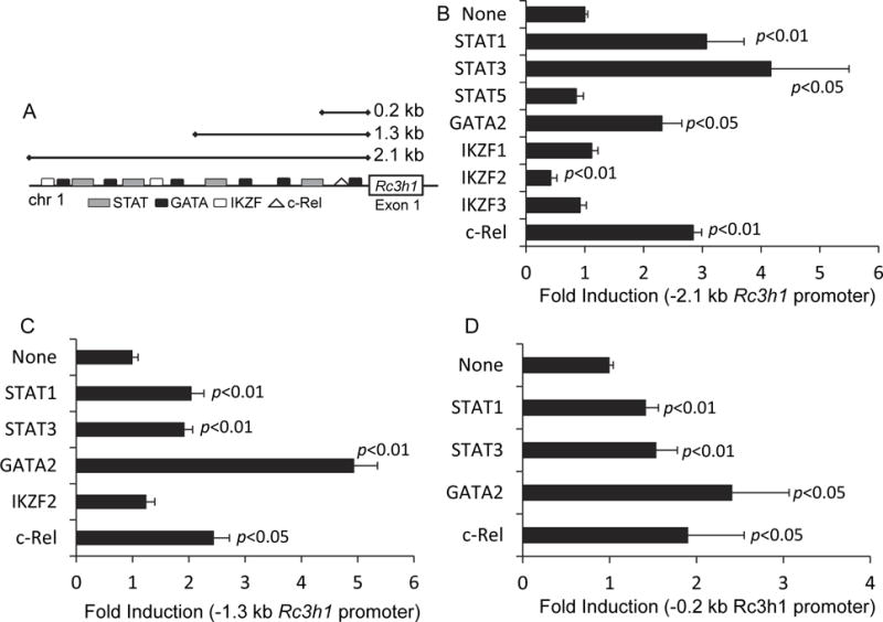
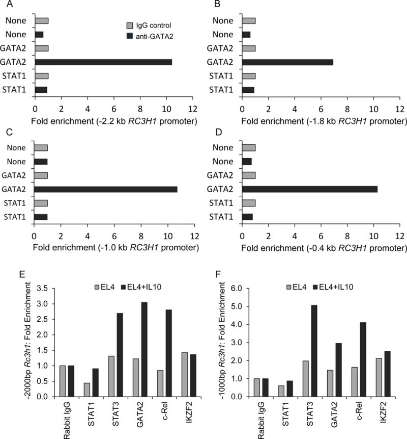
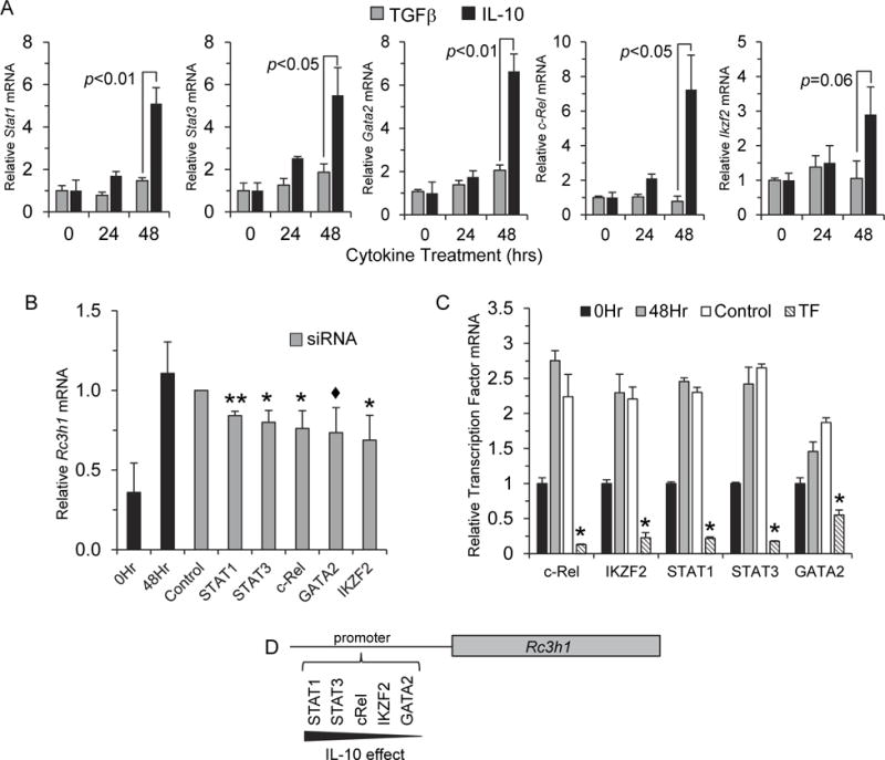
Roquin-1, a RING finger E3 ubiquitin ligase, functions as a modulator of inflammation; however, nothing is known about how Rc3h1 expression is regulated. Here, we describe an opposing relationship between Roquin-1 and the IL-17 proinflammatory cytokine by demonstrating that enforced expression of Rc3h1 restricts Il17a expression, and that exposure of T cells to IL-10, a cytokine with immunosuppressive activity, increases Rc3h1 expression. Luciferase reporter assays conducted using eight transcription factor plasmids (STAT1, STAT3, STAT5, GATA2, c-Rel, IKZF1, IKZF2, and IKZF3) demonstrated that STAT1, STAT3, GATA2, and c-Rel increased Rc3h1 promoter activity, whereas IKZF2 decreased activity. Gene expression of those five transcription factors increased in T cells exposed to IL-10. Transcription factor-specific siRNAs suppressed the IL-10 effect on Rc3h1 transcription. These findings identify a role for IL-10 in regulating Rc3h1 transcription, and they have implications for understanding how Roquin-1 controls the immune response.

Keywords: Cytokine; Gene expression; IL-10; Luciferase; Promoter; Transcription factor; siRNA.


Conflict of interest statement

Conflict of interest

The authors declare no conflict of interest.

Figures

Fig. 1
Fig. 1
Opposing relationship between Roquin-1 and IL-17. (A) MLN T cells 48 hr after stimulation using the Th17 induction regimen described in the Materials and Methods. (B) MLN T cells 48 hr after CD3 stimulation in the absence of Th17 induction. Cells were gated onto the CD4+ cell population. Representative histograms from three experiments. (C, D) Increased Il17a and decreased Rc3h1 transcription in MLN T cells following 24 and 48 hr of Th17 induction. Control cultures consisted of plate-bound anti-hamster antibody with normal hamster IgG. (E) Increased Rc3h1 transcription in EL4 cells following exposure to IL-10. (F) Exposure of EL4 cells to IL-10 resulted in increased Socs3 transcription. (G) Western blot showing increased levels of Roquin-1 protein expression in EL4 cells transfected with the Roquin-1 pEF6 expression plasmid. Ponceau S staining was done to compare sample protein levels. (H, I) Roquin-1 pEF6 transfection of EL4 cells resulted in increased Rc3h1 transcription and decreased Il17a transcription. Data are mean values ± SEM of at least three experiments.
Fig. 2
Fig. 2
Transcriptional regulation of Rc3h1. (A) A web-based search for transcriptional factors that bind to the Rc3h1 promoter identified binding sites for STATs, GATA, IKZF and c-Rel. Luciferase reporter assays were used to evaluate the activational effects of eight transcription factors on the Rc3h1 promoter regions. Using a (B) -2.1 kb Rc3h1 promoter region, STAT1, STAT3, GATA2, and c-Rel resulted in a significant increase in luciferase activity; IKZF2 resulted in a significant decrease. STAT1, STAT3, GATA2, and c-Rel also resulted in increased luciferase activity using the (C) -1.3 kb and (D) -0.2 kb Rc3h1 promoter regions. Values are fold induction of luciferase activity of transcription factor-transfected cells relative to non-transcription factor transfected cells. Data are mean values ± SEM of 3-4 independent experiments.
Fig. 3
Fig. 3
ChIP assays showing GATA2 interactions with: (A) -2.2 kb, (B) -1.8 kb, (C) -1.0 kb, and (D) -0.4 kb RC3H1 promoter regions. Data in each panel represent one ChIP experiment; the average values of all four data entries for the GATA2 ChIP assays (anti-GATA2 antibody) was significantly greater (p<0.001) than that of ChIP assay results done with None (mock) transfections, or STAT1 transcription factor transfections. (E, F) ChIP assays showing the effect of IL-10 on transcription factor interactions with the Rc3h1 promoter.
Fig. 4
Fig. 4
IL-10 increased Rc3h1 transcription factor expression. (A) EL4 cells were cultured in the presence of mouse rIL-10, or rTGFβ as a control cytokine. Gene expression was measured at 0, 24, and 48 hr. Compared to the rTGFβ control cytokine, exposure of cells to IL-10 resulted in a significant increase in gene expression of STAT1, STAT3, GATA2, and c-Rel, as well as a non-statistical increase in IKZF2 48 hr post-IL-10 treatment. Data are mean values ± SEM of 3 experiments. (B) EL4 cells were transfected with siRNAs to either STAT1, STAT3, GATA2, c-Rel, IKZF2, a scramble control siRNA, or no siRNA for 4 hr followed by exposure to rIL-10 as described in the Materials and Methods. Cells were harvested and Rc3h1 expression was measured by qRT-PCR. All transcription factor-specific siRNAs decreased Rc3h1 expression relative to the control siRNA-transfected cells. For STAT1 and STAT3, this occurred in a statistically-significant manner. (C) Efficiency of knockdown of transcription factor expression. Data are mean values ± SEM of 3 experiments. ** p<0.01, * p<0.05, ◆ p<0.1 and >0.05. (D) A model whereby IL-10 exerts an effect on the transcriptional regulation of Rc3h1.

References

    1. Akimova T, Beier UH, Wang L, Levine MH, Hancock WW. Helios expression is a marker of T cell activation and proliferation. PloS one. 2011;6:e24226. - PMC - PubMed
    1. Athanasopoulos V, Barker A, Yu D, Tan AH, Srivastava M, Contreras N, Wang J, Lam KP, Brown SH, Goodnow CC, Dixon NE, Leedman PJ, Saint R, Vinuesa CG. The ROQUIN family of proteins localizes to stress granules via the ROQ domain and binds target mRNAs. The FEBS journal. 2010;277:2109–2127. - PubMed
    1. Bertossi A, Aichinger M, Sansonetti P, Lech M, Neff F, Pal M, Wunderlich FT, Anders HJ, Klein L, Schmidt-Supprian M. Loss of Roquin induces early death and immune deregulation but not autoimmunity. J Exp Med. 2011;208:1749–1756. - PMC - PubMed
    1. Cai G, Nie X, Zhang W, Wu B, Lin J, Wang H, Jiang C, Shen Q. A regulatory role for IL-10 receptor signaling in development and B cell help of T follicular helper cells in mice. J Immunol. 2012;189:1294–1302. - PubMed
    1. Glasmacher E, Hoefig KP, Vogel KU, Rath N, Du L, Wolf C, Kremmer E, Wang X, Heissmeyer V. Roquin binds inducible costimulator mRNA and effectors of mRNA decay to induce microRNA-independent post-transcriptional repression. Nat Immunol. 2010;11:725–733. - PubMed

Publication types

LinkOut - more resources