. 2015 Jan;307(1):57-72.
doi: 10.1007/s00403-014-1489-z. Epub 2014 Jul 26.

Melanogenesis inhibitory effect of aerial part of Pueraria thunbergiana in vitro and in vivo

Affiliations

Melanogenesis inhibitory effect of aerial part of Pueraria thunbergiana in vitro and in vivo

EunByeol Han et al. Arch Dermatol Res. 2015 Jan.

Abstract

Melanin is major factor that determines skin color as well as one of the defense systems that prevent the UV-induced damage. In case of abnormal concentration of melanin, skin diseases or problems occur such as albinism, leukoplakia, melasma, freckles, moles, and lentigo. With the lifespan of humans has been extended, importance of 'life quality' has been increased. White and clean skin is very important part of the satisfaction of appearance, especially for Asia women. The aim of this study was to find an anti-melanogenesis activity for which the aerial part of Pueraria thunbergiana can be utilized based on the increase in demands for cosmetics, particularly natural products. We demonstrated anti-pigmentation effects of aerial part of P. thunbergiana by measuring melanin content and through staining in the B16F10 melanoma cell line. The aerial part of P. thunbergiana decreased tyrosinase activity significantly in B16F10 cell cultures, while there is no direct effect on enzyme in cell-free conditions. To define the mechanisms, real-time PCR, western blot, glucosidase activity and antioxidant activity assay were implemented. As results, we demonstrated that aerial part of P. thunbergiana has anti-melanogenesis activity via two mechanisms. One is downgrading microphthalmia-associated transcription factor by activating Akt/GSK-3β. Consequently, transcription of tyrosinase and tyrosinase-related protein 1 is decreased. Another is interrupting maturation of tyrosinase through inhibiting α-glucosidase. Furthermore, aerial part of P. thunbergiana showed great efficacy on pigmentation in vivo. These results suggest that aerial part of P. thunbergiana can be used as an anti-melanogenic agent.


Figures

Fig. 1
Fig. 1
Extraction and partition of fractions from aerial part of P. thunbergiana
Fig. 2
Fig. 2
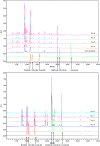
HPLC chromatogram showing peaks corresponding to isoflavones in extracts of the aerial part of P. thunbergiana, with absorbance evaluated at 260 nm
Fig. 3
Fig. 3
Chemical structure of isoflavones in extracts of the aerial part of P. thunbergiana
Fig. 4
Fig. 4
Concentration-dependent effects of aerial part of P. thunbergiana on B16F10 cell viability. Cytotoxicity of P. thunbergiana extracts in B16F10 melanoma cells was evaluated by treating the cells treated with aqueous or EtOAc fraction of extracts for 48 h. Cell viability was determined using the MTT assays, and results are expressed as percent viability relative to untreated cells
Fig. 5
Fig. 5
Effect of aerial part of P. thunbergiana on melanogenesis in B16F10 cells. B16F10 cells were cultured, and following adherence, the cells were treated with α-MSH. After 1 day, the cells were incubated with combinations of α-MSH and kojic acid or P. thunbergiana extract Nos. 1–8. Melanogenesis was quantified 2 days later, and the results expressed relative to controls. **p < 0.01, and ***p < 0.001 compared to cells stimulated with α-MSH alone
Fig. 6
Fig. 6
Effect of EtOAc fractions on melanogenesis in B16F10 cells. B16F10 cells were cultured, and following adherence, the cells were treated with α-MSH. The cells were treated with a combination of α-MSH with kojic acid, arbutin, or a range of concentrations of extract Nos. 5–8 (5, 10, 50, and 100 μg/mL). The results were expressed relative to untreated cells. *p < 0.05, **p < 0.01, and ***p < 0.001 compared to α-MSH-stimulated cells
Fig. 7
Fig. 7
Optical images (magnification ×400) showing melanin content with Fontana–Masson staining in B16F10 cells. After 24 h of α-MSH pre-incubation, B16F10 cells were treated with a combination of α-MSH and extract No. 6 or 7, arbutin, or kojic acid for 48 h. Pigmentation observed upon Fontana–Masson staining (magnification ×400)
Fig. 8
Fig. 8
Comparison of cell-free tyrosinase activity of aerial part of P. thunbergiana. Reactions were performed in potassium phosphate buffer with l-DOPA and mushroom tyrosinase. After 2 min, tyrosinase activity was assessed by measuring the absorbance at 475 nm and expressed relative to the controls. Kojic acid was used as a positive control
Fig. 9
Fig. 9
Inhibitory effects of aerial part of P. thunbergiana on cellular tyrosinase activity. B16F10 cells were pre-cultured with α-MSH for 24 h, and incubated for 48 h more in a medium containing several concentrations (10, 50, and 100 µg/mL) of extract No. 6 (a) or No. 7 (b). The cellular tyrosinase activity was measured and the results are expressed relative to controls. *p < 0.05, **p < 0.01, and ***p < 0.001 compared to α-MSH-stimulated cells
Fig. 10
Fig. 10
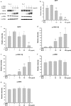
Effects of aerial part of P. thunbergiana on mRNA and proteins expression of tyrosinase. B16F10 cells were treated with α-MSH and No. 6 or No. 7 for the indicated concentration. mRNA levels (a) and expression levels (b) of tyrosinase were estimated. GAPDH was used as an internal standard. cf Quantitation of the western blots by using GAPDH as the loading control. *p < 0.05, **p < 0.01, and ***p < 0.001 compared to α-MSH-stimulated cells. ### p < 0.001 compared to nonstimulated cells
Fig. 11
Fig. 11
Inhibitory effects on α-glucosidase activity. α-Glucosidase was incubated with acarbose, Nos. 6, 7 (500 µg/mL) and PNPG. Each percentage value of α-glucosidase activity is reported relative to control. *p < 0.05, **p < 0.01, and ***p < 0.001 compared to control
Fig. 12
Fig. 12
Effects of aerial part of P. thunbergiana on mRNA and proteins expression of TRP1. B16F10 cells were treated with α-MSH and No. 6 or No. 7 for the indicated concentration. mRNA levels (a) and expression levels (b) of TRP1 were estimated. GAPDH was used as an internal standard. cd Quantitation of the western blots by using GAPDH as the loading control. *p < 0.05 and ***p < 0.001 compared to α-MSH-stimulated cells. ### p < 0.001 compared to nonstimulated cells
Fig. 13
Fig. 13
Effects of aerial part of P. thunbergiana on expression of melanogenesis-related proteins. B16F10 cells were treated with α-MSH and No. 6 or No. 7 for the indicated concentration. Western blot assay were performed to estimate and expression levels of total MITF, p-ERK and p-GSK-3β. Protein loading amounts were confirmed by GAPDH expression. bg Quantitation of the western blots by using GAPDH as the loading control. *p < 0.05, **p < 0.01, and ***p < 0.001 compared to α-MSH-stimulated cells
Fig. 14
Fig. 14
Effects of aerial part of P. thunbergiana on regulation of Akt/GSK-3β signal. Melanin contents were evaluated. Cells were pretreated in the absence (−) or presence (+) of LY294002 (20 µM) for 1 h and then cultured without (−) or with (+) 50 µg/mL of No. 6 (a) and 7 (b) for 48 h. ***p < 0.001 compared to sample-treated only
Fig. 15
Fig. 15
Effects of aerial part of P. thunbergiana on pigmentation in UV-irradiated animal. a Melanin possessing hairless mice were treated with 100 mg of cream base, kojic acid 1 % cream, No. 6 1 and 3 % cream on dorsal skins every day. UVB irradiation was performed according to the indicated schedules. The lightness (L* value) of skin was measured before applying cream on each day. b ΔL* values of each animal were calculated; values of days 17, 18, 19 minus values of days 2, 3, 4. Data represent mean ± SEM (n = 3). # p < 0.05, ## p < 0.01, and ### p < 0.001 compared with CON. *p < 0.05, **p < 0.01, and ***p < 0.001 compared with UVB group. c Microscopic images of skin sections. Melanogenesis of dorsal skin was highlighted by Fontana–Masson staining. Arrows indicate pigmentation
Fig. 16
Fig. 16
The multiple mechanisms of anti-melanogenesis effects of the aerial part of P. thunbergiana. The aerial part of P. thunbergiana has multi-action mediating inhibition of melanogenesis. The suppression in expression of tyrosinase and TRP1 is associated with MITF which is downgraded by Akt/GSK-3β signal. Maturation of tyrosinase is interrupted by inhibitory effect on α-glucosidase of the aerial part of P. thunbergiana. Antioxidant activity also inhibits melanogenesis

References

    1. Ando H, Kondoh H, Ichihashi M, Hearing VJ. Approaches to identify inhibitors of melanin biosynthesis via the quality control of tyrosinase. J Invest Dermatol. 2007;127(4):751–761. doi: 10.1038/sj.jid.5700683. - DOI - PubMed
    1. Aoki H, Moro O. Involvement of microphthalmia-associated transcription factor (MITF) in expression of human melanocortin-1 receptor (MC1R) Life Sci. 2002;71(18):2171–2179. doi: 10.1016/S0024-3205(02)01996-3. - DOI - PubMed
    1. Bolling BW, Blumberg JB, Chen CY. Extraction methods determine the antioxidant capacity and induction of quinone reductase by soy products in vitro. Food Chem. 2009;116(1):351–355. doi: 10.1016/j.foodchem.2009.01.087. - DOI - PMC - PubMed
    1. Branza-Nichita N, Petrescu AJ, Negroiu G, Dwek RA, Petrescu SM. N-glycosylation processing and glycoprotein folding-lessons from the tyrosinase-related proteins. Chem Rev. 2000;100(12):4697–4712. doi: 10.1021/cr990291y. - DOI - PubMed
    1. Chakraborty AK, Funasaka Y, Slominski A, Ermak G, Hwang J, Pawelek JM, Ichihashi M. Production and release of proopiomelanocortin (POMC) derived peptides by human melanocytes and keratinocytes in culture: regulation by ultraviolet B. Biochim Biophys Acta. 1996;1313(2):130–138. doi: 10.1016/0167-4889(96)00063-8. - DOI - PubMed

Publication types

MeSH terms

LinkOut - more resources