. 2014 Aug;141(16):3255-65.
doi: 10.1242/dev.105593. Epub 2014 Jul 25.

Visualisation of chicken macrophages using transgenic reporter genes: insights into the development of the avian macrophage lineage

Affiliations

Visualisation of chicken macrophages using transgenic reporter genes: insights into the development of the avian macrophage lineage

Adam Balic et al. Development. 2014 Aug.

Abstract

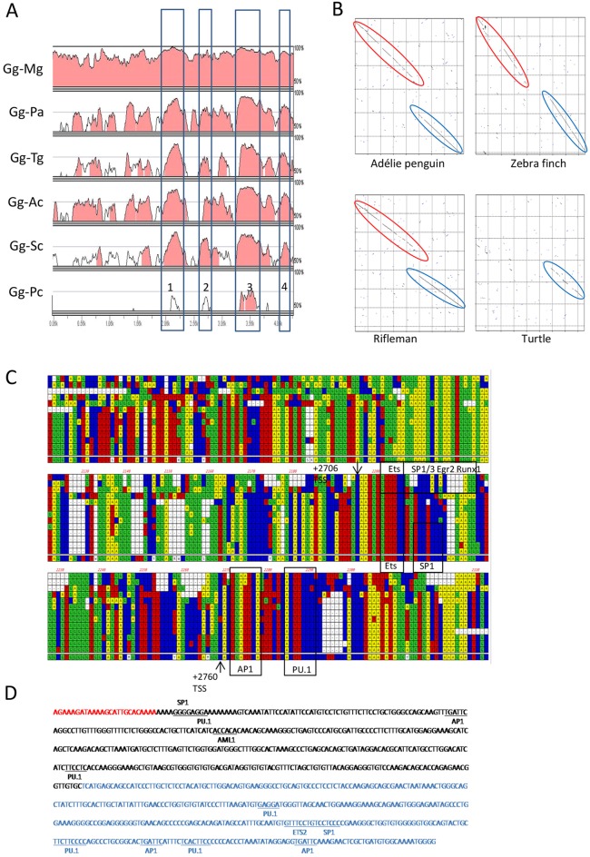
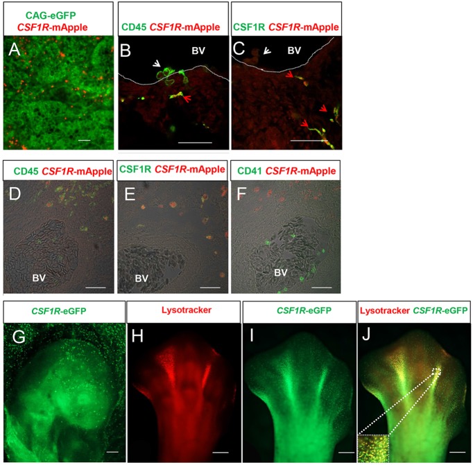
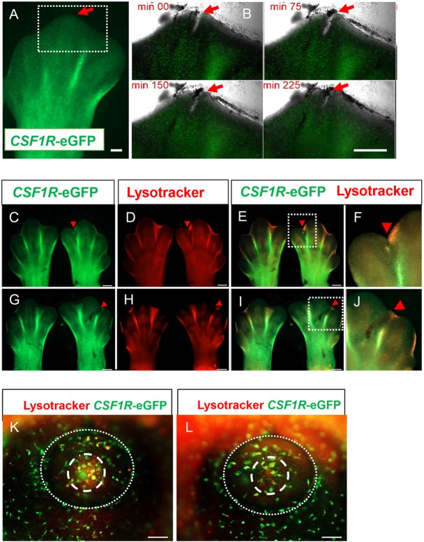
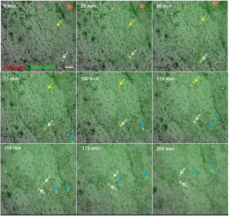
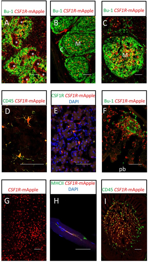
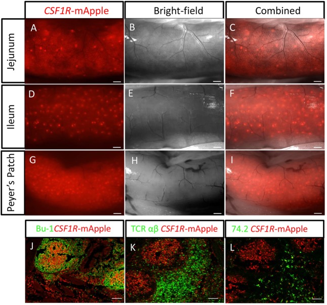
We have generated the first transgenic chickens in which reporter genes are expressed in a specific immune cell lineage, based upon control elements of the colony stimulating factor 1 receptor (CSF1R) locus. The Fms intronic regulatory element (FIRE) within CSF1R is shown to be highly conserved in amniotes and absolutely required for myeloid-restricted expression of fluorescent reporter genes. As in mammals, CSF1R-reporter genes were specifically expressed at high levels in cells of the macrophage lineage and at a much lower level in granulocytes. The cell lineage specificity of reporter gene expression was confirmed by demonstration of coincident expression with the endogenous CSF1R protein. In transgenic birds, expression of the reporter gene provided a defined marker for macrophage-lineage cells, identifying the earliest stages in the yolk sac, throughout embryonic development and in all adult tissues. The reporter genes permit detailed and dynamic visualisation of embryonic chicken macrophages. Chicken embryonic macrophages are not recruited to incisional wounds, but are able to recognise and phagocytose microbial antigens.

Keywords: Chicken; Dendritic cells; Embryonic development; Immunity; Macrophages; Transgenics.


Figures

Fig. 1.
Fig. 1.
Identification of putative macrophage lineage-specific regulatory elements in the first intron of the chicken CSF1R gene. (A) mVista alignment (http://gsd.lbl.gov/vista/) of the CSF1R first intron comparing chicken (Gg) with turkey (Mg), Adélie penguin (Pa), zebrafinch (Tg), rifleman (Ac), ostrich (Sc) and Chinese softshell turtle (Pc). Conserved regions (>70% homology over 100 bp window) are shaded. The positions of four major conserved non-coding elements (CNEs) are boxed and numbered. (B) Pustell DNA matrix alignment of the avian/reptile CSF1R CNE2 and CNE3. The unbroken diagonal lines represent regions of high sequence conservation, and the broken and offset lines indicate that an insertion has occurred in the chicken/turkey lineage in comparison with the other species shown here. The avian-specific CNE2 is highlighted in red; the CNE-3, which is conserved in birds and turtle sequence, is highlighted in blue. (C) Alignment of mammalian Fms-intronic regulatory element (FIRE) with the CSF1R CNE3 region in birds/reptiles. Species sequences from top to bottom are human, mouse, platypus, turtle, alligator, Adélie penguin, budgerigar, ostrich, rifleman, zebrafinch, duck, turkey, chicken and consensus sequence. Arrows indicate the location of the two murine FIRE transcription start sites (Sauter et al., 2013) and conserved transcription factor binding sites are also shown. (D) Sequence of the chicken macrophage lineage-specific regulatory element used in this study: binding sites for PU.1, C/EBP, AP1, SP1 and AML1 are identified. The avian-specific CNE2 is highlighted in red and the avian-reptile-mammal conserved CNE3 is in blue.
Fig. 2.
Fig. 2.
CSF1R-transgene expression is restricted to macrophages in MacReporter embryos. (A) CSF1R-mApple+ cells (red) are restricted to the lumen of primitive blood vessels in ubiquitous CAG-eGFP-expressing HH13 stage embryos (green). (B,C) Confocal analysis of transgene expression in HH21 stage CSF1R-mApple embryos indicates that transgene expression is restricted to CD45+ (B, green), CSF1R+ (C, green) cells in the mesenchyme (red arrowheads) and not CD45+ cells budding from the epithelial layer of the dorsal aorta (white arrowheads). Dotted lines mark the blood vessel (BV) lumen. Scale bars in A-C: 100 µm. (D-F) Confocal analysis of CSF1R staining (green) of CSF1R-mApple transgene-expressing cells (red) in the mesenchyme tissue of a HH29 embryo. The transgene is expressed in cells (red) that are CD45+ (D, green) and CSF1R+ (E, green), but are CD41/61 (F, green). Scale bars in D-F: 100 µm. BV, blood vessel lumen. (G) Scattered eGFP+ cells are found in the embryonic (Emb.) and extra-exbryonic (Ex-Emb) tissues of HH15 MacGreen embryos. Scale bar: 200 µm. (H-J) Colocalization of eGFP+ cells with LysoTracker Red-stained lysosomes in HH33 embryo footplate and in the interdigit region. Inset in J shows the boxed area in more detail. Scale bars in G-J: 200 µm.
Fig. 3.
Fig. 3.
Embryonic macrophages are not recruited to wounds. (A,B) Time-lapse imaging of embryonic macrophage response to incisional wounding in the footpad of HH31 stage embryos in vitro. The tip of the central digit of a footpad (A, red arrow) was wounded with an ultrafine tungsten needle. Scale bar: 200 µm. Subsequent panels (B) focus on the behaviour of macrophages in the region of the incisional wound (boxed area). (B) No recruitment of macrophages to the wound (red arrows) is observed. Scale bar: 500 µm. (C-L) In ovo macrophage response to wounding. LysoTrackerRed (LyTRd) staining of CSF1R-eGFP embryonic limb buds 24 h after incisional (C-F) or crush (G-J) wounding of HH31 embryonic limb buds. Wounded limb buds are on the right of each panel and control contralateral limb buds are shown on the left. Red arrowheads indicate site of wounding and boxed areas (E,I) show details of the wound site in F,J. Compared with the contralateral control limb bud, there is no accumulation of macrophages at the wound site (red arrowheads), and diminishment of macrophage accumulation in the interdigit region adjacent to the wound is apparent (E,F,I,J). Scale bars: 500 µm. (K,L) LyTRd staining of eye primordium of CSF1R-eGFP embryos wounded in the eye primodium at HH16 in ovo. There is no obvious recruitment of macrophages with lysosomes in the wounded (L) compared with unwounded (K) eye primordium, although LyTRd staining indicates a region of cell death (dashed circle) in the centre of the lens vesicle (dotted line). Scale bars: 100 µm.
Fig. 4.
Fig. 4.
Macrophages associated with the embryonic vasculature are highly motile and phagocytic, and undergo local division. Time-lapse imaging of region above the vitelline artery near the embryo proper. The aorta of CSF1R-eGFP embryos was injected with Texas Red-labelled zymosan 1 h prior to the beginning of imaging. Most zymosan particles adhered to the blood vessel walls (yellow arrows). eGFP+ macrophages are highly motile. Between 100 and 125 min from the start of filming, a zymosan particle (yellow arrow) becomes associated with a macrophage; this macrophage re-enters the circulation, removing the zymosan particle by 150 min. At 0 min, a zymosan particle is contained within a macrophage (white arrow); from 0-75 min this macrophage is both motile and exhibits changes in morphology. At 100 min, this macrophage (white arrow) no longer exhibits movement and does not extend any cellular processes. A similar macrophage without a phagocytised zymosan particle (blue arrow) exhibits identical behaviour. At 100-150 min, both undergo division (white and blue arrows), and daughter cells resume active patrolling of the vasculature. Scale bar: 50 µm.
Fig. 5.
Fig. 5.
Confocal analysis of MacRed chicken post-hatch mononuclear phagocyte populations. (A) Splenic mononuclear phagocytes (red) and Bu-1+ B-cells (green) from a 16-week-old MacRed chicken. Rings of transgene-expressing cells can clearly be seen surrounding the ellipsoid (asterisk). (B) Bursa of Fabricius from an 8-day-old MacRed chicken: Bu-1+ B cells (green) show arrangement of the B-cell follicles; mononuclear phagocytes (red) are present in the medulla (M) and interfollicular region (red arrow), but not in the cortex (C) of B-cell follicles in the bursa of Fabricius. (C) Caecal tonsil B-cell follicle from a 10-week-old MacRed chicken, showing location of mononuclear phagocytes (red) and Bu-1+ B-cells (green). Transgene-expressing cells concentrated in the medulla region (M) of the B-cell follicle are a dense network of FDC. (D) Microglial cells (red) in the cerebellum of an 8-day-old MacRed chicken showing colocalisation with CD45 staining (green). (E) Kupffer cells (red) showing colocalisation with CSF1R (green) from a 13-week-old MacRed chicken liver. (F) Lung mononuclear phagocytes (red) and Bu-1+ B-cells (green) in the interstitial tissue of the parabronchial wall from a 16-week-old MacRed chicken. The parabronchial lumen (pb) is indicated. (G) Epidermal mononuclear phagocyte cells (red) in epidermal sheet preparation from a 10-week-old MacRed chicken. (H) Breast muscle mononuclear phagocytes (red) from a 16-week-old MacRed chicken co-expressing MHCII (green). (I) Feather pulp mononuclear phagocytes from an 8-day-old MacRed chicken (red) co-stained with CD45 (green). Scale bars: 50 µm.
Fig. 6.
Fig. 6.
F distribution of lymphoid aggregates in the MacRed chicken gut. (A-I) External views of different regions of a 1-year-old MacRed chicken showing several scattered lymphoid aggregates in the jejunum (A-C), numerous scattered lymphoid aggregates in the ileum (D-F) and a high concentration of lymphoid aggregates in the ileum Peyer's Patch. Scale bars in A-I: 500 µm. (J-L) Immunofluorescence staining of Peyer's patches showing organisation of CSF1R-mApple-expressing cells (red) in relation to: (J) Bu-1+ B-cells (green), (K) TCR αβ (Vβ1)+ T-cells (green) and (L) CVI-ChNL-74.2+ macrophages (green). Scale bars in J-L: 100 µm.

References

    1. Al-Roubaie S., Hughe J. H., Filla M. B., Lansford R., Lehoux S., Jones E. A. V. (2012). Time-lapse microscopy of macrophages during embryonic vascular development. Dev. Dyn. 241, 1423-1431 10.1002/dvdy.23835 - DOI - PubMed
    1. Auffray C., Fogg D., Garfa M., Elain G., Join-Lambert O., Kayal S., Sarnacki S., Cumano A., Lauvau G., Geissmann F. (2007). Monitoring of blood vessels and tissues by a population of monocytes with patrolling behavior. Science 317, 666-670 10.1126/science.1142883 - DOI - PubMed
    1. Befus A. D., Johnston N., Leslie G. A., Bienenstock J. (1980). Gut-associated lymphoid tissue in the chicken. I. Morphology, ontogeny, and some functional characteristics of Peyer's patches. J. Immunol. 125, 2626-2632. - PubMed
    1. Beug H., von Kirchbach A., Döderlein G., Conscience J.-F., Graf T. (1979). Chicken hematopoietic cells transformed by seven strains of defective avian leukemia viruses display three distinct phenotypes of differentiation. Cell 18, 375-390 10.1016/0092-8674(79)90057-6 - DOI - PubMed
    1. Bollerot K., Romero S., Dunon D., Jaffredo T. (2005). Core binding factor in the early avian embryo: cloning of Cbfbeta and combinatorial expression patterns with Runx1. Gene Expr. Patterns 6, 29-39 10.1016/j.modgep.2005.05.003 - DOI - PubMed

Publication types

MeSH terms

Substances