. 2014 Sep 1;193(5):2469-82.
doi: 10.4049/jimmunol.1303370. Epub 2014 Jul 25.

ADAM9 is a novel product of polymorphonuclear neutrophils: regulation of expression and contributions to extracellular matrix protein degradation during acute lung injury

Affiliations

ADAM9 is a novel product of polymorphonuclear neutrophils: regulation of expression and contributions to extracellular matrix protein degradation during acute lung injury

Robin Roychaudhuri et al. J Immunol. .

Abstract

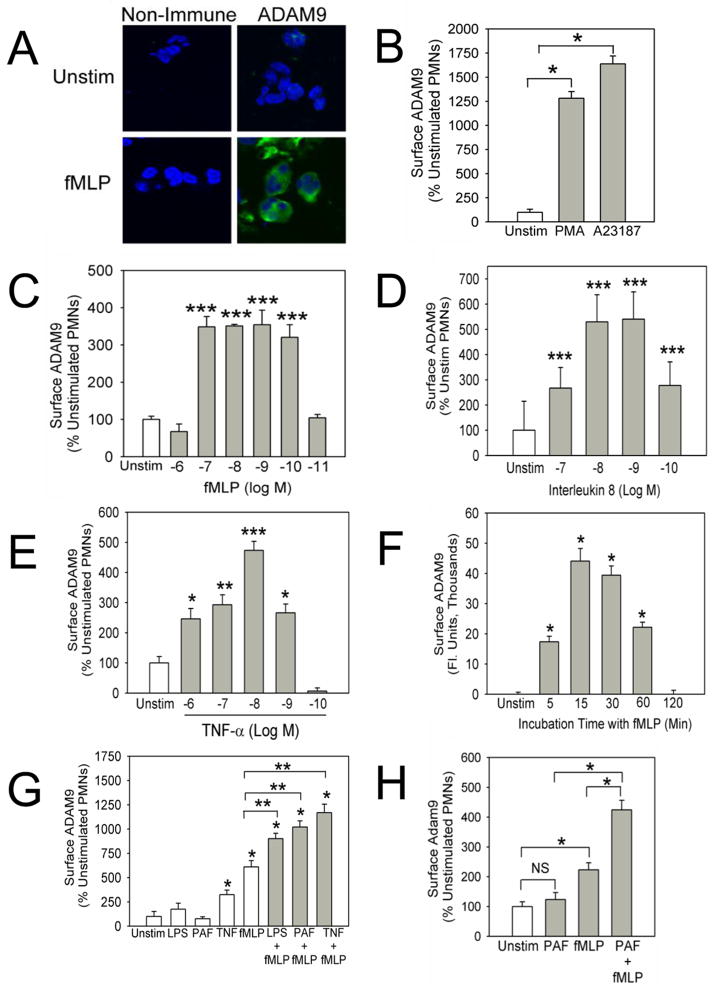
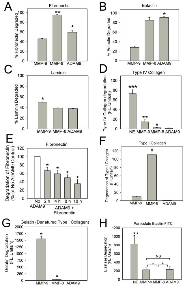
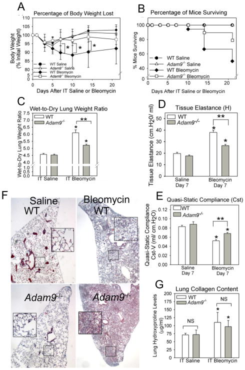
A disintegrin and a metalloproteinase domain (ADAM) 9 is known to be expressed by monocytes and macrophages. In this study, we report that ADAM9 is also a product of human and murine polymorphonuclear neutrophils (PMNs). ADAM9 is not synthesized de novo by circulating PMNs. Rather, ADAM9 protein is stored in the gelatinase and specific granules and the secretory vesicles of human PMNs. Unstimulated PMNs express minimal quantities of surface ADAM9, but activation of PMNs with degranulating agonists rapidly (within 15 min) increases PMN surface ADAM9 levels. Human PMNs produce small quantities of soluble forms of ADAM9. Surprisingly, ADAM9 degrades several extracellular matrix (ECM) proteins, including fibronectin, entactin, laminin, and insoluble elastin, as potently as matrix metalloproteinase-9. However, ADAM9 does not degrade types I, III, or IV collagen or denatured collagens in vitro. To determine whether Adam9 regulates PMN recruitment or ECM protein turnover during inflammatory responses, we compared wild-type and Adam9(-/-) mice in bacterial LPS- and bleomycin-mediated acute lung injury (ALI). Adam9 lung levels increase 10-fold during LPS-mediated ALI in wild-type mice (due to increases in leukocyte-derived Adam9), but Adam9 does not regulate lung PMN (or macrophage) counts during ALI. Adam9 increases mortality, promotes lung injury, reduces lung compliance, and increases degradation of lung elastin during LPS- and/or bleomycin-mediated ALI. Adam9 does not regulate collagen accumulation in the bleomycin-treated lung. Thus, ADAM9 is expressed in an inducible fashion on PMN surfaces where it degrades some ECM proteins, and it promotes alveolar-capillary barrier injury during ALI in mice.


Figures

Figure 1
Figure 1. Pro-inflammatory mediators up-regulate surface ADAM9 levels on human and murine PMNs
In A, PMNs were isolated from healthy human volunteers, incubated with or without 10−7 M fMLP at 37°C for 30 min, and fixed. PMNs were then immunostained with Alexa-488 for surface ADAM9 or incubated with an isotype-matched non-immune primary antibody and examined using confocal microscopy. Images shown are representative of 6 separate experiments (magnification is x 400). In B–D, human PMNs were incubated at 37°C for 30 min with or without 10−7 M PMA or 10−6 M A23187 (B), 10−6–10−11 M fMLP (C), 10−7–10−10 M IL-8 (D), 10−6–10−10 M TNF-α (E). In F, PMNs were incubated for up to 120 min at 37°C with or without 10−7 M fMLP (F). In G, PMNs were incubated for 30 min at 37°C with or without 100 ng/ml LPS, 10−7 M PAF, 100 U/ml TNF-α, or 10−7 M fMLP alone (white bars), or PMNs were incubated at 37°C for 15 min with 100 ng/ml LPS, 10−7 M PAF, or 100 U/ml TNF-α and then activated for 30 min at 37°C with 10−7 M fMLP (grey bars). In B–G, cells were fixed, immunostained for surface ADAM9, and surface ADAM9 levels were quantified as described in Methods. In B and F, * indicates p < 0.001 versus unstimulated cells. In C–E, * indicates p < 0.04, ** p = 0.004, and *** p < 0.0001 versus unstimulated PMNs. In G, * indicates p < 0.001 compared with unstimulated cells and ** p < 0.001 vs. each agonist when tested alone. In H, PMNs were isolated from unchallenged C57BL/6 WT mice, and incubated at 37°C without agonists for 45 min (unstim), or with 10−6 M fMLP or 10−6 M PAF for 30 min, or with 10−6 M PAF for 15 min and then 10−6 M fMLP for 30 min. Cells were immunostained for surface Adam9 as described in Methods. Asterisk indicates p < 0.001. Data are expressed as mean ± SEM as a percentage of surface staining associated with unstimulated cells (B–E, and G–H) or in arbitrary fluorescence units (F); n = 150–300 cells per group. Results are representative of 3–4 separate experiments.
Figure 2
Figure 2. ADAM9 is stored in the tertiary and specific granules and secretory vesicles of PMNs
In A, unstimulated human PMNs were fixed, permeabilized with methanol, and then immunostained with Alexa-488 for intracellular ADAM9 (top right images) and with Alexa-546 for markers of the tertiary granules (MMP9), specific granules (lactoferrin; LF), or azurophil granules (myeloperoxidase; MPO) in the bottom left images, and nuclei were counter-stained with DAPI (top left images). Cells were examined using a confocal microscope and merged images are shown in the bottom right images. Images representative of three separate experiments are shown. As a control, PMNs were stained with isotype-matched non-immune primary antibodies [rabbit IgG (Rb IgG) and goat IgG (Gt IgG) or murine IgG (Ms IgG)] shown in the bottom right quadrant in A. In B, PMNs were incubated for 15 min at 37°C with or without PMA and then subjected to subcellular fractionation as described in Methods. The PMN granule fractions [including the azurophil (AZ), specific (Sp), and gelatinase (Gel) granules and combined plasma membrane and secretory vesicle (MV) fraction], were detergent solubilized, and along with the cytosolic fraction (Cy) subjected to western blot analysis using an antibody to ADAM9 (in B). ADAM9 levels were also quantified in each of these PMN fractions [and PMN supernatants (SN)] using an ELISA and results were normalized to total protein levels (C). Results shown in B are representative of PMN preparations from two donors. In C, data are mean ± SD (n = 2 PMN preparations).
Figure 3
Figure 3. Soluble ADAM9 ectodomain degrades several basement membrane proteins and insoluble elastin
Equimolar (25 nM) concentrations of human soluble active ADAM9, MMP-8, or MMP-9 versus buffer alone were incubated with 2 μg of purified human fibronectin (in A), entactin (in B) or laminin (in C) at 37°C for 18 h at pH 7.4. The reaction products were separated on 4–20% Tris-HCl SDS-PAGE gels which were stained with Coomassie Blue dye. Densitometry was used to quantify intact ECM proteins incubated without versus with proteinases. The results are expressed as % of ECM protein that was degraded by each proteinase. Data are mean ± SEM; n = 3–6 experiments. In A, * indicates p = 0.031 and ** p < 0.001 versus the % of fibronectin cleaved by MMP-9. In B, * indicates p < 0.001 versus the % of entactin degraded by MMP-9. In C, * indicates p < 0.001 versus the % of laminin that was degraded by MMP-8 and ADAM9. In E, 25 nM ADAM9 or buffer alone were incubated with 2 μg of fibronectin for up to 18 h at 37°C and degradation of fibronectin measured at intervals as outlined above. Data are mean ± SD (n = 4 experiments) and * indicates p < 0.05 versus fibronectin incubated without ADAM9. In D, F, G, and H, equimolar (25 nM) concentrations of human soluble active MMP-9, ADAM9, and MMP-8 versus buffer alone were incubated with 50 μg/ml DQ-FITC-conjugated type IV collagen (in D), 50 μg/ml DQ-FITC-conjugated type I collagen (in F), 50 μg/ml DQ-FITC-conjugated gelatin (in G), or 20 mg/ml particulate elastin-FITC (in H) at 37°C for 18 h at pH 7.4 (in D and H, human soluble active neutrophil elastase [NE] was used as an additional control). Cleavage of each substrate was quantified in fluorescence units using fluorimetry. Results are mean ± SEM; n =3–7 experiments. In D, * indicates p = 0.041; ** p = 0.009; and *** p <0.001 versus the amount of type IV collagen degraded by ADAM9. In F, * indicates p < 0.001 vs. the amount of type I collagen degraded by ADAM9 or MMP-9. In G, * indicates p < 0.001 vs. the amount of gelatin degraded by ADAM9. In H, * indicates p = 0.033 vs. the amounts of elastin degraded by MMP-8 and ** p < 0.001 vs. the amount of elastin degraded by all other proteinases.
Figure 4
Figure 4. Surface ADAM9 on activated PMNs has similar ECM protein-degrading activity as sADAM9
We incubated equal numbers of PAF- and fMLP-activated and fixed WT, Mmp-8−/−, Mmp-9−/−, or Adam-9−/− PMNs versus buffer alone at 37°C for 18 h at pH 7.4 with 50 μg/ml DQ-FITC-conjugated type I collagen (in A), or 50 μg/ml quenched DQ-FITC-conjugated gelatin (B) or 20 mg/ml FITC-conjugated particulate elastin (in C), and quantified metalloproteinase-mediated cleavage of the substrates by surface proteinases, as described in Methods. The results for proteinase-deficient PMNs are expressed as a percentage of the MP activity associated with the surface of WT PMNs. Data are mean ± SEM; n = 3–4 experiments. In A * indicates p= 0.008; in B, * indicates p < 0.001; and in C, * indicates p = 0.006 and ** p < 0.001.
Figure 5
Figure 5. Adam9 is upregulated in the lung during ALI in mice
In A, we delivered 10 μg of LPS or PBS to C57BL/6 WT mice by the IT route, and 4 h and 24 h later measured steady state Adam9, Adam10, and Adam17 mRNA levels in whole lung samples using qRT-RT-PCR. Data are mean ± SEM; n = 6 mice/group. Asterisk indicates p < 0.001. In B, we immunostained lungs sections from unchallenged C57BL/6 WT mice (Unchal) or C57BL/6 WT mice harvested 24 h after 10 μg of LPS was delivered by the IT route using rabbit anti-Adam9 IgG or non-immune rabbit IgG and the immunoperoxidase method. Images shown are representative of 4 mice per group (magnification is x 400 and x 1000). Black arrows indicate PMNs staining positively for Adam9 and yellow arrows indicate macrophage staining positively for Adam9. In C and D, sections of lungs from unchallenged mice (in C) or lungs harvested 24 h after LPS was instilled by the IT route (in D) were immunostained with Alexa-488 for Adam9 (second columns), and Alexa-546 for markers of epithelial cells (pancytokeratin; Pck), PMNs (Ly6G), or macrophages (Mac-3). Sections were then counterstained with DAPI (first columns). Lung sections were examined using confocal microscopy, and merged images are shown in the fourth columns (magnification is X 200 for Pck-stained sections and X 500 for Ly6G and Mac3-stained sections). No staining was detected in lung sections stained with isotype matched control primary antibodies (data not shown).
Figure 6
Figure 6. ADAM9 is not required for PMN recruitment into the lung but promotes LPS-mediated ALI in mice
In A–F, we delivered 10 μg of LPS or PBS by the IT route to WT vs. Adam9−/− mice. Four hours to 1 week later, we performed BAL or removed lungs from the mice. We counted total leukocytes (A) and PMNs (B) in BAL samples. In A and B, data are mean ± SEM; n = 5–8 PBS-treated mice and n = 15–18 LPS-treated mice. Asterisk indicates p ≤ 0.05 compared with PBS-treated mice belonging to the same genotype at the same time point in A and B. In C, wet-to-dry lung weight ratios were measured 24–72 h after LPS or PBS were delivered by the IT route to WT and Adam9−/− mice. Data are mean ± SEM; n = 4–9 PBS-treated mice, n = 9–25 LPS-treated WT mice, and n = 7–16 LPS-treated Adam9−/− mice. Asterisk indicates p = 0.016 and **, p ≤ 0.002. In D–E, total protein (D) and hemoglobin levels [in E, as a marker of leakage of erythrocytes across the alveolar-capillary barrier (41)] were measured in BALF samples 24 h after instilling PBS or LPS in mice. In D–E, data are mean ± SEM; n = 4 PBS-treated mice and n = 5–12 LPS-treated mice. In D, asterisk indicates p < 0.05 and ** p = 0.018. In E, asterisk indicates p < 0.035 and **, p < 0.035 versus PBS-treated mice belonging to the same genotype. In F, desmosine levels were measured in BALF samples 24 h after PBS or LPS were instilled by the IT route. Data are mean ± SEM; n = 4–9 PBS-treated mice and n = 8–16 LPS-treated mice. Asterisk indicates p = 0.016 and ** p = 0.021 versus PBS-treated WT mice.
Figure 7
Figure 7. ADAM9 promotes weight loss, mortality, and ALI but does not regulate lung collagen accumulation in bleomycin-treated mice
In A–B, we delivered 30 mU of bleomycin vs. saline by the IT route to WT vs. Adam9−/− mice and measured changes in body weight relative to baseline body weight (A) and recorded survival of the mice (B) over 21 days. In A, n = 4–5 saline-treated mice and n = 22 bleomycin-treated mice were studied. Asterisk indicates p ≤ 0.044 for bleomycin-treated WT versus bleomycin-treated Adam9−/− mice. In B, n = 5–8 saline-treated mice and n = 9–15 bleomycin-treated mice were studied. In C–E, we delivered 100 mU of bleomycin vs. saline by the IT route to WT vs. Adam9−/− mice and measured wet-to-dry lung weight ratios and respiratory mechanics using a FlexiVent device. In C, 7–17 saline-treated mice and 8–10 bleomycin-treated mice were studied per group. Asterisk indicates p < 0.001 when compared with saline-treated mice belonging to the same genotype and ** indicates p < 0.001. In D–E, 4–6 saline-treated mice and 5–9 bleomycin-treated mice were studied per group. Asterisk indicates p < 0.001 for saline-treated mice belonging to the same genotype and ** indicates p < 0.001. In F–G, 30 mU of bleomycin or saline was instilled by the IT route to WT vs. Adam9−/− mice and 21 days later, the right lungs were inflated, removed, fixed, and stained with Masson’s trichrome stain. Representative images of lung sections from saline- and bleomycin-treated WT and Adam9−/− mice are shown in F (magnification is X100 and magnification of insets is x 400). In G, lung collagen levels were assessed using hydroxyproline assays performed on hydrolysates of left lungs removed after 21 days. In G, data are mean ± SEM; n = 5 saline-treated mice and n = 9–15 bleomycin-treated mice. Asterisk indicates p ≤ 0.043 when compared with saline-treated mice belonging to the same genotype.

References

    1. Blobel CP. ADAMs: key components in EGFR signalling and development. Nat Rev Mol Cell Biol. 2005;6:32–43. - PubMed
    1. Owen CA. Leukocyte cell surface proteinases: regulation of expression, functions, and mechanisms of surface localization. Int J Biochem Cell Biol. 2008;40:1246–1272. - PMC - PubMed
    1. Namba K, Nishio M, Mori K, Miyamoto N, Tsurudome M, Ito M, Kawano M, Uchida A, Ito Y. Involvement of ADAM9 in multinucleated giant cell formation of blood monocytes. Cell Immunol. 2001;213:104–113. - PubMed
    1. Izumi Y, Hirata M, Hasuwa H, Iwamoto R, Umata T, Miyado K, Tamai Y, Kurisaki T, Sehara-Fujisawa A, Ohno S, Mekada E. A metalloprotease-disintegrin, MDC9/meltrin-gamma/ADAM9 and PKCdelta are involved in TPA-induced ectodomain shedding of membrane-anchored heparin-binding EGF-like growth factor. EMBO J. 1998;17:7260–7272. - PMC - PubMed
    1. Zigrino P, Nischt R, Mauch C. The disintegrin-like and cysteine-rich domains of ADAM-9 mediate interactions between melanoma cells and fibroblasts. J Biol Chem. 2011;286:6801–6807. - PMC - PubMed

Publication types

MeSH terms