. 2014 Sep;15(9):839-45.
doi: 10.1038/ni.2948. Epub 2014 Jul 27.

The SKIV2L RNA exosome limits activation of the RIG-I-like receptors

Affiliations

The SKIV2L RNA exosome limits activation of the RIG-I-like receptors

Sterling C Eckard et al. Nat Immunol. 2014 Sep.

Abstract

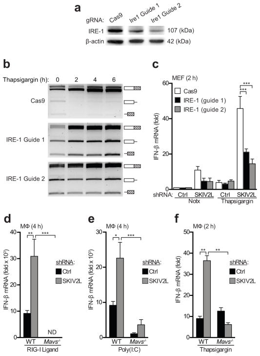
Sensors of the innate immune system that detect intracellular nucleic acids must be regulated to prevent inappropriate activation by endogenous DNA and RNA. The exonuclease Trex1 regulates the DNA-sensing pathway by metabolizing potential DNA ligands that trigger it. However, an analogous mechanism for regulating the RIG-I-like receptors (RLRs) that detect RNA remains unknown. We found here that the SKIV2L RNA exosome potently limited the activation of RLRs. The unfolded protein response (UPR), which generated endogenous RLR ligands through the cleavage of cellular RNA by the endonuclease IRE-1, triggered the production of type I interferons in cells depleted of SKIV2L. Humans with deficiency in SKIV2L had a type I interferon signature in their peripheral blood. Our findings reveal a mechanism for the intracellular metabolism of immunostimulatory RNA, with implications for specific autoimmune disorders.


Conflict of interest statement

COMPETING FINANCIAL INTERESTS

The authors declare no competing financial interests.

Figures

Figure 1
Figure 1. SKIV2L is a negative regulator of the RIG-I-like receptor-mediated antiviral response
(a) SKIV2L and XRN1 were stably knocked down in primary macrophages, and the respective protein levels were evaluated by immunoblotting. (b,c) Primary mouse macrophages, stably transduced as indicated with shRNA knockdown constructs, were treated with an HCV RIG-I ligand (b) or Poly (I:C) (c) and assessed for IFN-β production by quantitative RT-PCR. (d–f) RIG-I ligand-stimulated macrophages with the indicated knockdown constructs were evaluated by quantitative RT-PCR for expression of IFNa4 (d), CXCL10 (e) and IL-6 (f). (g,h) Macrophages were stimulated with DMXAA to activate STING directly, and with LPS to activate TLR4. *P < 0.05; **P < 0.001; ***P < 0.0005; ****P < 0.0001 (2-way ANOVA, b–f). Data are representative of at least three independent experiments with biological triplicates (mean + s.d., a–h).
Figure 2
Figure 2. The UPR activates an antiviral response in SKIV2L-depleted cells
(a) SV40 Large T antigen-immortalized mouse embryonic fibroblasts were transduced with the indicated knockdown constructs and treated with thapsigargin (TG). Ifnb mRNA induction was evaluated using quantitative RT-PCR at the indicated time points. Data are representative of 3 experiments. (b) Primary mouse macrophages were transduced and treated as in (a). (c) Endonuclease activity of IRE-1 is responsible for removal of an intron in XBP-1 mRNA, as well as destructive cleavage of ER-localized RNAs. The kinetics of UPR activation were assessed using a splicing assay for XBP-1 mRNA. The illustration shows the PstI digest site in the intron of unspliced XBP-1 mRNA used to distinguish spliced (S) and unspliced (U1, U2) cDNAs. (d–e) Primary mouse macrophages were treated with thapsigargin (TG; e) or tunicamycin (TM; f) treatment. *P < 0.05; **P < 0.0001 (2-way ANOVA, b–e). Data are representative of at least three independent experiments with biological triplicates (mean + s.d., b–e).
Figure 3
Figure 3. SKIV2L specifically regulates the MAVS-dependent antiviral response
(a) Immortalized MEFs were transduced with lenti-CRISPR constructs encoding CAS9 alone or two different guide RNAs targeting Ern1. IRE-1α protein was measured by immunoblot. (b) CAS9 control and IRE-1α-targeted cells were treated with thapsigargin to induce the UPR, and IRE-1- mediated XBP-1 mRNA splicing was quantitated as in Fig. 2d. (c) Control and IRE-1-targeted cells were treated with thapsigargin, and IFN-β mRNA was measured by quantitative RT-PCR at the indicated time points. Data are representative of one experiment with biological triplicates testing two independent guide RNAs (mean + s.d., a–c). (d–f) Bone marrow macrophages of the indicated genotypes were transduced with a control or SKIV2L shRNA, treated with RIG-I ligand (d), Poly I:C (e), or thapsigargin (f), and harvested for analysis at the indicated time points. *P < 0.001; **P < 0.0005; ***P < 0.0001; (2-way ANOVA, c–f). Data are representative of at least three independent experiments with biological triplicates (mean + s.d., d–f).
Figure 4
Figure 4. SKIV2L-deficient humans have a robust type I IFN signature
(a) SKIV2L protein was evaluated by immunoblot in control and THES (c.1635insA/c.1635insA) patient lymphoblastoid cell lines. (b) Quantitative RT-PCR measuring six human interferon-stimulated genes (ISGs) in cDNA prepared from whole blood. For each gene, we present values from 29 AGS control patients (black closed circles) together with 3 controls with heterozygous THES mutations (red filled squares), 82 AGS patients (black open triangles), 2 THES patients with SKIV2L mutations (red filled triangles), and 3 THES patients with TTC37 mutations (blue open circles). Horizontal bars represent the median relative quantification (RQ) value for each ISG probe in each group, normalized to HPRT and 18S rRNA expression within each sample. Note that the data from AGS controls and AGS patients were recently published and are reproduced here for comparison to the THES controls and THES patients. (c) Interferon scores were calculated using the median fold change in RQ value for all of the six interferon-stimulated genes in each individual as described. Horizontal bars show the mean interferon scores in patients and controls. (Note: Cannot provide P value, as there are only 2 SKIV2L THES patient samples)
Figure 5
Figure 5. Differential role of SKIV2L and TTC37 in RLR regulation
(a) Immunoblots of primary BMDM lysates showing TTC37 protein expression in knockdown and control cells. Data are representative of two independent experiments. (b,c) Cells were transduced with the indicated lentiviral shRNA constructs and treated with thapsigarin or RIG-I ligands and assessed for IFN-β production by quantitative RT-PCR. RIG-I ligand-treated samples were harvested at 4 h and thapsigargin-treated samples were harvested at 2 h. *P < 0.01; **P < 0.005; ***P < 0.0001; (one-way ANOVA, b–c). Data are representative of two independent experiments with biological triplicates (mean + s.d., b–c).

Comment in

References

    1. Barbalat R, Ewald SE, Mouchess ML, Barton GM. Nucleic acid recognition by the innate immune system. Annu Rev Immunol. 2011;29:185–214. - PubMed
    1. Pichlmair A, Reis e Sousa C. Innate recognition of viruses. Immunity. 2007;27:370–383. - PubMed
    1. Kato H, et al. Length-dependent recognition of double-stranded ribonucleic acids by retinoic acid-inducible gene-I and melanoma differentiation-associated gene 5. J Exp Med. 2008;205:1601–1610. - PMC - PubMed
    1. Stetson DB, Medzhitov R. Recognition of cytosolic DNA activates an IRF3-dependent innate immune response. Immunity. 2006;24:93–103. - PubMed
    1. Xiao TS, Fitzgerald KA. The cGAS-STING Pathway for DNA Sensing. Mol Cell. 2013;51:135–139. doi: 10.1016/j.molcel.2013.07.004. - DOI - PMC - PubMed

Publication types

MeSH terms

Supplementary concepts