

doi: 10.1007/978-1-4939-1215-5_16.
Generation of transgenic mouse fluorescent reporter lines for studying hematopoietic development
Affiliations
- PMID: 25064110
- PMCID: PMC4418647
- DOI: 10.1007/978-1-4939-1215-5_16
Item in Clipboard
Generation of transgenic mouse fluorescent reporter lines for studying hematopoietic development
Methods Mol Biol.
2014.
Abstract
During the development of the hematopoietic system, at least eight distinct lineages are generated in the mouse embryo. Transgenic mice expressing fluorescent proteins at various points in the hematopoietic hierarchy, from hematopoietic stem cell to multipotent progenitors to each of the final differentiated cell types, have provided valuable tools for tagging, tracking, and isolating these cells. In this chapter, we discuss general considerations in designing a transgene and survey available fluorescent probes and methods for confirming and analyzing transgene expression in the hematopoietic systems of the embryo, fetus, and postnatal/adult animal.
Figures
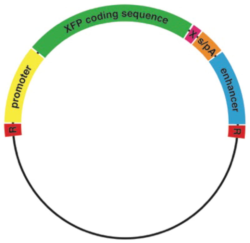
"R" marks the restriction enzyme sites used for removal of the bacterial backbone before transgene injection. s/pA represents the splicing and polyadenylation signals. A stop codon (denoted by "X") should also be included in the construct design. The enhancer may be positioned upstream or downstream from the promoter and sequences encoding the FP and regulatory signals. For additional details, see text.
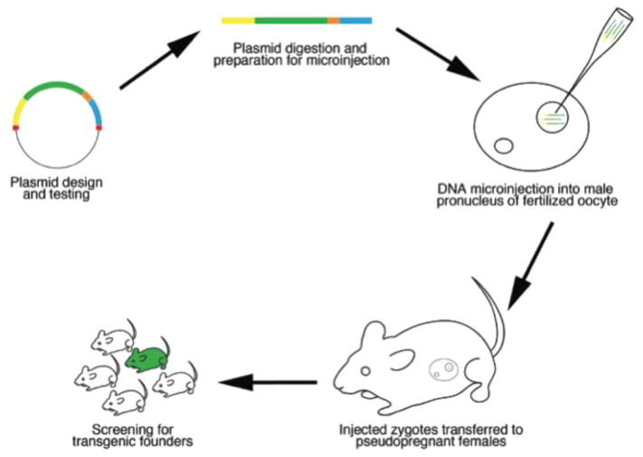
Pronuclear injection of the transgene is shown in this cartoon but transgenic mouse lines can also be generated by blastocyst injection or embryo aggregation with genetically modified ES cells (see text).
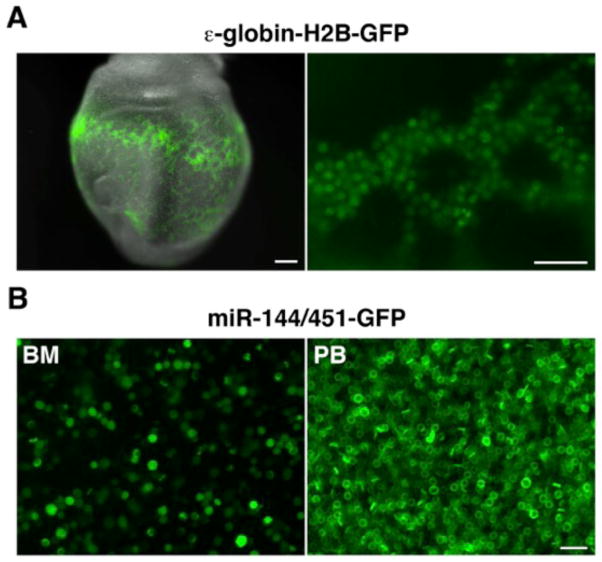
(A) GFP expression in the primitive erythroid cells of an embryonic stage (E)8.5 ε-globin-H2B-GFP embryo (24) (left panel, whole embryo, scale bar 200 μm; right panel, magnified view of yolk sac, scale bar, 50μm). Embryos were photographed on a Zeiss Lumar V12 stereomicroscope equipped with epifluorescence illumination and a NeoLumar S 1.5X FWD 30 mm objective. (B) Wet preparation of green fluorescent erythroid cells from the bone marrow (BM) and peripheral blood (PB) of an adult miR-144/451-GFP knock-in mouse (26). The cells were photographed on a Zeiss Axio Observer Z1 inverted microscope with epifluorescence illumination and a Plan-Apochromat 20X/0.8 objective. Scale bar, 20μm.
References
-
- Prasher DC, Eckenrode VK, Ward WW, Prendergast FG, Cormier MJ. Primary structure of the Aequorea victoria green-fluorescent protein. Gene. 1992 Feb 15;111(2):229–33. - PubMed
-
- Shaner NC, Steinbach PA, Tsien RY. A guide to choosing fluorescent proteins. Nat Methods. 2005 Dec;2(12):905–9. - PubMed
-
- Chudakov DM, Matz MV, Lukyanov S, Lukyanov KA. Fluorescent proteins and their applications in imaging living cells and tissues. Physiol Rev. 2010 Jul;90(3):1103–63. - PubMed
Publication types
MeSH terms
Substances
Grants and funding
LinkOut - more resources
Full Text Sources
Other Literature Sources
