. 2014 Aug;20(8):919-26.
doi: 10.1038/nm.3599. Epub 2014 Jul 27.

An epithelial circadian clock controls pulmonary inflammation and glucocorticoid action

Affiliations

An epithelial circadian clock controls pulmonary inflammation and glucocorticoid action

Julie Gibbs et al. Nat Med. 2014 Aug.

Abstract

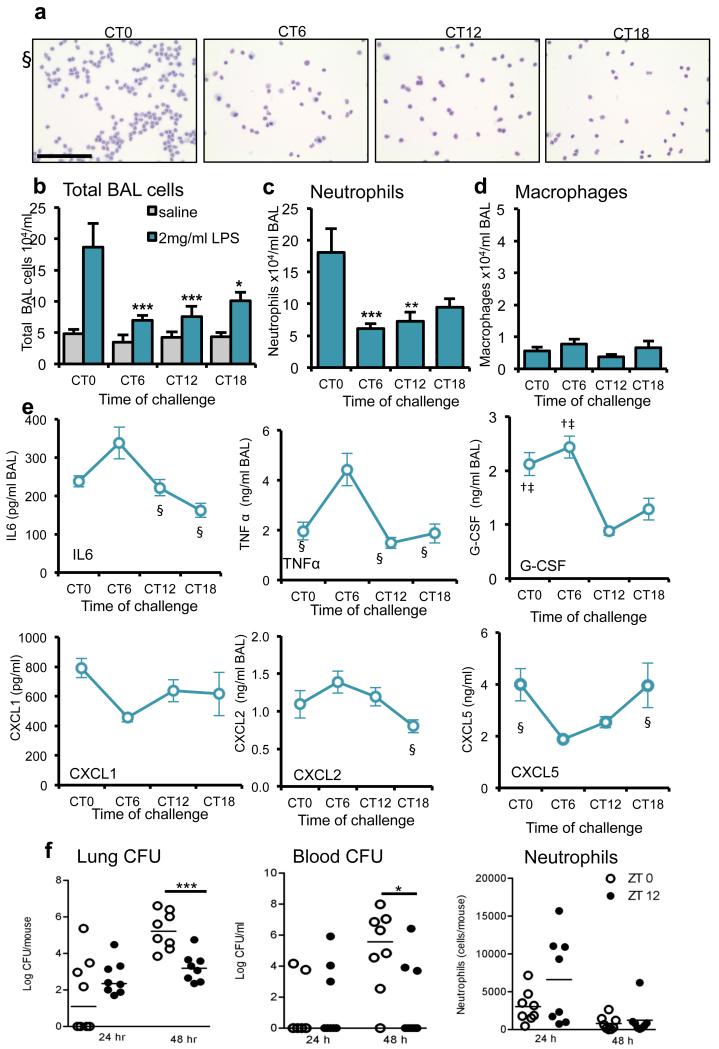
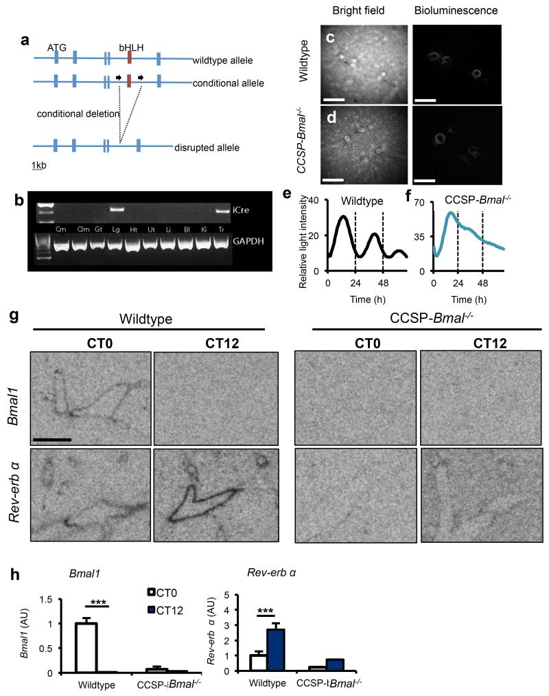
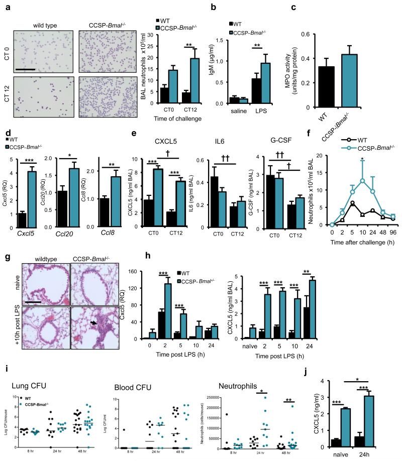
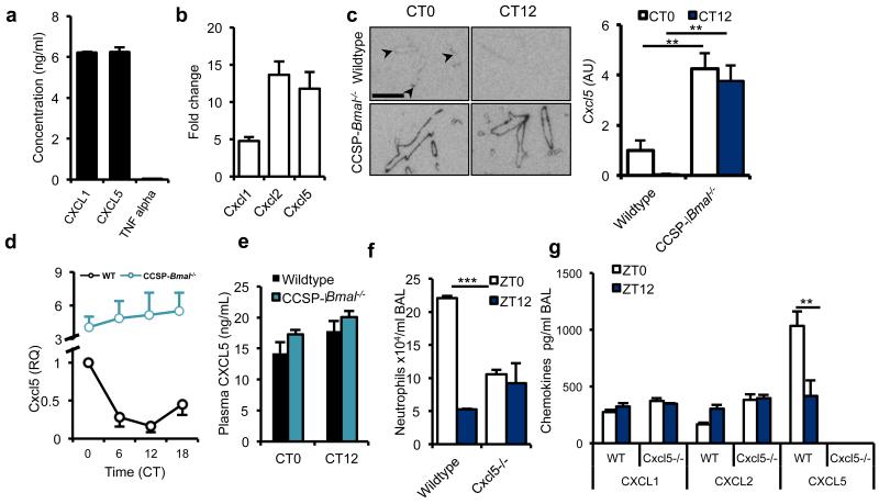
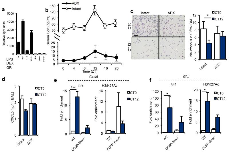
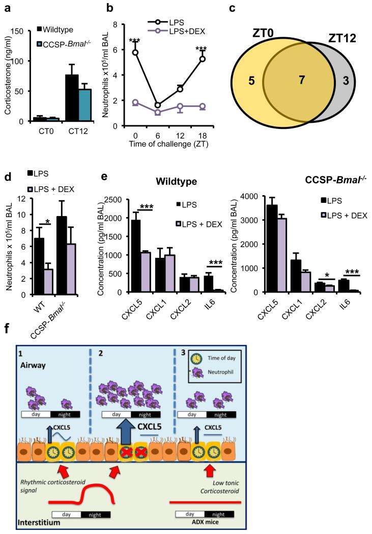
The circadian system is an important regulator of immune function. Human inflammatory lung diseases frequently show time-of-day variation in symptom severity and lung function, but the mechanisms and cell types underlying these effects remain unclear. We show that pulmonary antibacterial responses are modulated by a circadian clock within epithelial club (Clara) cells. These drive circadian neutrophil recruitment to the lung via the chemokine CXCL5. Genetic ablation of the clock gene Bmal1 (also called Arntl or MOP3) in bronchiolar cells disrupts rhythmic Cxcl5 expression, resulting in exaggerated inflammatory responses to lipopolysaccharide and an impaired host response to Streptococcus pneumoniae infection. Adrenalectomy blocks rhythmic inflammatory responses and the circadian regulation of CXCL5, suggesting a key role for the adrenal axis in driving CXCL5 expression and pulmonary neutrophil recruitment. Glucocorticoid receptor occupancy at the Cxcl5 locus shows circadian oscillations, but this is disrupted in mice with bronchiole-specific ablation of Bmal1, leading to enhanced CXCL5 expression despite normal corticosteroid secretion. The therapeutic effects of the synthetic glucocorticoid dexamethasone depend on intact clock function in the airway. We now define a regulatory mechanism that links the circadian clock and glucocorticoid hormones to control both time-of-day variation and the magnitude of pulmonary inflammation and responses to bacterial infection.


Figures

Figure 1
Figure 1
The pulmonary inflammatory response to LPS administration is gated by the circadian clock. (a) Staining of cytospins of BAL fluid from LPS-treated C57Bl/6 mice. Scale bar, 100 μm. (b) Total cell counts in BAL samples collected after LPS challenge at CT0 (n = 6), CT6 (n = 7) CT12 (n = 8) and CT18 (n = 6) or vehicle (n = 4/time point), two-way analysis of variance (ANOVA) and post hoc Bonferroni) (c) Neutrophil and (d) macrophage numbers in the same samples (one-way ANOVA and post hoc Bonferroni) (e) Cytokine levels in these BAL samples were compared between time points using one-way ANOVA and post hoc Bonferroni) § significantly different from CT6 (P≤0.05); † significantly different from CT12 (P≤0.05); ‡ significantly different from CT18 (P≤0.05).; G-CSF, granulocyte colony–stimulating factor. (f) Bacterial load in the lung and blood, and neutrophil counts in BAL, 48h after infection of C57Bl/6 mice with S. pneumoniae (n = 8/time point) at dawn (ZT0) and dusk (ZT12) (median values marked, Mann-Whitney U-test). Data are expressed as mean ± s.e.m and *P ≤ 0.05, **P ≤ 0.01, ***P ≤ 0.005.
Figure 2
Figure 2
Targeted ablation of Bmal1 in CCSP expressing cells disrupts circadian rhythmicity in whole lung. (a) Schematic illustrating the region surrounding the basic helix-loop-helix (bHLH) domain of the mouse Bmal1 locus, the conditional floxed allele and the disrupted allele. (b) Expression of iCre (and the housekeeping gene Gapdh) in tissues harvested from CCSP-Bmal−/− (Bmal1fl/fl; CCSP-icre+/−) mice (Cm, cerebrum; Clm, cerebellum; Gt, gut; Lg, lung; Ht, heart; Ut, uterus; Li, liver; Bl, bladder; Ki, kidney; Tr, trachea). (c,d) Ectopic lung slices from Bmal1fl/fl; CCSP-icre+/− (CCSP-Bmal−/−) or CCSP-icre negative littermate controls (wildtype) on a Per2-luc background were placed under a bioluminescence camera. Scale bars, 500 μm. (e,f) Bioluminescence intensity from bronchioles quantified and plotted as a function of time. Data are representative of 3 independent trials. (g, h) Quantification of clock genes Bmal1 and Nr1d1 (rev-erb α) expression in the bronchioles of lungs at CT0 (n=4/genotype) and CT12 (n=3/genotype) (scale bar, 5 mm, two-way ANOVA and post hoc Bonferroni. Data are expressed as mean ± s.e.m and *P ≤ 0.05, **P ≤ 0.01, ***P ≤ 0.005.
Figure 3
Figure 3
Loss of the club cell clock results in enhanced neutrophil responses to LPS. (a) Neutrophil recruitment after aerosolized LPS challenge in CCSP-Bmal−/− and wild-type littermates at CT0 (n=8/genotype) and CT12 (n=8 wild-type, n=10 CCSP-Bmal−/−) (two-way ANOVA and post hoc Bonferroni; scale bar, 100 μm). (b) IgM levels in BAL fluid after saline (n = 6/genotype) or LPS exposure (n=11 CCSP-Bmal−/−, n = 9 wild-type littermates) (two-way ANOVA and post hoc Bonferroni). (c) Myeloperoxidase (MPO) activity levels in lung tissue from CCSP-Bmal−/− (n = 10) and wild-type littermates (n = 8) mice challenged with LPS at CT12. (d) Confirmation of a quantitative PCR (qPCR) array, measuring transcription of Cxcl5, Ccl20 and Ccl8 in CCSP-Bmal−/− (n = 12) and wild-type littermate (n = 7) lung tissue 2 h after LPS challenge, qPCR data normalized to wild-type littermate controls and values expressed as relative quantification (RQ) to wild-type levels, Student’s t-test). (e) Diurnal variation in LPS-induced cytokine secretion in CCSP-Bmal−/− mice (n = 8 at CT0, n = 10 at CT12) and wild-type littermates (n = 7/time point) (two-way ANOVA and post hoc Bonferroni; genotype differences ***P ≤ 0.005, time-of-day differences †P ≤ 0.05 and ††P ≤ 0.01,). (f) Quantification of BAL neutrophils at timed intervals after aerosolized LPS at ZT4 in CCSP-Bmal−/− mice (0h: n = 3 and 2h-96h: n = 4) and wild-type littermates (0h–10h: n = 4 and 24-96h: n=5); two-way ANOVA and post hoc Bonferroni). (g) H&E staining of histological sections of lung collected 10h after LPS illustrating neutrophil infiltration (arrow) (representative of n = 4; scale bar, 100 μm). (h) Cxcl5 transcript and CXCL5 protein levels in LPS-stimulated lungs from CCSP-Bmal−/− mice (0h: n = 3 and 2h-24h: n = 4)) and wild-type litttermates (n = 4); qPCR data normalized to wild-type naive lung; two-way ANOVA and post hoc Bonferroni). (i) Bacterial load in the lung and blood, and neutrophil counts in BAL after infection with S. pneumonia in CCSP-Bmal−/− mice (8h – 24h: n = 9; 48h: n = 19) and wild-type littermates (8h: n = 8; 24h: n = 10; and 48h: n = 14), (median values marked, Mann-Whitney U-test). (j) CXCL5 levels in BAL fluid from naïve, and 24h post bacterial infection in CCSP-Bmal−/− mice (naïve: n = 4 and infected: n = 6) and wild-type littermates (n = 5), two-way ANOVA and post hoc Bonferonni). Data are expressed as mean ± s.e.m and *P ≤ 0.05, **P ≤ 0.01, ***P ≤ 0.005.
Figure 4
Figure 4
BMAL1 regulates CXCL5 expression in CCSP positive cells. (a) Secretion of CXCL1, CXCL5 and TNF-α by cultured mouse club cells (representative of 3 independent trials). (b) Expression of Cxcl1, Cxcl2 and Cxcl5 by primary human normal bronchial epithelial cells in response to IL-1β stimulation (values normalized to expression levels in unstimulated epithelial cells, n = 3). (c) Localization of Cxcl5 expression in the lungs of CCSP-Bmal−/− and wild-type littermates at CT0 (n = 4) and CT12 (n = 3); two-way ANOVA and post hoc Bonferroni; arrowheads mark bronchioles where expression is low; scale bar, 5 mm). (d) Expression of Cxcl5 mRNA over circadian time in lung from CCSP-Bmal−/− mice (n = 4) and wild-type littermates (n = 4), normalized to wild-type littermate value at CT0. (e) Circulating levels of CXCL5 in plasma determined at two opposing time points by ELISA in CCSP-Bmal−/− (CT0: n = 3 and CT12: n = 4) and wild-type littermates (CT0: n = 4 and CT12: n = 5). (f,g) Neutrophil counts and chemokine levels in BAL from Cxcl5−/− mice exposed to LPS at two opposing time points, n = 3, one-way ANOVA and post hoc Bonferroni. Data are expressed as mean ± s.e.m and *P ≤ 0.05, **P ≤ 0.01, ***P ≤ 0.005.
Figure 5
Figure 5
Endogenous glucocorticoid rhythms regulate rhythmic repression of Cxcl5. (a) The effects of LPS, glucocorticoids and GR over-expression on activity of the Cxcl5-luciferase promoter in RAT-1 cells, as assessed by luciferase activity (representative of 4 independent trials). (b) Concentration of circulating glucocorticoids (Cort) in intact (n = 5/time point) and adrenalectomized (ADX, n = 8/time point) C57Bl/6 mice across the circadian day; dotted line represents lower limit of detection. (c) LPS-induced neutrophilia in ADX mice (CT0: n = 11; CT12: n = 13) and intact counterparts (n = 12) (scale bar, 100 μm Student’s t-test with Bonferroni correction for multiple comparisons) (d) LPS induced CXCL5 production in ADX mice (CT0: n = 6; CT12: n = 7) and intact counterparts (CT0: n = 6; CT12: n = 5). (e,f) Chromatin immunoprecipitation (ChIP) analysis in CCSP-Bmal1−/− and littermate controls (WT) of GR binding and H3/K27Ac at CT0 and CT12 to a hypersensitive region on the Cxcl5 promoter (e) and the Glul promoter (f) (n = 3; two-way ANOVA and post hoc Bonferroni). Data are expressed as mean ± s.e.m and *P ≤ 0.05, **P ≤ 0.01, ***P ≤ 0.005.
Figure 6
Figure 6
Anti-inflammatory effects of glucocorticoids depend on a bronchiolar clock. a) Endogenous circadian rhythms of circulating corticosterone in CCSP-Bmal−/− mice (CT0: n = 11; CT12: n = 10) and wild-type littermates (CT0: n = 10; CT12: n = 11). (b) Effects of pretreatment with DEX (1 mg per kg body weight) on pulmonary neutrophilia in C57Bl/6 mice across the circadian day (n = 6 per time point; two-way ANOVA, post hoc Bonferonni;). (c) Venn diagram illustrating the numbers of cytokines significantly repressed by DEX at ZT0 (yellow), ZT12 (gray) or both, n = 6 per time point. (d,e) BAL neutrophil numbers after indicated treatments in CCSP-Bmal−/− (LPS: n = 6; + DEX: n = 7) and littermate (WT) controls (LPS: n = 3; + DEX: n = 6).; Student’s t-test; *P ≤ 0.05) (d) and local cytokine and chemokine production in the same study; one-way ANOVA and post hoc Bonferroni (e). (f) Schematic illustrating how a local bronchiolar clock and circulating rhythmic glucocorticoids regulate the response to timed endotoxin and bacterial challenge. In the normal state, CXCL5 is regulated by interaction of a local circadian bronchiolar clock and systemic repressive glucocorticoid signals of adrenal origin, resulting in clock-regulated responses to endotoxin and bacterial infection (1). Ablation of Bmal1 in the epithelial club cells leads to disrupted GR signaling, non-rhythmic expression of CXCL5 and neutrophilia, despite a persistent glucocorticoid rhythm (2). In this state, neutrophil chemotaxis may be impaired, leading to reduced efficiency of bacterial clearance. In adrenalectomized mice, loss of the rhythmic repressive adrenal signal leads to loss of circadian gating of CXCL5 and an increased neutrophilic response to endotoxin (3). Data are expressed as mean ± s.e.m and *P ≤ 0.05, **P ≤ 0.01, ***P ≤ 0.005.

Comment in

References

    1. Martinez FJ, Donohue JF, Rennard SI. The future of chronic obstructive pulmonary disease treatment–difficulties of and barriers to drug development. Lancet. 2011;378:1027–1037. Medline CrossRef</jrn>. - PubMed
    1. Scheiermann C, et al. Adrenergic nerves govern circadian leukocyte recruitment to tissues. Immunity. 2012;37:290–301. - PMC - PubMed
    1. Scheiermann C, Kunisaki Y, Frenette PS. Circadian control of the immune system. Nat. Rev. Immunol. 2013;13:190–198. - PMC - PubMed
    1. Keller M, et al. A circadian clock in macrophages controls inflammatory immune responses. Proc. Natl. Acad. Sci. USA. 2009;106:21407–21412. - PMC - PubMed
    1. Gibbs JE, et al. The nuclear receptor REV-ERBα mediates circadian regulation of innate immunity through selective regulation of inflammatory cytokines. Proc. Natl. Acad. Sci. USA. 2012;109:582–587. - PMC - PubMed

Publication types

MeSH terms