Review
. 2014 Jul;195(2):82-7.
doi: 10.1016/j.molbiopara.2014.07.006. Epub 2014 Jul 23.

The role of PfEMP1 adhesion domain classification in Plasmodium falciparum pathogenesis research

Affiliations
Review

The role of PfEMP1 adhesion domain classification in Plasmodium falciparum pathogenesis research

Joseph D Smith. Mol Biochem Parasitol. 2014 Jul.

Abstract

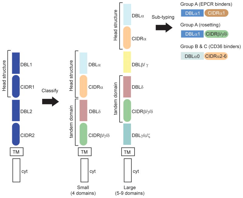
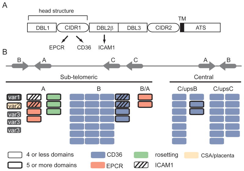
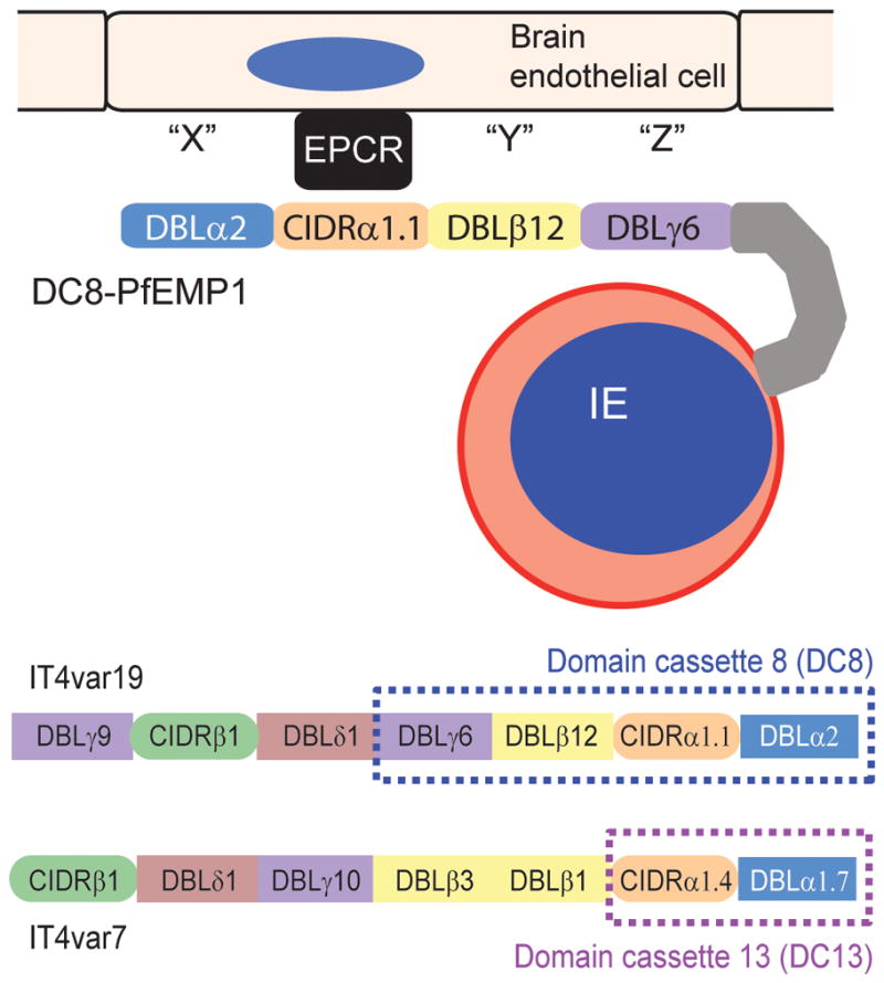
The Plasmodium falciparum erythrocyte membrane protein 1 (PfEMP1) family has a key role in parasite survival, transmission, and virulence. PfEMP1 are exported to the erythrocyte membrane and mediate binding of infected erythrocytes to the endothelial lining of blood vessels. This process aids parasite survival by avoiding spleen-dependent killing mechanisms, but it is associated with adhesion-based disease complications. Switching between PfEMP1 proteins enables parasites to evade host immunity and modifies parasite tropism for different microvascular beds. The PfEMP1 protein family is one of the most diverse adhesion modules in nature. This review covers PfEMP1 adhesion domain classification and the significant role it is playing in deciphering and deconvoluting P. falciparum cytoadhesion and disease.

Keywords: Antigenic variation; Malaria; Pathogenesis; Plasmodium falciparum; Var.


Figures

Fig 1
Fig 1
Adhesion domain classification of PfEMP1 proteins. The blue PfEMP1 shows a typical arrangement of PfEMP1 domains. The first arrow indicates how adhesion domain classification reveals higher domain organization in PfEMP1. Specific DBL and CIDR domain types form preferential tandem domain arrangements (DBLα-CIDRα and DBLδ-CIDRβ/γ/δ). The same tandem arrangements are present in small (4-domain) and large (5 or more domain) PfEMP1, but large proteins contain unique DBL subtypes (β or γ) that are not present in small proteins. The second arrow indicates that further sub-classification of adhesion domains (e.g. CIDRα1) distinguishes three different PfEMP1 head structure types and functional differences between group A, B, and C proteins. TM is transmembrane, cyt is cytoplasmic tail.
Fig 2
Fig 2
The genome organization and predicted PfEMP1 binding properties of the 3D7 genome reference isolate. (A) Schematic representation of PfEMP1 domain architecture. The CIDR domain in the PfEMP1 head structure has diverged into CD36 (group B and C) and EPCR (group A) binding variants; binding is highly predictable by adhesion domain classification. ICAM1 binding has been mapped to specific DBLβ subtypes following the PfEMP1 head structure. (B) Group A and B var genes are located in sub-telomeric regions and group C var in central chromosome regions [4]. The predicted binding properties for the 3D7 genome reference isolate are shown based on adhesion domain classification and recombinant protein binding studies. Blue proteins encode CD36-binding CIDR domains [9], red proteins encode EPCR-binding CIDR domains [23], green proteins encode head structures associated with rosetting [27], striped proteins encode DBLβ3 or DBLβ5 domains associated with ICAM1 binding [–39], and the beige protein (VAR2CSA) is linked to CSA binding in the placenta [7]. Large PfEMP1 proteins with 5 or more domains are distinguished by a thick border.
Fig 3
Fig 3
EPCR-binding PfEMP1 variants. The top shows a schematic of a DC8-expressing infected erythrocyte binding to brain endothelial cells. All four domains in the DC8 cassette encode binding activity for brain endothelial cells [44], EPCR binding maps to the CIDRα1.1 [23]. The bottom compares the protein architectures of a DC8 (IT4var19) and a DC13 (IT4var7) PfEMP1 variant, both of which have been shown to be selected on brain endothelial cells [42;44] and to encode EPCR binding activity in the orange colored CIDR domain [23]. Differences in domain composition between DC8, DC13, and other EPCR binding PfEMP1 variants have potential implications for the overall PfEMP1 binding specificities and parasite binding tropism for brain and other microvascular beds.

References

    1. Miller LH, Baruch DI, Marsh K, Doumbo OK. The pathogenic basis of malaria. Nature. 2002;415:673–9. - PubMed
    1. White NJ, Turner GD, Day NP, Dondorp AM. Lethal malaria: Marchiafava and Bignami were right. J Infect Dis. 2013;208:192–8. - PMC - PubMed
    1. Kraemer SM, Smith JD. A family affair: var genes, PfEMP1 binding, and malaria disease. Current Opinion in Microbiology. 2006;9:374–80. - PubMed
    1. Gardner MJ, Hall N, Fung E, White O, Berriman M, Hyman RW, et al. Genome sequence of the human malaria parasite Plasmodium falciparum. Nature. 2002;419:498–511. - PMC - PubMed
    1. Kraemer SM, Kyes SA, Aggarwal G, Springer AL, Nelson SO, Christodoulou Z, et al. Patterns of gene recombination shape var gene repertoires in Plasmodium falciparum: comparisons of geographically diverse isolates. BMC Genomics. 2007;8:45. - PMC - PubMed

Publication types

MeSH terms

Substances

LinkOut - more resources