

Liver regeneration after living donor transplantation: adult-to-adult living donor liver transplantation cohort study
- PMID: 25065488
- PMCID: PMC4276514
- DOI: 10.1002/lt.23966
Liver regeneration after living donor transplantation: adult-to-adult living donor liver transplantation cohort study
Abstract
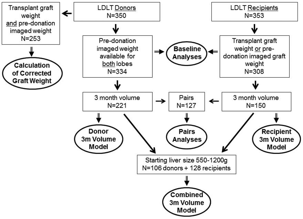
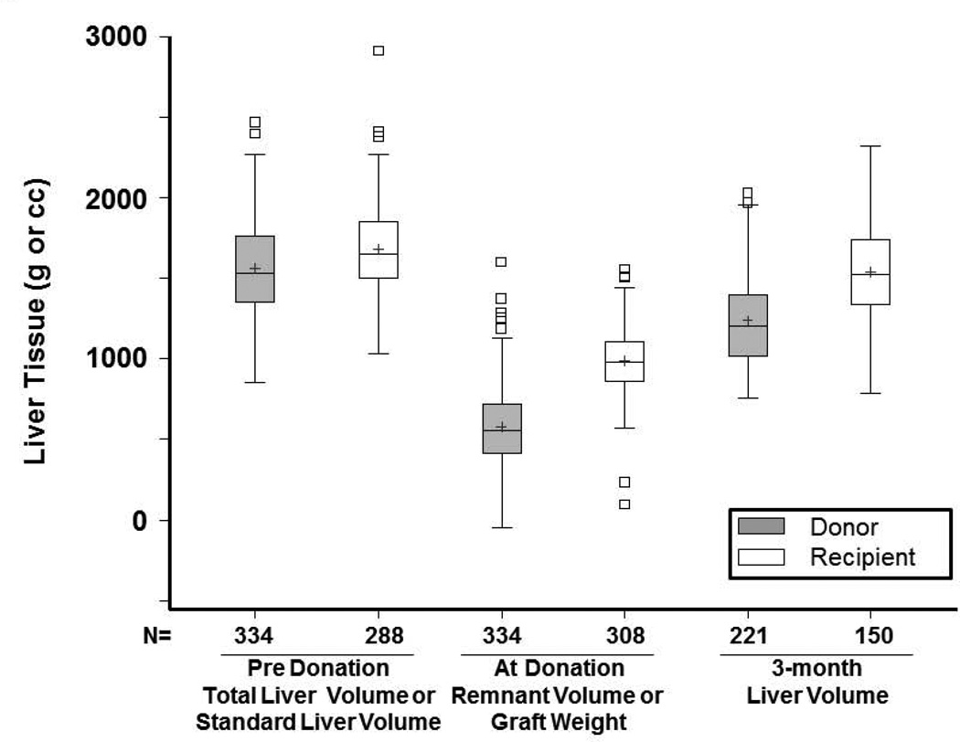
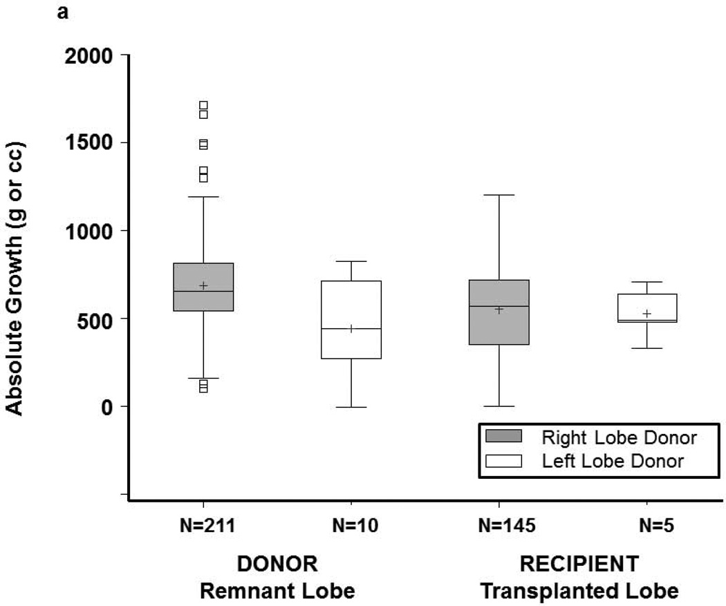
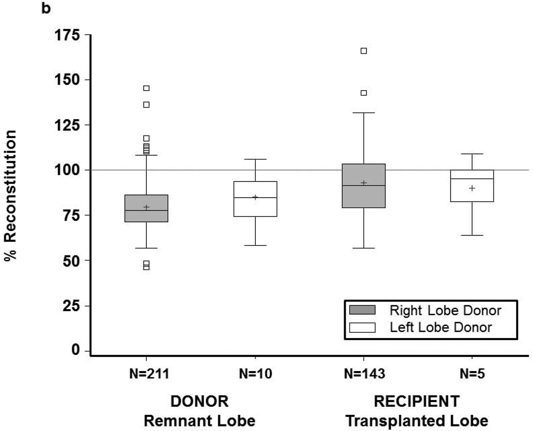
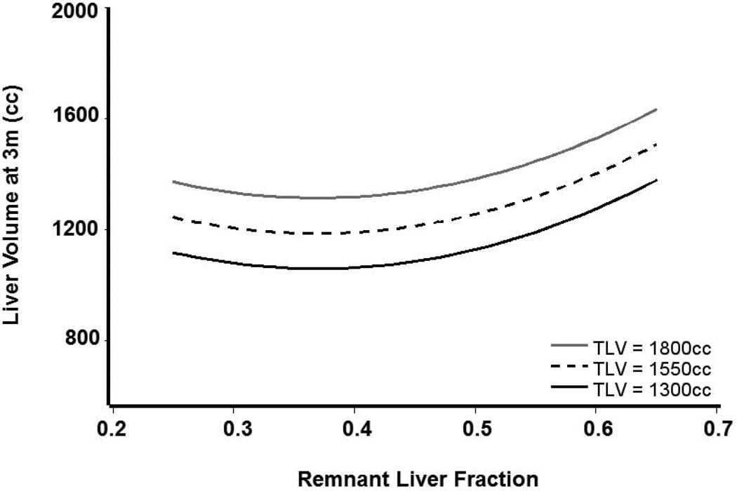
Adult-to-adult living donors and recipients were studied to characterize patterns of liver growth and identify associated factors in a multicenter study. Three hundred and fifty donors and 353 recipients in the Adult-to-Adult Living Donor Liver Transplantation Cohort Study (A2ALL) receiving transplants between March 2003 and February 2010 were included. Potential predictors of 3-month liver volume included total and standard liver volumes (TLV and SLV), Model for End-Stage Liver Disease (MELD) score (in recipients), the remnant and graft size, remnant-to-donor and graft-to-recipient weight ratios (RDWR and GRWR), remnant/TLV, and graft/SLV. Among donors, 3-month absolute growth was 676 ± 251 g (mean ± SD), and percentage reconstitution was 80% ± 13%. Among recipients, GRWR was 1.3% ± 0.4% (8 < 0.8%). Graft weight was 60% ± 13% of SLV. Three-month absolute growth was 549 ± 267 g, and percentage reconstitution was 93% ± 18%. Predictors of greater 3-month liver volume included larger patient size (donors and recipients), larger graft volume (recipients), and larger TLV (donors). Donors with the smallest remnant/TLV ratios had larger than expected growth but also had higher postoperative bilirubin and international normalized ratio at 7 and 30 days. In a combined donor-recipient analysis, donors had smaller 3-month liver volumes than recipients adjusted for patient size, remnant or graft volume, and TLV or SLV (P = 0.004). Recipient graft failure in the first 90 days was predicted by poor graft function at day 7 (HR = 4.50, P = 0.001) but not by GRWR or graft fraction (P > 0.90 for each). Both donors and recipients had rapid yet incomplete restoration of tissue mass in the first 3 months, and this confirmed previous reports. Recipients achieved a greater percentage of expected total volume. Patient size and recipient graft volume significantly influenced 3-month volumes. Importantly, donor liver volume is a critical predictor of the rate of regeneration, and donor remnant fraction affects postresection function. Liver Transpl 21:79-88, 2015. © 2014 AASLD.
© 2014 American Association for the Study of Liver Diseases.
Conflict of interest statement
The authors have no conflicts of interest to disclose.
Figures
Similar articles
-
Increasing the recipient benefit/donor risk ratio by lowering the graft size requirement for living donor liver transplantation.Liver Transpl. 2012 Sep;18(9):1078-82. doi: 10.1002/lt.23433. Liver Transpl. 2012. PMID: 22927142
-
Donor age affects liver regeneration during early period in the graft liver and late period in the remnant liver after living donor liver transplantation.World J Surg. 2012 May;36(5):1102-1111. doi: 10.1007/s00268-012-1496-1. World J Surg. 2012. PMID: 22374540
-
Right lobe living donors ages 55 years old and older in liver transplantation.Liver Transpl. 2017 Oct;23(10):1305-1311. doi: 10.1002/lt.24823. Liver Transpl. 2017. PMID: 28734130
-
Status of Adult Living Donor Liver Transplantation in the United States: Results from the Adult-To-Adult Living Donor Liver Transplantation Cohort Study.Gastroenterol Clin North Am. 2018 Jun;47(2):297-311. doi: 10.1016/j.gtc.2018.01.004. Gastroenterol Clin North Am. 2018. PMID: 29735025 Review.
-
Outcomes of adult patients adopting small-for-size grafts in living donor liver transplantation: A systematic review and meta-analysis.Hepatobiliary Pancreat Dis Int. 2019 Jun;18(3):206-213. doi: 10.1016/j.hbpd.2019.03.007. Epub 2019 Mar 29. Hepatobiliary Pancreat Dis Int. 2019. PMID: 30952435
Cited by
-
Patterns of Early Allograft Dysfunction in Adult Live Donor Liver Transplantation: The A2ALL Experience.Transplantation. 2016 Jul;100(7):1490-9. doi: 10.1097/TP.0000000000001240. Transplantation. 2016. PMID: 27326811 Free PMC article.
-
Impact of donor age on liver regeneration and function following adult living donor liver transplantation.Exp Ther Med. 2019 May;17(5):3965-3970. doi: 10.3892/etm.2019.7454. Epub 2019 Mar 29. Exp Ther Med. 2019. PMID: 31007739 Free PMC article.
-
Management of the middle hepatic vein in right lobe living donor liver transplantation: A meta-analysis.J Huazhong Univ Sci Technolog Med Sci. 2015 Aug;35(4):600-605. doi: 10.1007/s11596-015-1477-3. Epub 2015 Jul 31. J Huazhong Univ Sci Technolog Med Sci. 2015. PMID: 26223934
-
4D Flow MRI in the portal venous system: imaging and analysis methods, and clinical applications.Radiol Med. 2022 Nov;127(11):1181-1198. doi: 10.1007/s11547-022-01553-x. Epub 2022 Sep 19. Radiol Med. 2022. PMID: 36123520 Free PMC article. Review.
-
Short-Term Monitoring of Graft Regeneration in Partial Liver Transplantation Recipients.Ann Transplant. 2023 Dec 12;28:e941444. doi: 10.12659/AOT.941444. Ann Transplant. 2023. PMID: 38083825 Free PMC article.
References
-
- Fausto N, Campbell JS, Riehle KJ. Liver regeneration. Hepatology. 2006;43:S45–S53. - PubMed
-
- Pomfret EA, Pomposelli JJ, Gordon FD, Erbay N, Lyn Price L, Lewis WD, et al. Liver regeneration and surgical outcome in donors of right-lobe liver grafts. Transplantation. 2003;76:5–10. - PubMed
-
- Humar A, Kosari K, Sielaff TD, Glessing B, Gomes M, Dietz C, et al. Liver regeneration after adult living donor and deceased donor split-liver transplants. Liver Transpl. 2004;10:374–378. - PubMed
-
- Gruttadauria S, Parikh V, Pagano D, Tuzzolino F, Cintorino D, Miraglia R, et al. Early regeneration of the remnant liver volume after right hepatectomy for living donation: a multiple regression analysis. Liver Transpl. 2012;18:907–913. - PubMed
Publication types
MeSH terms
Grants and funding
- U01 DK062496/DK/NIDDK NIH HHS/United States
- U01 DK062536/DK/NIDDK NIH HHS/United States
- U01-DK62444/DK/NIDDK NIH HHS/United States
- U01-DK62536/DK/NIDDK NIH HHS/United States
- U01-DK62505/DK/NIDDK NIH HHS/United States
- U01 DK062498/DK/NIDDK NIH HHS/United States
- U01 DK062484/DK/NIDDK NIH HHS/United States
- U01-DK62483/DK/NIDDK NIH HHS/United States
- U01 DK062483/DK/NIDDK NIH HHS/United States
- U01 DK062467/DK/NIDDK NIH HHS/United States
- U01-DK62467/DK/NIDDK NIH HHS/United States
- U01 DK062531/DK/NIDDK NIH HHS/United States
- U01 DK062444/DK/NIDDK NIH HHS/United States
- U01-DK62531/DK/NIDDK NIH HHS/United States
- U01-DK62484/DK/NIDDK NIH HHS/United States
- U01 DK062505/DK/NIDDK NIH HHS/United States
- U01-DK62494/DK/NIDDK NIH HHS/United States
- U01-DK62498/DK/NIDDK NIH HHS/United States
- U01-DK62496/DK/NIDDK NIH HHS/United States
- U01 DK062494/DK/NIDDK NIH HHS/United States