. 2014 Nov;158(5):875-91.
doi: 10.1016/j.ajo.2014.07.025. Epub 2014 Jul 24.

Retinoblastoma. Fifty years of progress. The LXXI Edward Jackson Memorial Lecture

Affiliations

Retinoblastoma. Fifty years of progress. The LXXI Edward Jackson Memorial Lecture

Hans E Grossniklaus. Am J Ophthalmol. 2014 Nov.

Abstract

Purpose: To review the progress made in understanding the genetic basis, molecular pathology, and treatment of retinoblastoma since the previous Jackson lecture on the topic was published 50 years ago.

Design: Perspective based on personal experience and the literature.

Methods: The literature regarding retinoblastoma was reviewed since 1963. Advances in understanding the biology and treatment of retinoblastoma provided context through the author's clinical, pathologic, and research experiences.

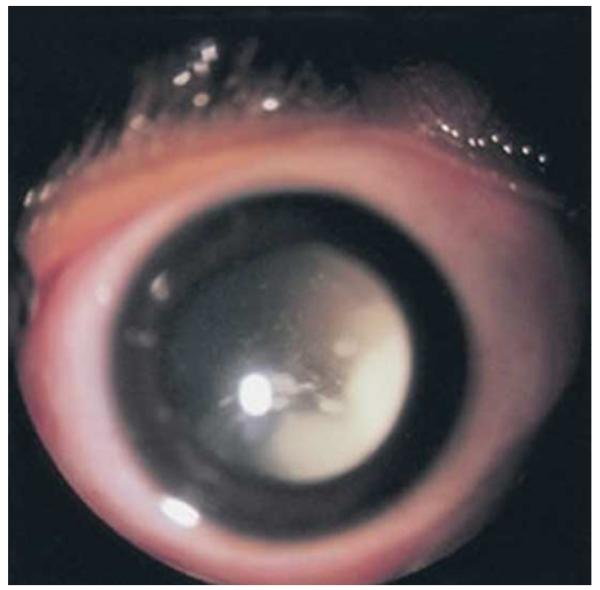
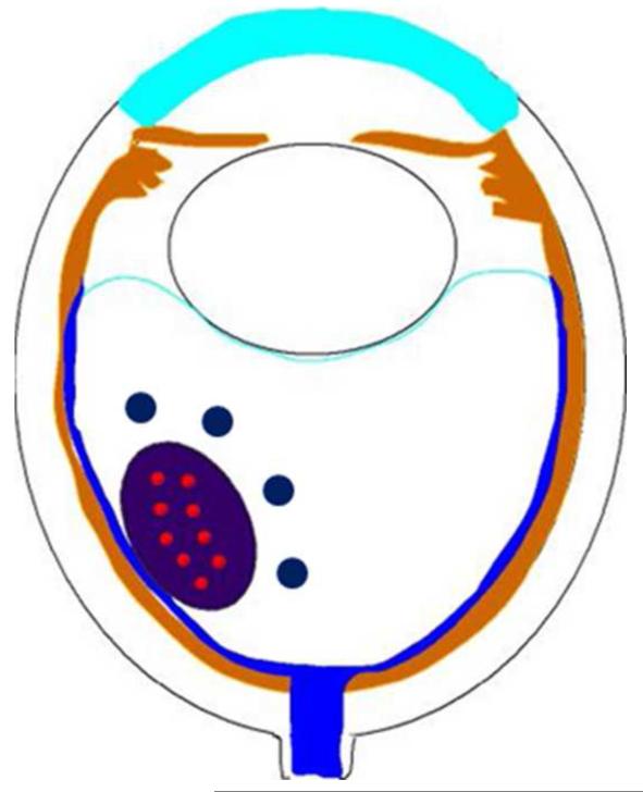
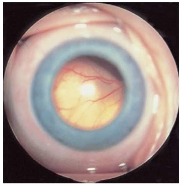
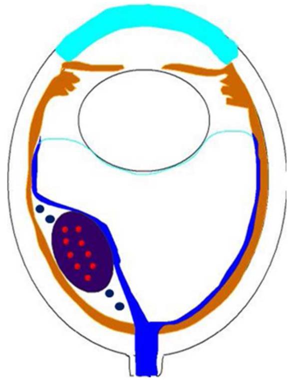
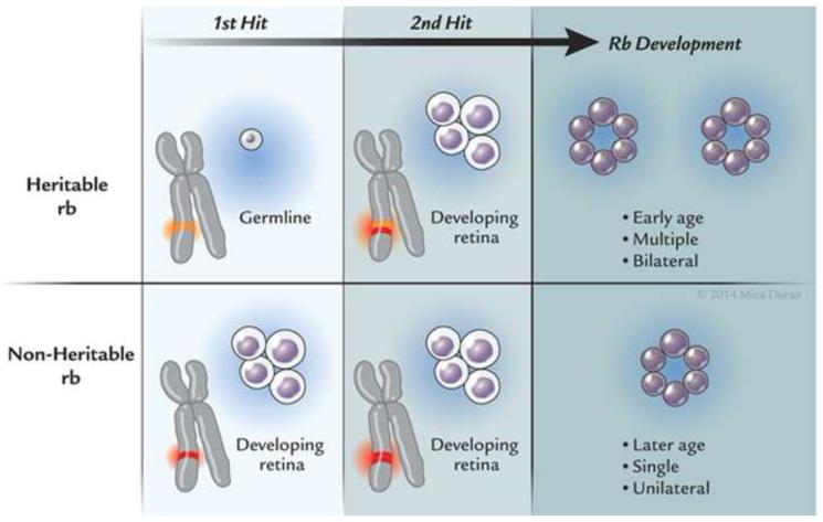
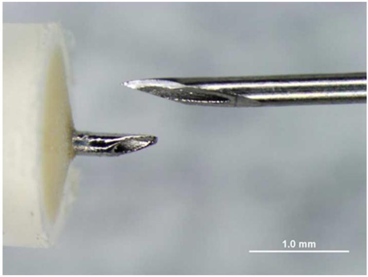
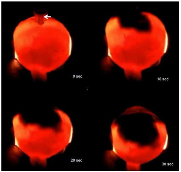
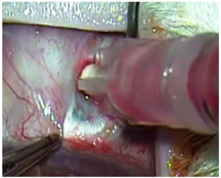
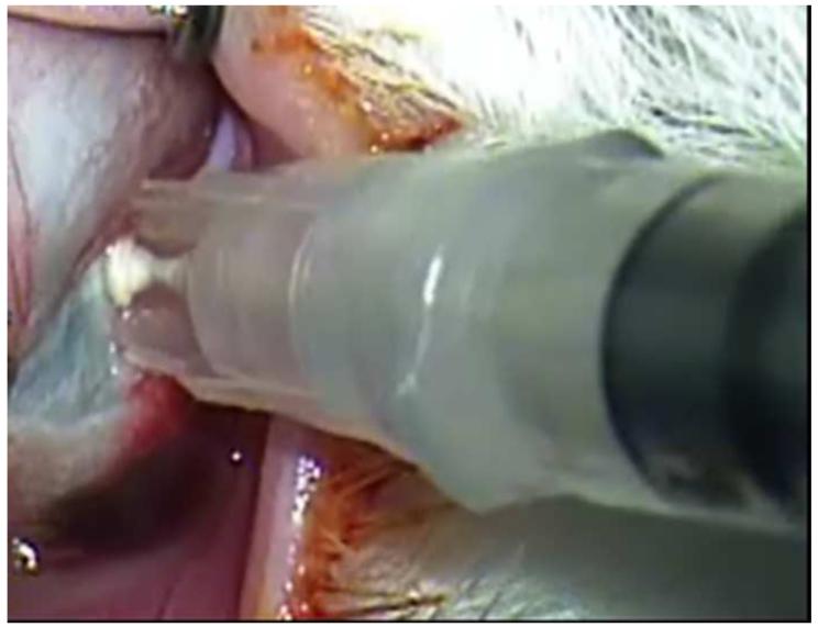
Results: Retinoblastoma was first identified in the 1500s and defined as a unique clinicopathologic entity in 1809. Until the mid-1900s, knowledge advanced sporadically, with technological developments of ophthalmoscopy and light microscopy, and with the introduction of surgical enucleation, chemotherapy, and radiation therapy. During the last 50 years, research and treatment have progressed at an unprecedented rate owing to innovations in molecular biology and the development of targeted therapies. During this time period, the retinoblastoma gene was discovered; techniques for genetic testing for retinoblastoma were developed; and plaque brachytherapy, chemoreduction, intra-arterial chemotherapy, and intraocular injections of chemotherapeutic agents were successfully introduced.

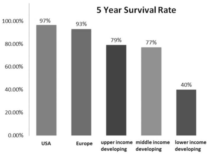
Conclusions: Nearly all patients with retinoblastoma in developed countries can now be cured of their primary cancer--a remarkable achievement for a childhood cancer that once was uniformly fatal. Much of this success is owed to deciphering the role of the Rb gene, and the benefits of targeted therapies, such as chemoreduction with consolidation as well as intra-arterial and intravitreal chemotherapies. Going forward, the main challenge will be ensuring that access to care is available for all children, particularly those in developing countries.


Figures

Figure 1
Figure 1
Endophytic retinoblastoma. Left. Endophytic retinoblastoma extends from the retina into the vitreous, thus resulting in a white mass associated with tumor shedding into the vitreous (i.e. seeds). Right. The primary tumor is vascularized (red) whereas the vitreous seeds (blue) are avascular.
Figure 1
Figure 1
Endophytic retinoblastoma. Left. Endophytic retinoblastoma extends from the retina into the vitreous, thus resulting in a white mass associated with tumor shedding into the vitreous (i.e. seeds). Right. The primary tumor is vascularized (red) whereas the vitreous seeds (blue) are avascular.
Figure 2
Figure 2
Exophytic retinoblastoma. Left. Exophytic retinoblastoma extends from the retina into the subretinal space and clinically appears as a white to slightly yellow subretinal mass. Right. The primary tumor is vascularized (red) and subretinal seeds (blue) are avascular.
Figure 2
Figure 2
Exophytic retinoblastoma. Left. Exophytic retinoblastoma extends from the retina into the subretinal space and clinically appears as a white to slightly yellow subretinal mass. Right. The primary tumor is vascularized (red) and subretinal seeds (blue) are avascular.
Figure 3
Figure 3
Anterior diffuse retinoblastoma. Left. Anterior diffuse retinoblastoma results in a white pseudohypopyon. Right. The primary tumor arises as a small mass in the area of the ora serrate and seeds into the aqueous in front of the anterior hyaloid, spreads through the pupil, and accumulates in the peripheral iris.
Figure 3
Figure 3
Anterior diffuse retinoblastoma. Left. Anterior diffuse retinoblastoma results in a white pseudohypopyon. Right. The primary tumor arises as a small mass in the area of the ora serrate and seeds into the aqueous in front of the anterior hyaloid, spreads through the pupil, and accumulates in the peripheral iris.
Figure 4
Figure 4
Retinocytoma. This patient has two calcified peripheral retinal lesion that represent retinocytoma, once confused with spontaneously regressed retinoblastoma. The patient’s daughter developed retinoblastoma. (courtesy of G. Baker Hubbard III MD)
Figure 5
Figure 5
Retinoblastoma pathology. Upper left. Retinoblasotmas have a gross white, fleshy to cottage-cheese appearance. Upper right. Retinoblastoma is a small round blue cell tumor, forming sheets of cells. Lower left. Retinoblastoma may exhibit rosette formation, including Homer Wright rosettes with neuropil in the lumen (arrows), or Flexner-Wintersteiner rosettes with a clear lumen (arrowheads). Lower right. Retinoblasotma may contain well differentiated areas, exhibiting cells with small nuclei, rather abundant cytoplasm, and fleurettes with photoreceptor outer segments in the lumen. (upper right, lower left and lower right, hematoxylin and eosin, 25X)
Figure 5
Figure 5
Retinoblastoma pathology. Upper left. Retinoblasotmas have a gross white, fleshy to cottage-cheese appearance. Upper right. Retinoblastoma is a small round blue cell tumor, forming sheets of cells. Lower left. Retinoblastoma may exhibit rosette formation, including Homer Wright rosettes with neuropil in the lumen (arrows), or Flexner-Wintersteiner rosettes with a clear lumen (arrowheads). Lower right. Retinoblasotma may contain well differentiated areas, exhibiting cells with small nuclei, rather abundant cytoplasm, and fleurettes with photoreceptor outer segments in the lumen. (upper right, lower left and lower right, hematoxylin and eosin, 25X)
Figure 6
Figure 6
Retinoblastoma inheritance, Heritable retinoblastoma requires two mutations: one that is inherited in the patient’s germline and the other in the developing retina. These tumors are usually bilateral, multiple, and occur at an early age. Non-heritable (acquired) retinoblastoma also requires two mutations, although both of these mutations occur in the developing retina. These tumors are usually single, unilateral, and occur at a later age.
Figure 7
Figure 7
Rb in progression to retinoblastoma. 1. Retinoblastoma (Rb) recruits proteins such as HDAC and BRG1 to E2F sites resulting in chromatin structure alterations and prevention of access to transcriptional machinery. 2. Phosphorylation by CDKs release HDAC and BRG1, thus releasing histone from DNA, allowing access to transcriptional machinery and resulting in cell division. Mutations in both alleles of Rb result in uncontrolled proliferation/retinocytoma formation, and further events including P16Ink4a, P14/Arf, or BRCA2 mutations, SV40 interactions with Rb, or Zeb1 activation by inactivated pRb allow progression to retinoblastoma. 3. The cell cycle may be exited under normal conditions when Rb is hypophosphorylated via MITF induction of p16, thus allowing HDCAC and BRG1 to rebind to Rb via E2F sites and suppress transcription. 4. Alternative cell cycle exit occurs under stress or hyperpoliferation; Rb is hyperphosphorylated, releases E2f and undergoes Tp53 mediated apoptosis, which may be suppressed by BRCA1 mutations. Thus, Rb mutations initiate the process and retinoblastoma results through progressive mutational events involving chromosome 9, 13 and 17.
Figure 8
Figure 8
Retinoblastoma cell of origin. Gene expression profiling (GEP) of retinoblastoma has resulted in two general phenotypes supporting a primitive retinal precursor cell (RPC) origin of retinoblastoma. This cell type is present in the outer nuclear zone of the developing retina. If mutational events occur early, before further differentiation of the RPC, group 1 retinoblastoma occurs; if the mutational event occurs later, group 2 retinoblastoma occurs. Group 1 retinoblastoma expresses RPC genes and group 2 retinoblastoma expresses cone genes. Group 1 retinoblastoma has a more primitive phenotype and group 2 retinoblastoma may exhibit photoreceptor differentiation, including rosette and fleurette formation.
Figure 9
Figure 9
Grading anaplasia in retinoblastoma anaplasia. Upper left. No anaplasia (retinoctyoma) exhibits small, compact nuceli, abundant eosinophilic cytoplasm, and photoreceptor differentiation. Upper right. Mild anaplasia is characterized by tumor cells with round, hyperchromatic nuclei, increased nuclear to cytoplasmic ratios, and a moderate amount of eosinophilic cytoplasm. Lower left. Moderate anaplasia is characterized by cells with larger nuclei, high nuclear to cytoplasmic ratios, and scanty eosinophilic cytoplasm. Lower right. Severe anaplasia is characterized by cells with larger, hyperchromatic nuclei, nuclear molding/angulation, and virtually no cytoplasm. (hematoxylin and eosin, 100X)
Figure 9
Figure 9
Grading anaplasia in retinoblastoma anaplasia. Upper left. No anaplasia (retinoctyoma) exhibits small, compact nuceli, abundant eosinophilic cytoplasm, and photoreceptor differentiation. Upper right. Mild anaplasia is characterized by tumor cells with round, hyperchromatic nuclei, increased nuclear to cytoplasmic ratios, and a moderate amount of eosinophilic cytoplasm. Lower left. Moderate anaplasia is characterized by cells with larger nuclei, high nuclear to cytoplasmic ratios, and scanty eosinophilic cytoplasm. Lower right. Severe anaplasia is characterized by cells with larger, hyperchromatic nuclei, nuclear molding/angulation, and virtually no cytoplasm. (hematoxylin and eosin, 100X)
Figure 10
Figure 10
Retinoblastoma survival based on anaplasia grade. This Kaplan-Meir plot no deaths or metastases associated with mildly anaplastic tumors, an approximate 97% 10 year survival/no metastases in patients with moderately anaplastic tumors, and an approximate 75% 10 year survival/no metastases in patients with severely anaplastic tumors (P<0.01).
Figure 11
Figure 11
Chemotherapeutic agents used in treating retinoblastoma. In general, there are four classes of chemotherapeutic agents used to treat retinoblastoma. Alkylating agents, such as melphalan, cause DNA intrastrand linking and cross-linking, resulting in damaged DNA that is not transcribed. Topoisomerase inhibitors, such as etoposide and topotecan, work by capping free double strands of DNA caused topoisomerase cleavage. The capped, damaged DNA is unable to be repaired and is degraded via apoptosis. Platinum based antineoplastic agents, such as carboplatin, bind DNA bases, thus resulting in kinked DNA; this damaged DNA is then degraded via apoptosis. Vinca alkyloids, such as vincristine, bind tubulin dimers, thus blocking mitotic spindle formation and inhibiting mitosis. Typically, combinations of chemotherapeutic agents with different mechanisms of action are used to treat retinoblastoma via systemic chemoreduction, such as Vincristine, Etopopside, and Carboplatin (VEC) therapy, which is given over 5 to 6 cycles.
Figure 12
Figure 12
Intravitreal injection of topotecan in rabbit model of retinoblastoma. Upper left. There are vitreous seeds of retinoblastoma (*) in the rabbit model. Upper right. The 32 gauge 4 mm needle (arrow) is being inserted at the pars plana. Lower left. The needle (arrow) is inserted to its hub and topotecan is injected into the vitreous. Lower right. After 3 weekly injections of 20μg of topotecan, vitreous seeds have disappeared, the retina/optic nerve appear normal and there is no spread of extraocular tumor.
Figure 12
Figure 12
Intravitreal injection of topotecan in rabbit model of retinoblastoma. Upper left. There are vitreous seeds of retinoblastoma (*) in the rabbit model. Upper right. The 32 gauge 4 mm needle (arrow) is being inserted at the pars plana. Lower left. The needle (arrow) is inserted to its hub and topotecan is injected into the vitreous. Lower right. After 3 weekly injections of 20μg of topotecan, vitreous seeds have disappeared, the retina/optic nerve appear normal and there is no spread of extraocular tumor.
Figure 13
Figure 13
Suprachoroidal injection of fluorospheres. Upper left. Comparison of microneedle (left) with standard 30 gauge needle; note that the microneedle is the same length as the bevel of the 30 gauge needle. Upper right. India ink injected over ora seratta migrates posteriorly in suprachoroidal space over 30 seconds. Lower left. 10 micron diameter pink fluorospheres in hub of 1 cc syringe with microneedle inserted into suprachoroidal space in a rabbit. Lower right. After injection, a volume of 200 microliters of flourospheres are in the suprachoroidal space and not in the hub of the needle.
Figure 13
Figure 13
Suprachoroidal injection of fluorospheres. Upper left. Comparison of microneedle (left) with standard 30 gauge needle; note that the microneedle is the same length as the bevel of the 30 gauge needle. Upper right. India ink injected over ora seratta migrates posteriorly in suprachoroidal space over 30 seconds. Lower left. 10 micron diameter pink fluorospheres in hub of 1 cc syringe with microneedle inserted into suprachoroidal space in a rabbit. Lower right. After injection, a volume of 200 microliters of flourospheres are in the suprachoroidal space and not in the hub of the needle.
Figure 14
Figure 14
Regional 5-year survival of children with retinoblastoma. Five-year survival rates for children with retinoblastoma vary among countries by their socioeconomic rank, ranging from nearly 97% in the United States to 40% in low income developing countries. Modified from Canturk et al

References

    1. Liesegang TJ, Hoskins HD, Jensen AD. The significance of the Edward Jackson lecture. Am J Ophthalmol. 2005;139(3):530–532. - PubMed
    1. Dunphy EB. The story of retinoblastoma. The XX Edward Jackson Memorial Lecture. Am J Ophthalmol. 1964;58(4):539–552. - PubMed
    1. Albert DM. Historic review of retinoblastoma. Ophthalmology. 1987;94(6):654–662. - PubMed
    1. Pawius P. Observationes anatomical. 1657;16:336. Cited by Bartolini, Bartolini Hafniae.
    1. Wardrop J. Observations on the Fungus Haematodes or Soft Cancer. George Ramsay and Co; Edinburgh: 2013. p. 1809.

Publication types

LinkOut - more resources