. 2014 Oct 1;91(3):369-79.
doi: 10.1016/j.bcp.2014.07.013. Epub 2014 Jul 24.

GABAB receptor phosphorylation regulates KCTD12-induced K⁺ current desensitization

Affiliations

GABAB receptor phosphorylation regulates KCTD12-induced K⁺ current desensitization

Lisa Adelfinger et al. Biochem Pharmacol. .

Abstract

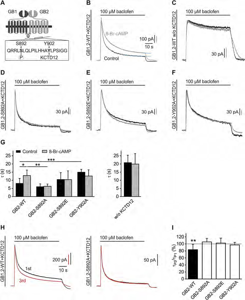
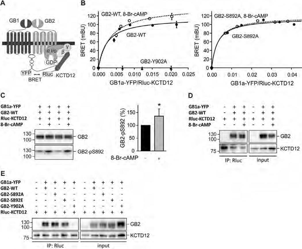
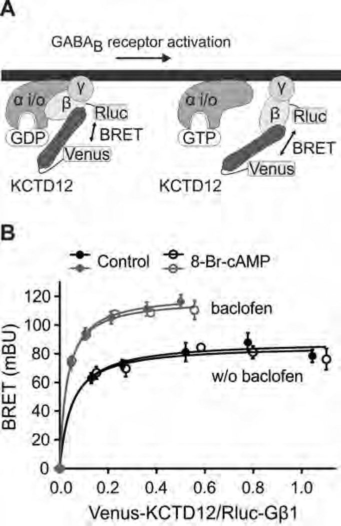
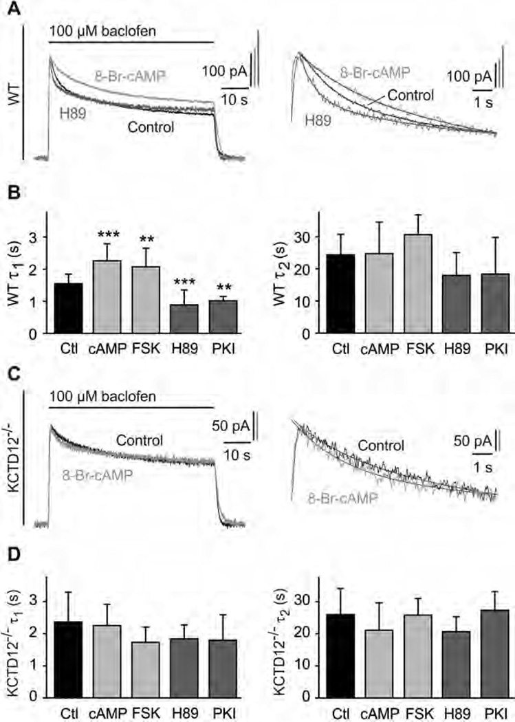
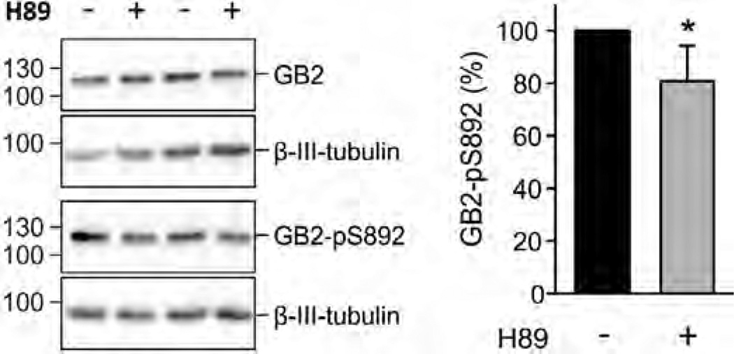
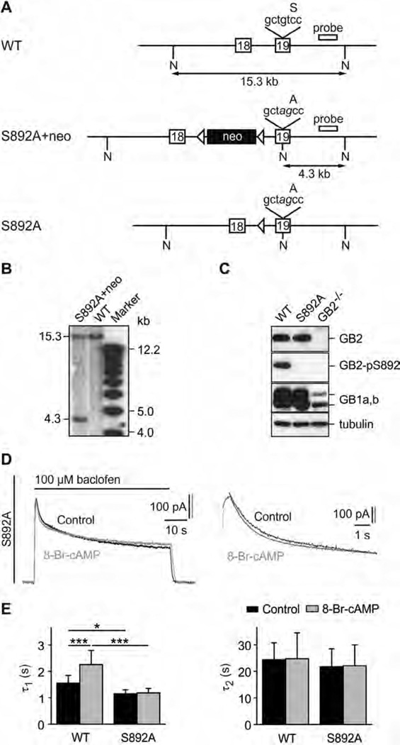
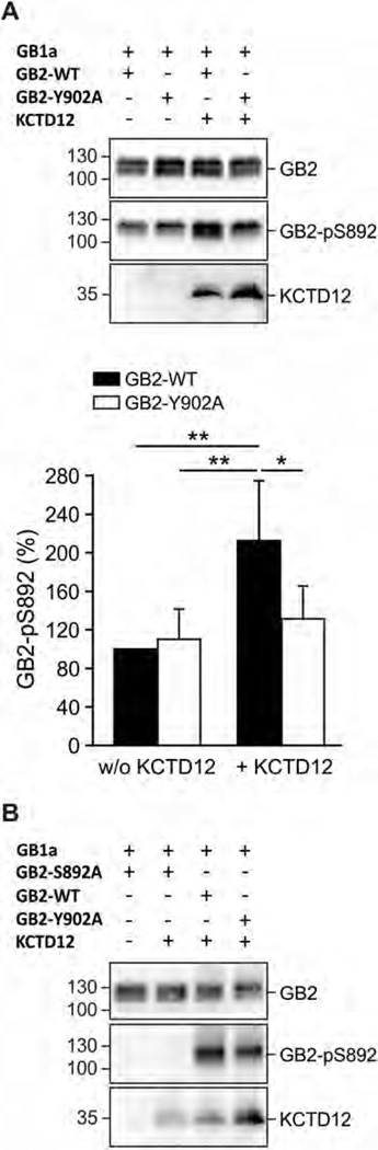
GABAB receptors assemble from GABAB1 and GABAB2 subunits. GABAB2 additionally associates with auxiliary KCTD subunits (named after their K(+) channel tetramerization-domain). GABAB receptors couple to heterotrimeric G-proteins and activate inwardly-rectifying K(+) channels through the βγ subunits released from the G-protein. Receptor-activated K(+) currents desensitize in the sustained presence of agonist to avoid excessive effects on neuronal activity. Desensitization of K(+) currents integrates distinct mechanistic underpinnings. GABAB receptor activity reduces protein kinase-A activity, which reduces phosphorylation of serine-892 in GABAB2 and promotes receptor degradation. This form of desensitization operates on the time scale of several minutes to hours. A faster form of desensitization is induced by the auxiliary subunit KCTD12, which interferes with channel activation by binding to the G-protein βγ subunits. Here we show that the two mechanisms of desensitization influence each other. Serine-892 phosphorylation in heterologous cells rearranges KCTD12 at the receptor and slows KCTD12-induced desensitization. Likewise, protein kinase-A activation in hippocampal neurons slows fast desensitization of GABAB receptor-activated K(+) currents while protein kinase-A inhibition accelerates fast desensitization. Protein kinase-A fails to regulate fast desensitization in KCTD12 knock-out mice or knock-in mice with a serine-892 to alanine mutation, thus demonstrating that serine-892 phosphorylation regulates KCTD12-induced desensitization in vivo. Fast current desensitization is accelerated in hippocampal neurons carrying the serine-892 to alanine mutation, showing that tonic serine-892 phosphorylation normally limits KCTD12-induced desensitization. Tonic serine-892 phosphorylation is in turn promoted by assembly of receptors with KCTD12. This cross-regulation of serine-892 phosphorylation and KCTD12 activity sharpens the response during repeated receptor activation.

Keywords: G-protein coupled receptor; GABA-B; GPCR; Kir3; PKA.


Figures

Fig. 1
Fig. 1
S892 phosphorylation slows KCTD12-induced desensitization of baclofen-activated K+ currents. (A) Scheme highlighting the close proximity of the PKA phosphorylation-site S892 and the KCTD12 binding-site Y902 in the C-terminal domain of GB2. (B) Representative traces of GIRK currents activated by baclofen (100 µM) and recorded at −50 mV from CHO-K1 cells expressing GB2-WT, GB1b, GIRK1/2 and KCTD12. Cells were pre-incubated without (Control, black trace) or with 8-Br-cAMP(1 mM, 30 min; grey trace). The desensitization time constants τ1 and τ2 were derived from double-exponential fits (blue trace) to the decay phase of K+ currents during baclofen application. (C) Representative traces as in (B) from CHO-K1 cells lacking KCTD12. (D–F) Representative traces as in (B) from CHO-K1 cells expressing the GB2 mutants S892A (D), S892E (E) or Y902A (F). (G) Bar graph summarizing the time constant τ (amplitude-weighted mean time constant calculated from τ1 and τ2) of baclofen-induced K+ current desensitization. Data are means ± SD, n = 5–16. *,p < 0.05; **,p < 0.01; ***,p < 0.001; Sidak’s multiple comparison test. (H) Repeated activation of GIRK currents in CHO-K1 cells expressing WT GABAB receptors (GB1,2-WT) together with KCTD12 results in a sharpening of the current response (left; 1st response black, 3rd response red). No sharpening of the current response upon repeated activation is observed with mutated GABAB receptors (GB1,2-S892A) that cannot be phosphorylated at S892 (right). Baclofen applications were for 60-s in intervals of 5 min. (I) Bar graph summarizing experiments as shown in (H). In experiments with GB2-WT, the τ of the 3rd response (τP3) was significantly reduced compared to the τ of the 1st response (τP1). No change in the desensitization of the current response upon repeated activation is seen (i) when S892 cannot be phosphorylated (S892A), (ii) with the phospho-mimetic S892E and (iii) when KCTD12 cannot bind to GB2 (Y902A). Data are means ± SD, n = 7–12. **, p < 0.01; t test.
Fig. 2
Fig. 2
S892 phosphorylation rearranges the receptor/KCTD12 complex. (A) Scheme of the BRET Rluc-KCTD12 donor and GB1a-YFP acceptor pair. (B) BRET donor saturation curves from HEK293T cells expressing fixed amounts of GB2-WT, GB2-Y902A (both left) or GB2-S892A (right) together with Rluc-KCTD12 and increasing amounts of GB1a-YFP. Cells were pre-incubated with 8-Br-cAMP(1 mM, 30 min; open circles) to activate PKA. BRET is expressed as milli BRET units (mBU) determined as net BRET × 1000. Data points are means ± SD of technical quadruplicates of a representative experiment, n = 4–7. (C) Western blot of lysates of HEK293T cells expressing GB1a-YFP, GB2-WT and Rluc-KCTD12 in the presence and absence of 8-Br-cAMP(1 mM, 30 min pre-incubation). Activation of PKA does not affect the total amount of GB2 but increases phosphorylation at S892 (GB2-pS892). GB2 runs as two bands on SDS–PAGE, likely due to differences in posttranslational modification. The higher molecular weight band appears to be more heavily phosphorylated at S892. Blots are representative of six independent experiments. Bar graph summarizing the amount of S892 phosphorylation normalized to the amount of GB2 on the same blot (GB2-pS892 in %). Data are means ± SD, n = 6. *, p < 0.05; t test. (D) Co-immunoprecipitation of GABAB receptors with KCTD12 from HEK293T cells expressing GB1a-YFP, GB2-WT and Rluc-KCTD1 2 in the presence and absence of 8-Br-cAMP(1 mM, 30 min pre-incubation). Equivalent amounts of GABAB receptors were co-precipitated. Blots are representative of five independent experiments. (E) Co-immunoprecipitation of GABAB receptors with KCTD12 from HEK293T cells expressing GB1a-YFP, GB2 (WT, S892A, S892E or Y902A) and Rluc-KCTD12. Y902A does not bind KCTD12 and was used as a negative control. Blots are representative of three independent experiments.
Fig. 3
Fig. 3
Binding of KCTD12 to the G-protein βγ subunits is modulated by receptor activation but not by PKA activation. (A) Scheme of the BRET Rluc-Gβ1 donor and Venus-KCTD12 acceptor pair. (B) BRET donor saturation curves from CHO-K1 cells stably expressing GB1 and GB2 and transiently expressing fixed amounts of Rluc-Gβ1 and Gγ2 together with increasing amounts of Venus-KCTD12. Following receptor activation with baclofen (100 µM, 5 min pre-incubation; grey traces) the BRET curves were shifted towards higher BRETmax values. Activation of PKA with 8-Br-cAMP (1 mM, 30 min pre-incubation; open circles) had no effect on the BRET curves. BRET is expressed as milli BRET units (mBU) determined as net BRET × 1000. Data points are means ± SD of technical quadruplicates of a representative experiment, n = 3.
Fig. 4
Fig. 4
PKA activation slows fast desensitization of baclofen-activated K+ currents in cultured hippocampal neurons of WT but not of KCTD12−/− mice. (A) Representative baclofen-activated K+ currents recorded at −50 mV from WT hippocampal neurons. PKA activity was modulated by pre-incubation for 30 min with 8-Br-cAMP (1 mM; bright grey trace) or H89 (2 µM; dark grey trace). Controls represent recordings from untreated neurons (black trace). The desensitization time constants τ1 and τ2 were derived from double-exponential fits to the decay phase of K+ currents during baclofen application (enlarged on the right). (B) Bar graph summarizing the time constants τ1 and τ2 of baclofen-induced K+ current desensitization for the indicated treatments. Data are means ± SD, n = 7–35. **, p < 0.01; ***, p < 0.001; Dunnett’s multiple comparison test, compared to Ctl. Ctl, control; FSK, forskolin; cAMP, 8-Br-cAMP. (C) Representative baclofen-activated K+ current responses from hippocampal KCTD12−/− neurons pre-incubated with 8-Br-cAMP (grey trace) or untreated KCTD12−/− neurons (control, black trace) as in (A). Traces were fitted as in (A). (D) Bar graph summarizing the time constants τ1 and τ2 of baclofen-induced K+ current desensitization for the indicated treatments. Data are means ± SD, n = 5–17, Dunnett’s multiple comparison test, compared to Ctl.
Fig. 5
Fig. 5
Basal phosphorylation of S892 is decreased by inhibiting PKA in hippocampal neurons of WT mice. Western blot of cultured neurons in the presence and absence of H89 (10 µM, 2 h pre-incubation; left). Inhibition of PKA does not affect the amount of GB2 protein but decreases the amount of S892 phosphorylation (GB2-pS892). β-III-Tubulin was used as a loading control. Bar graph summarizing the amount of phosphorylated S892 (GB2-pS892 in %; right). The total amount of GB2 and GB2-pS892 were normalized to β-III-tubulin on the same blot. Data are means ± SD, n = 6. *, p < 0.05; t test.
Fig. 6
Fig. 6
Lack of S892 phosphorylation in S892A knock-in mice accelerates fast desensitization of GABAB-activated K+ currents. (A) WT and mutated GB2 alleles. The S892 to alanine mutation (tcc → gcc) and a silent diagnostic NheI restriction site (gctagc) were introduced into exon 19 using homologous recombination in Balb/c embryonic stem (ES) cells. Mutated nucleotides are shown in italic. A neomycin marker (neo) flanked by loxP sites (arrowheads) was used for selection of ES cells. Correctly targeted ES cells (S892A + neo allele) were injected into C57BL/6 blastocysts. A founder mouse was crossed with a Balb/c mouse expressing Cre-recombinase to excise the neomycin cassette, leaving one loxP site behind (S892A allele). The hybridization probe used in the Southern blot in (B) is indicated. A, alanine; N, NheI restriction sites; S, serine. (B) Southern blot of NheI cut genomic DNA from correctly targeted ES cells. The probe labels a 15.3 kb fragment for the WT allele and a 4.3 kb fragment for the S892A + neo allele. (C) Western blot analysis of brain extracts showing that S892A mice express normal levels of GB2, GB1a and GB1b proteins. S892 was phosphorylated in brain extracts of WT but not S892A mice as shown with an antibody specific for phosphorylated S892 (GB2-pS892). Brain extracts of GB2-deficient mice (GB2−/−) [38] confirm the specificity of the GB2 and GB2-pS892 antibodies. β-III-Tubulin was used as a loading control. (D) Representative GABAB-activated K+ currents recorded at −50 mV in response to baclofen application (100 µM) from cultured hippocampal neurons of S892A mice. PKA was activated by pre-incubation for 30 min with 8-Br-cAMP (1 mM; grey trace). Controls represent recordings from untreated neurons (black trace). The desensitization time constants τ1 and τ2 were derived from double-exponential fits to the decay phase of K+ currents during baclofen application (enlarged on the right). (E) Bar graph summarizing the time constants τ1 and τ2 of baclofen-induced K+ current desensitization in WT and S892A neurons. Data are means ± SD, n = 5–7. *,p < 0.05; ***, p < 0.001; Sidak’s multiple comparison test.
Fig. 7
Fig. 7
Binding of KCTD12 to GABAB receptors increases S892 phosphorylation. (A) Lysates of HEK293T cells expressing GB1a and either GB2-WT or GB2-Y902A in the presence and absence of KCTD12 (up). S892 phosphorylation (GB2-pS892) is increased in the presence of KCTD12 for GB2-WT, but not for GB2-Y902A. Blots are representative of five independent experiments. Bar graph summarizing the amount of phosphorylated S892 normalized to GB2 protein on the same blot (GB2-pS892 in %; bottom). Data are means ± SDn = 5. *, p < 0.05; **, p < 0.01; Sidak’s multiple comparison test. (B) Western blot of lysates from transfected HEK293T cells expressing the indicated proteins. Phosphorylation of S892 (GB2-pS892) is observed with GB2-WT and GB2-Y902A but not with GB2-S892A, showing that the pS892 antibody specifically detects phosphorylation of S892. Note increased phosphorylation of S892 in the presence of KCTD12 with WT receptors (GB2-WT) compared to mutant receptors that cannot bind KCTD12 (GB2-Y902A). Blots are representative of three independent experiments.

References

    1. Gassmann M, Bettler B. Regulation of neuronal GABAB receptor functions by subunit composition. Nat Rev Neurosci. 2012;13:380–394. - PubMed
    1. Vigot R, Barbieri S, Brauner-Osborne H, Turecek R, Shigemoto R, Zhang YP, et al. Differential compartmentalization and distinct functions of GABAB receptor variants. Neuron. 2006;50:589–601. - PMC - PubMed
    1. Bettler B, Kaupmann K, Mosbacher J, Gassmann M. Molecular structure and physiological functions of GABAB receptors. Physiol Rev. 2004;84:835–867. - PubMed
    1. Couve A, Moss SJ, Pangalos MN. GABAB receptors: a new paradigm in G protein signaling. Mol Cell Neurosci. 2000;16:296–312. - PubMed
    1. Calver AR, Robbins MJ, Cosio C, Rice SQ, Babbs AJ, Hirst WD, et al. The C-terminal domains of the GABAB receptor subunits mediate intracellular trafficking but are not required for receptor signaling. J Neurosci. 2001;21:1203–1210. - PMC - PubMed

Publication types

MeSH terms