

Cxcr4 is transiently expressed in both epithelial and mesenchymal compartments of nascent hair follicles but is not required for follicle formation
- PMID: 25066162
- PMCID: PMC4372720
- DOI: 10.1111/exd.12523
Cxcr4 is transiently expressed in both epithelial and mesenchymal compartments of nascent hair follicles but is not required for follicle formation
Abstract
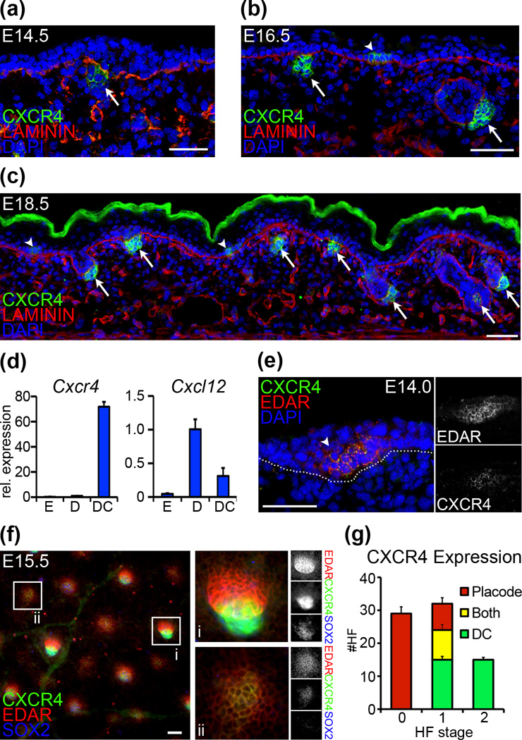
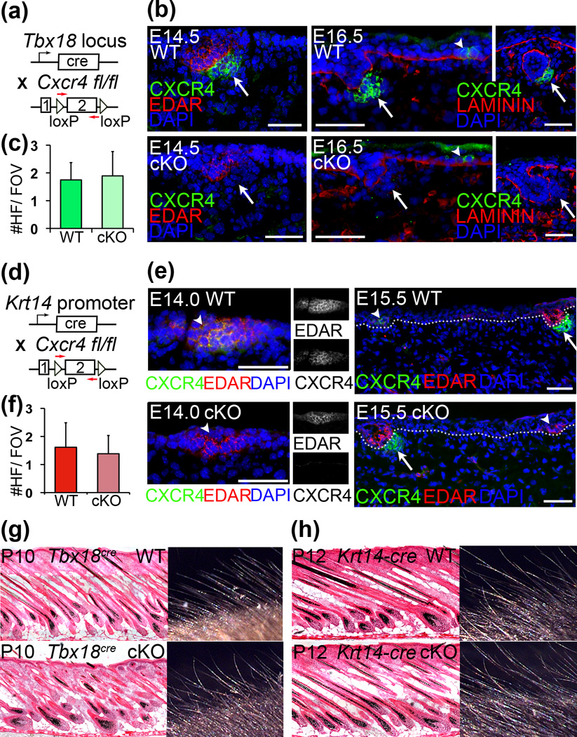
Hair follicle (HF) morphogenesis relies on the coordinated exchange of signals between mesenchymal and epithelial compartments of embryonic skin. Chemokine receptor Cxcr4 expression was recently identified in dermal condensates (DCs) of nascent HFs, but its role in promoting HF morphogenesis remains unknown. Our analyses confirmed Cxcr4 expression in condensate cells, and additionally revealed transient Cxcr4 expression in incipient epithelial hair placodes. Placodal Cxcr4 appeared prior to detection in DCs, representing a switch of expression between epithelial and mesenchymal compartments. To explore the functional role of this receptor in both compartments for early HF formation, we conditionally ablated Cxcr4 with condensate-targeting Tbx18(cre) knock-in and epidermis-targeting Krt14-cre transgenic mice. Conditional knockouts for both crosses were viable throughout embryogenesis and into adulthood. Morphological and biochemical marker analyses revealed comparable numbers of HFs forming in knockout embryos compared to wild-type littermate controls in both cases, suggesting that neither dermal nor epithelial Cxcr4 expression is required for early HF morphogenesis. We conclude that Cxcr4 expression and chemokine signaling through this receptor in embryonic mouse skin is dispensable for HF formation.
Keywords: dermal papilla cells; hair follicle morphogenesis; hair follicle stem cells; mesenchymal-epithelial interactions; stem cell niche.
© 2014 John Wiley & Sons A/S. Published by John Wiley & Sons Ltd.
Conflict of interest statement
The authors declare no conflict of interest.
Figures
References
-
- Mikkola ML. Genetic basis of skin appendage development. Semin Cell Dev Biol. 2007;18:225–236. - PubMed
-
- Millar SE. Molecular mechanisms regulating hair follicle development. J Invest Dermatol. 2002;118:216–225. - PubMed
-
- Kucia M, Jankowski K, Reca R, et al. CXCR4-SDF-1 signaling, locomotion, chemotaxis and adhesion. J Mol Histol. 2004;35:233–245. - PubMed
Publication types
MeSH terms
Substances
Grants and funding
LinkOut - more resources
Full Text Sources
Other Literature Sources
Molecular Biology Databases
Research Materials
Miscellaneous
