

Volumetric spectroscopic imaging of glioblastoma multiforme radiation treatment volumes
- PMID: 25066215
- PMCID: PMC4346247
- DOI: 10.1016/j.ijrobp.2014.03.049
Volumetric spectroscopic imaging of glioblastoma multiforme radiation treatment volumes
Abstract
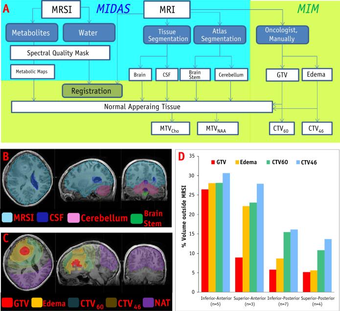
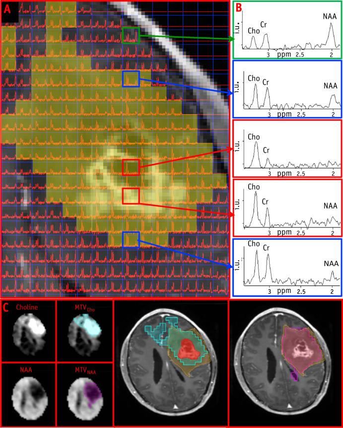
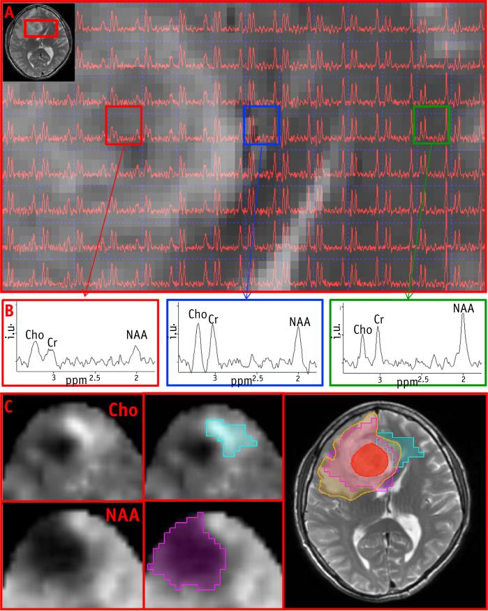
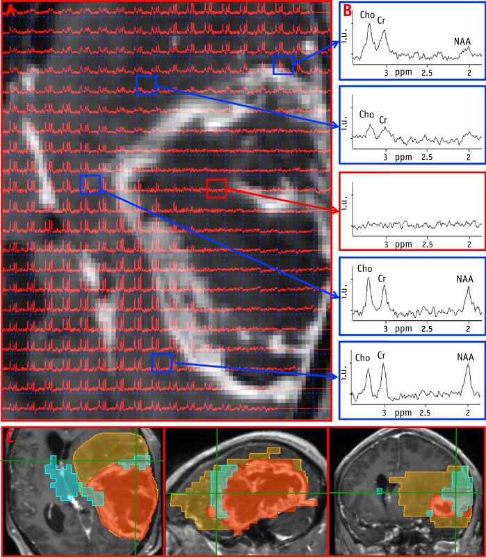
Purpose: Magnetic resonance (MR) imaging and computed tomography (CT) are used almost exclusively in radiation therapy planning of glioblastoma multiforme (GBM), despite their well-recognized limitations. MR spectroscopic imaging (MRSI) can identify biochemical patterns associated with normal brain and tumor, predominantly by observation of choline (Cho) and N-acetylaspartate (NAA) distributions. In this study, volumetric 3-dimensional MRSI was used to map these compounds over a wide region of the brain and to evaluate metabolite-defined treatment targets (metabolic tumor volumes [MTV]).
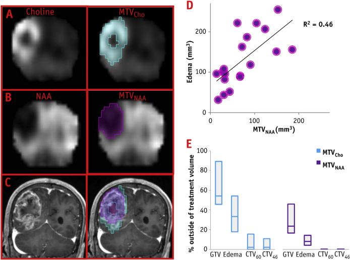
Methods and materials: Volumetric MRSI with effective voxel size of ∼1.0 mL and standard clinical MR images were obtained from 19 GBM patients. Gross tumor volumes and edema were manually outlined, and clinical target volumes (CTVs) receiving 46 and 60 Gy were defined (CTV46 and CTV60, respectively). MTVCho and MTVNAA were constructed based on volumes with high Cho and low NAA relative to values estimated from normal-appearing tissue.
Results: The MRSI coverage of the brain was between 70% and 76%. The MTVNAA were almost entirely contained within the edema, and the correlation between the 2 volumes was significant (r=0.68, P=.001). In contrast, a considerable fraction of MTVCho was outside of the edema (median, 33%) and for some patients it was also outside of the CTV46 and CTV60. These untreated volumes were greater than 10% for 7 patients (37%) in the study, and on average more than one-third (34.3%) of the MTVCho for these patients were outside of CTV60.
Conclusions: This study demonstrates the potential usefulness of whole-brain MRSI for radiation therapy planning of GBM and revealed that areas of metabolically active tumor are not covered by standard RT volumes. The described integration of MTV into the RT system will pave the way to future clinical trials investigating outcomes in patients treated based on metabolic information.
Copyright © 2014 Elsevier Inc. All rights reserved.
Figures
Similar articles
-
Association of Radiomics and Metabolic Tumor Volumes in Radiation Treatment of Glioblastoma Multiforme.Int J Radiat Oncol Biol Phys. 2017 Mar 1;97(3):586-595. doi: 10.1016/j.ijrobp.2016.11.011. Epub 2016 Nov 15. Int J Radiat Oncol Biol Phys. 2017. PMID: 28011044 Free PMC article.
-
3-Dimensional magnetic resonance spectroscopic imaging at 3 Tesla for early response assessment of glioblastoma patients during external beam radiation therapy.Int J Radiat Oncol Biol Phys. 2014 Sep 1;90(1):181-9. doi: 10.1016/j.ijrobp.2014.05.014. Epub 2014 Jun 28. Int J Radiat Oncol Biol Phys. 2014. PMID: 24986746 Free PMC article.
-
Evaluation of the lactate-to-N-acetyl-aspartate ratio defined with magnetic resonance spectroscopic imaging before radiation therapy as a new predictive marker of the site of relapse in patients with glioblastoma multiforme.Int J Radiat Oncol Biol Phys. 2014 Oct 1;90(2):385-93. doi: 10.1016/j.ijrobp.2014.06.009. Epub 2014 Aug 4. Int J Radiat Oncol Biol Phys. 2014. PMID: 25104068 Clinical Trial.
-
Proton magnetic resonance spectroscopy imaging in the study of human brain cancer.Q J Nucl Med Mol Imaging. 2009 Dec;53(6):618-30. Q J Nucl Med Mol Imaging. 2009. PMID: 20016453 Review.
-
Use of high-resolution volumetric MR spectroscopic imaging in assessing treatment response of glioblastoma to an HDAC inhibitor.AJR Am J Roentgenol. 2014 Aug;203(2):W158-65. doi: 10.2214/AJR.14.12518. AJR Am J Roentgenol. 2014. PMID: 25055291 Free PMC article. Review.
Cited by
-
The Role of Standard and Advanced Imaging for the Management of Brain Malignancies From a Radiation Oncology Standpoint.Neurosurgery. 2019 Aug 1;85(2):165-179. doi: 10.1093/neuros/nyy461. Neurosurgery. 2019. PMID: 30535032 Free PMC article. Review.
-
Three-dimensional echo planar spectroscopic imaging for differentiation of true progression from pseudoprogression in patients with glioblastoma.NMR Biomed. 2019 Feb;32(2):e4042. doi: 10.1002/nbm.4042. Epub 2018 Dec 17. NMR Biomed. 2019. PMID: 30556932 Free PMC article.
-
The Use of Quantitative Imaging in Radiation Oncology: A Quantitative Imaging Network (QIN) Perspective.Int J Radiat Oncol Biol Phys. 2018 Nov 15;102(4):1219-1235. doi: 10.1016/j.ijrobp.2018.06.023. Epub 2018 Jun 30. Int J Radiat Oncol Biol Phys. 2018. PMID: 29966725 Free PMC article. Review.
-
Defining occult disease in glioblastoma using spectroscopic MRI: implications for clinical target volume delineation.Radiat Oncol. 2025 May 22;20(1):86. doi: 10.1186/s13014-025-02666-z. Radiat Oncol. 2025. PMID: 40405247 Free PMC article.
-
Emerging MRI Techniques to Redefine Treatment Response in Patients With Glioblastoma.J Magn Reson Imaging. 2020 Oct;52(4):978-997. doi: 10.1002/jmri.27105. Epub 2020 Mar 19. J Magn Reson Imaging. 2020. PMID: 32190946 Free PMC article. Review.
References
-
- Hochberg FH, Pruitt A. Assumptions in the radiotherapy of glioblastoma. Neurology. 1980;30:907–911. - PubMed
-
- Chang EL, Akyurek S, Avalos T, et al. Evaluation of peritumoral edema in the delineation of radiotherapy clinical target volumes for glioblastoma. Int J Radiat Oncol Biol Phys. 2007;68:144–150. - PubMed
-
- Lee SW, Fraass BA, Marsh LH, et al. Patterns of failure following high-dose 3-D conformal radiotherapy for high-grade astrocytomas: A quantitative dosimetric study. Int J Radiat Oncol Biol Phys. 1999;43:79–88. - PubMed
-
- Oppitz U, Maessen D, Zunterer H, et al. 3D-recurrence-patterns of glioblastomas after CT-planned postoperative irradiation. Radiother Oncol. 1999;53:53–57. - PubMed
-
- Burger PC, Dubois PJ, Schold SC, Jr, et al. Computerized tomographic and pathologic studies of the untreated, quiescent, and recurrent glioblastoma multiforme. J Neurosurg. 1983;58:159–169. - PubMed
Publication types
MeSH terms
Substances
Grants and funding
LinkOut - more resources
Full Text Sources
Other Literature Sources
Medical
