. 2014 Mar 28:5:128-40.
doi: 10.1016/j.nicl.2014.03.012. eCollection 2014.

Prospective longitudinal MRI study of brain volumes and diffusion changes during the first year after moderate to severe traumatic brain injury

Affiliations

Prospective longitudinal MRI study of brain volumes and diffusion changes during the first year after moderate to severe traumatic brain injury

Veronika Brezova et al. Neuroimage Clin. .

Abstract

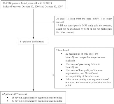
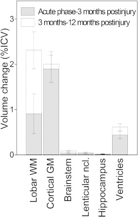
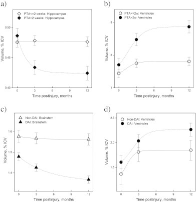
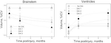
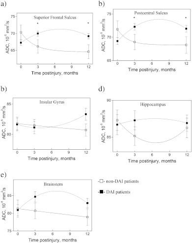
The objectives of this prospective study in 62 moderate-severe TBI patients were to investigate volume change in cortical gray matter (GM), hippocampus, lenticular nucleus, lobar white matter (WM), brainstem and ventricles using a within subject design and repeated MRI in the early phase (1-26 days) and 3 and 12 months postinjury and to assess changes in GM apparent diffusion coefficient (ADC) in normal appearing tissue in the cortex, hippocampus and brainstem. The impact of Glasgow Coma Scale (GCS) score at admission, duration of post-traumatic amnesia (PTA), and diffusion axonal injury (DAI) grade on brain volumes and ADC values over time was assessed. Lastly, we determined if MRI-derived brain volumes from the 3-month scans provided additional, significant predictive value to 12-month outcome classified with the Glasgow Outcome Scale-Extended after adjusting for GCS, PTA and age. Cortical GM loss was rapid, largely finished by 3 months, but the volume reduction was unrelated to GCS score, PTA, or presence of DAI. However, cortical GM volume at 3 months was a significant independent predictor of 12-month outcome. Volume loss in the hippocampus and lenticular nucleus was protracted and statistically significant first at 12 months. Slopes of volume reduction over time for the cortical and subcortical GGM were significantly different. Hippocampal volume loss was most pronounced and rapid in individuals with PTA > 2 weeks. The 3-month volumes of the hippocampus and lentiform nucleus were the best independent predictors of 12-month outcome after adjusting for GCS, PTA and age. In the brainstem, volume loss was significant at both 3 and 12 months. Brainstem volume reduction was associated with lower GCS score and the presence of DAI. Lobar WM volume was significantly decreased first after 12 months. Surprisingly DAI grade had no impact on lobar WM volume. Ventricular dilation developed predominantly during the first 3 months, and was strongly associated with volume changes in the brainstem and cortical GM, but not lobar WM volume. Higher ADC values were detected in the cortex in individuals with severe TBI, DAI and PTA > 2 weeks, from 3 months. There were no associations between ADC values and brain volumes, and ADC values did not predict outcome.

Keywords: ADC; Diffuse axonal injury; Glasgow Coma Scale; Outcome; Post-traumatic amnesia.


Figures

Fig. 1
Fig. 1
TBI patients with GCS ≤ 13 that could not be explained by other factors than traumatic head injury, admitted to the Neurosurgical Department, St. Olav's Hospital, Trondheim University Hospital, Norway in the period 2004–2007. Flow chart of the inclusion and exclusion criteria, and the final number of patients participating in the current study.
Fig. 2
Fig. 2
Successful NeuroQuant segmentation of repeated MRIs obtained in a 21-year-old patient with moderate traumatic brain injury at a) 9 days postinjury and b) 3 months and c) 12 months postinjury.
Fig. 3
Fig. 3
ROI placement for ADC measurements in a) formula image 
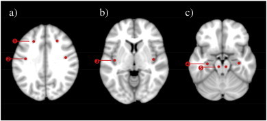
superior frontal sulcus ROI, formula image postcentral sulcus ROI; b) formula image insular gyrus ROI; c) formula image hippocampal ROI, formula image brainstem ROI. Circle-shaped ROIs with radius of 2.7 mm were positioned in apparently healthy tissue. In TBI patients with visible focal pathology, the ROI was moved to the closest apparently healthy GM tissue within the structure of interest.
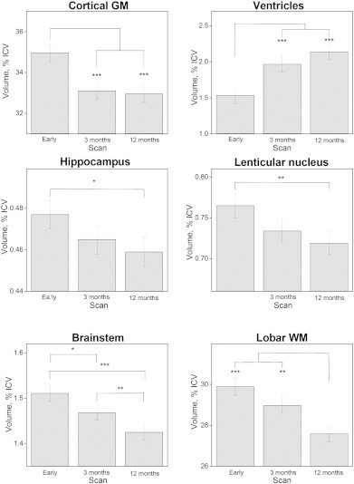
Fig. 4
Fig. 4
Longitudinal changes in mean ICV-corrected brain volumes with standard errors in the early phase (0–26 days post-injury, scan 1) and 3 months (scan 2), and 12 months (scan 3) postinjury. Volumes of the left and right sides of the structures were summed and evaluated together. ***p < 0.001, **p < 0.01, *p < 0.05.
Fig. 5
Fig. 5
Illustration of the volume changes over time. Volume changes (as %ICV) early phase–3 months and 3 months–12 months postinjury. The differences between different time points are calculated using linear mixed model. Means ± error bars are plotted.
Fig. 6
Fig. 6
Mean volumes of different brain structures plotted for: a) hippocampus and b) ventricles of patients with PTA > and ≤2 weeks. c) Brainstem and d) ventricles of DAI and non-DAI patients. For statistical evaluation of volume differences see Materials and methods and Results.
Fig. 7
Fig. 7
The association between the longitudinal changes in brainstem and ventricular ICV-corrected volumes for the non-DAI and the three DAI groups (DAI grades 1, 2, and 3). Figures show means and standard errors. Left and right side volumes were summed and evaluated together. Significant differences in brainstem and ventricular volumes between the non-DAI and the three DAI groups were found at all time points. Please note that the units on the y axis differ between the plots, and that the x axis does not cross the y axis at 0.
Fig. 8
Fig. 8
Longitudinal changes in mean ADC values with standard deviations in non-DAI (□) and DAI (▪) patients in the superior frontal sulcus (a), postcentral sulcus (b), insular gyrus  (c), hippocampus (d) and brainstem (e). Right and left side values were averaged. Statistical significant differences between non-DAI and DAI groups are marked with *. Significant differences were found between: a) DAI (1) and DAI (2), p = 0.009; non-DAI (2) and DAI (2), p = 0.028; non-DAI (3) and DAI (3), p = 0.001. b) DAI (1) and DAI (2), p = 0.006; non-DAI (2) and DAI (2), p = 0.020; non-DAI (3) and DAI (3), p = 0.004. The p-values were corrected for multiple comparisons. Please note that the units on the y axis differ between the plots, and that the x axis does not cross the y axis at 0.
Fig. 9
Fig. 9
The associations between longitudinal changes in mean ADC values for the non-DAI group (non-DAI = DAI 0) and the three DAI groups (DAI grades 1, 2, 3). Longitudinal analysis within each DAI group was performed separately for both ROIs using a linear mixed model. Significant F-values were followed by the Student's T-test. The resulting p-values were Bonferroni corrected separately for every ROI. At each time point, differences between DAI subgroups were evaluated using one-way ANOVA, and significant F-values were followed by independent samples T-tests, the resulting p-values were Bonferroni corrected for the three time points separately. Significant differences between different time points for each DAI group were confirmed and marked as: @—significantly different from scan 2; $—significantly different from scan 3 (***p < 0.001, **p < 0.01, *p < 0.05).

References

    1. Anderson C.V., Bigler E.D. Ventricular dilation, cortical atrophy, and neuropsychological outcome following traumatic brain injury. Journal of Neuropsychiatry and Clinical Neurosciences. 1995;7:42–48. 7711490 - PubMed
    1. Bendlin B.B., Ries M.L., Lazar M., Alexander A.L., Dempsey R.J., Rowley H.A. Longitudinal changes in patients with traumatic brain injury assessed with diffusion-tensor and volumetric imaging. Neuroimage. 2008;42:503–514. 18556217 - PMC - PubMed
    1. Bergsneider M., Hovda D.A., McArthur D.L., Etchepare M., Huang S., Sehati N. Metabolic recovery following human traumatic brain injury based on FDG-PET: time course and relationship to neurological disability. Journal of Head Trauma Rehabilitation. 2001;16:135–148. 11275575 - PubMed
    1. Bigler E.D., Blatter D.D., Anderson C.V., Johnson S.C., Gale S.D., Hopkins R.O. Hippocampal volume in normal aging and traumatic brain injury. AJNR. American Journal of Neuroradiology. 1997;18:11–23. 9010515 - PMC - PubMed
    1. Blatter D.D., Bigler E.D., Gale S.D., Johnson S.C., Anderson C.V., Burnett B.M. MR-based brain and cerebrospinal fluid measurement after traumatic brain injury: correlation with neuropsychological outcome. AJNR. American Journal of Neuroradiology. 1997;18:1–10. 9010514 - PMC - PubMed