

A fibril-like assembly of oligomers of a peptide derived from β-amyloid
- PMID: 25068693
- PMCID: PMC4183627
- DOI: 10.1021/ja505713y
A fibril-like assembly of oligomers of a peptide derived from β-amyloid
Abstract
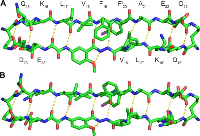
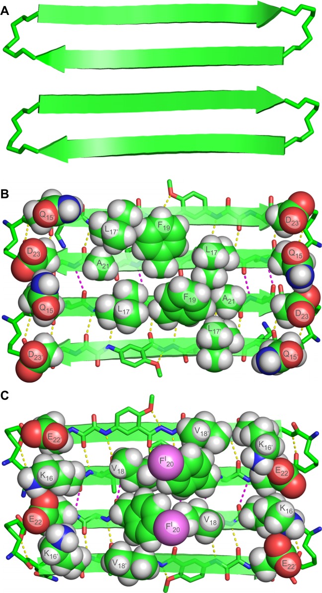
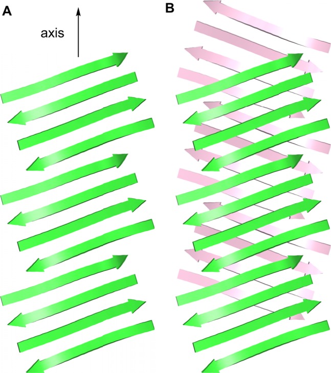
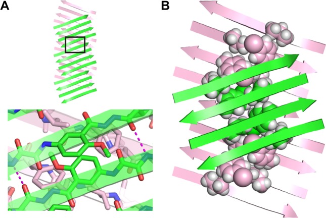
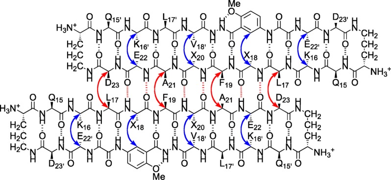
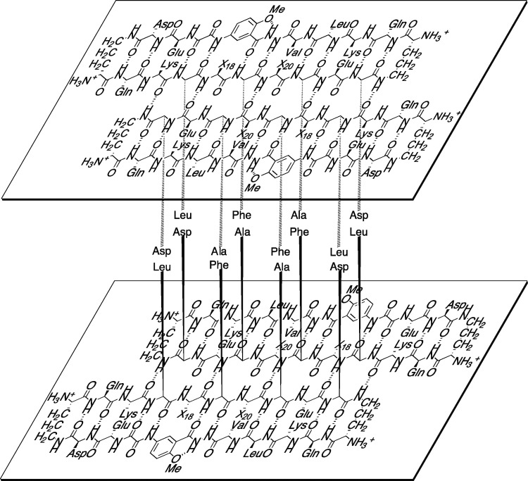
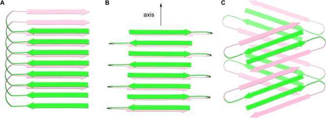
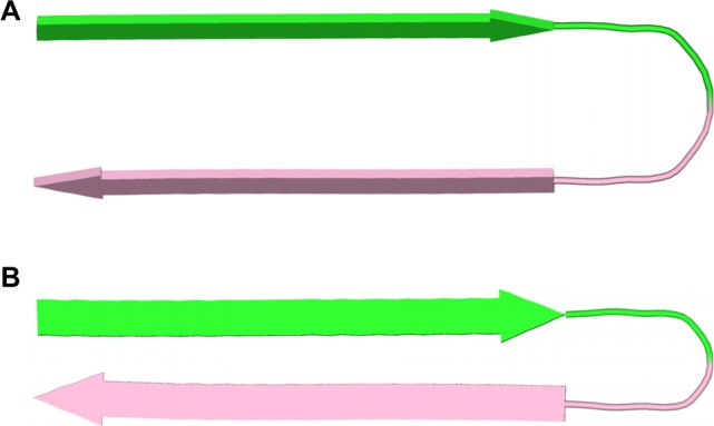
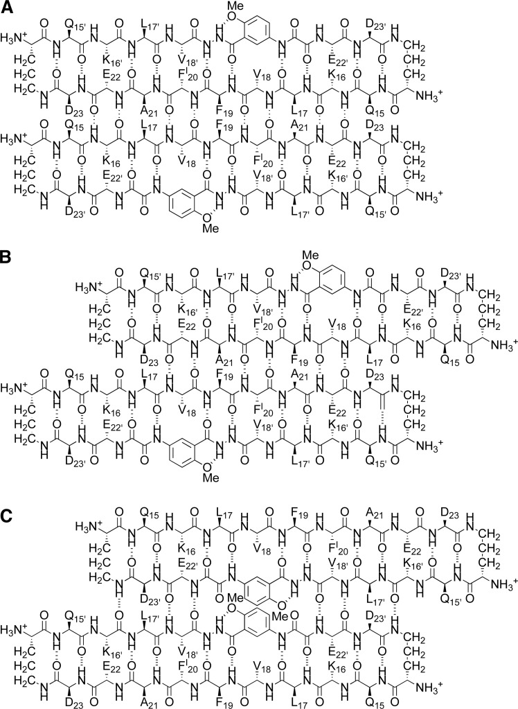
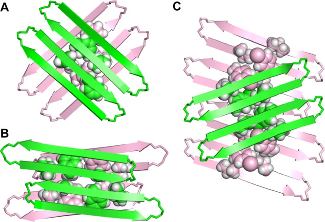
A macrocyclic β-sheet peptide containing two nonapeptide segments based on Aβ(15-23) (QKLVFFAED) forms fibril-like assemblies of oligomers in the solid state. The X-ray crystallographic structure of macrocyclic β-sheet peptide 3 was determined at 1.75 Å resolution. The macrocycle forms hydrogen-bonded dimers, which further assemble along the fibril axis in a fashion resembling a herringbone pattern. The extended β-sheet comprising the dimers is laminated against a second layer of dimers through hydrophobic interactions to form a fibril-like assembly that runs the length of the crystal lattice. The second layer is offset by one monomer subunit, so that the fibril-like assembly is composed of partially overlapping dimers, rather than discrete tetramers. In aqueous solution, macrocyclic β-sheet 3 and homologues 4 and 5 form discrete tetramers, rather than extended fibril-like assemblies. The fibril-like assemblies of oligomers formed in the solid state by macrocyclic β-sheet 3 represent a new mode of supramolecular assembly not previously observed for the amyloidogenic central region of Aβ. The structures observed at atomic resolution for this peptide model system may offer insights into the structures of oligomers and oligomer assemblies formed by full-length Aβ and may provide a window into the propagation and replication of amyloid oligomers.
Figures
References
-
- Hamley I. W. Chem. Rev. 2012, 112, 5147–5192. - PubMed
- Benilova I.; Karran E.; De Stooper B. Nat. Neurosci. 2012, 15, 349–357. - PubMed
- Querfurth H. W.; LaFerla F. H. N N. Engl. J. Med. 2010, 362, 329–344. - PubMed
- Larson M. E.; Lesné S. E. J. Neurochem. 2012, 192Suppl. 1125–139. - PMC - PubMed
- Fändrich M. J. Mol. Biol. 2012, 421, 427–440. - PubMed
-
- Benzinger T. L.; Gregory D. M.; Burkoth T. S.; Miller-Auer H.; Lynn D. G.; Botto R. E.; Meredith S. C. Proc. Natl. Acad. Sci. U.S.A. 1998, 95, 13407–13412. - PMC - PubMed
- Lührs T.; Ritter C.; Adrian M.; Riek-Loher D.; Bohrmann B.; Döbeli H.; Schubert D.; Riek R. Proc. Natl. Acad. Sci. U.S.A. 2005, 102, 17342–17347. - PMC - PubMed
- Petkova A. T.; Yau W.-M.; Tycko R. Biochemistry 2006, 45, 498–512. - PMC - PubMed
- Paravastu A. K.; Leapman R. D.; Yau W.-M.; Tycko R. Proc. Natl. Acad. Sci. U.S.A. 2008, 105, 18349–18354. - PMC - PubMed
- McDonald M.; Box H.; Bian W.; Kendall A.; Tycko R.; Stubbs G. J. Mol. Biol. 2012, 423, 454–461. - PMC - PubMed
-
- Yu L.; Edalji R.; Harlan J. E.; Holzman T. F.; Lopez A. P.; Labkovsky B.; Hillen H.; Barghorn S.; Ebert U.; Richardson P. L.; Miesbauer L.; Solomon L.; Bartley D.; Walter K.; Johnson R. W.; Hajduk P. J.; Olejniczak E. T. Biochemistry 2009, 48, 1870–1877. - PubMed
- Cerf E.; Sarroukh R.; Tamamizu-Kato S.; Breydo L.; Derclaye S.; Dufrênes Y. V.; Narayanaswami V.; Goormaghtigh E.; Ruysschaert J.-M.; Raussens V. Biochem. J. 2009, 421, 415–423. - PubMed
- Chimon S.; Shaibat M. A.; Jones C. R.; Calero D. C.; Aizezi B.; Ishii Y. Nat. Struc. Mol. Bio. 2010, 14, 1157–1164. - PubMed
- Streltsov V. A.; Varghese J. N.; Masters C. L.; Nuttall S. D. J. Neurosci. 2011, 31, 1419–1426. - PMC - PubMed
-
- Ma B.; Nussinov Proc. Natl. Acad. Sci. U.S.A. 2002, 99, 14126–14131. - PMC - PubMed
- Tarus B.; Straub J. E.; Thirumalai D. J. Mol. Biol. 2005, 345, 1141–1156. - PubMed
- Miller Y.; Ma B.; Nussinov R. Biophys. J. 2009, 97, 1168–1177. - PMC - PubMed
- Ahmed M.; Davis J.; Aucoin D.; Sato T.; Ahuja S.; Aimoto S.; Elliott J. I.; van Nostrand W. E.; Smith S. O. Nat. Struct. Mol. Biol. 2010, 17, 561–567. - PMC - PubMed
-
- Hilbich C.; Kisters-Woike B.; Reed J.; Masters C. L.; Beyreuther K. J. Mol. Biol. 1992, 228, 460–473. - PubMed
- Wood S. J.; Wetzel R.; Martin J. D.; Hurle M. R. Biochemistry 1995, 34, 724–730. - PubMed
- Tjernberg L. O.; Callaway D. J. E.; Tjernberg A.; Hahne S.; Lilliehöök C.; Terenius L.; Thyberg J.; Nordstedt C. J. Biol. Chem. 1999, 274, 12619–12625. - PubMed
Publication types
MeSH terms
Substances
Grants and funding
LinkOut - more resources
Full Text Sources
Other Literature Sources
