. 2014 Jul 28;9(7):e102978.
doi: 10.1371/journal.pone.0102978. eCollection 2014.

Isoflurane reversibly destabilizes hippocampal dendritic spines by an actin-dependent mechanism

Affiliations

Isoflurane reversibly destabilizes hippocampal dendritic spines by an actin-dependent mechanism

Jimcy Platholi et al. PLoS One. .

Abstract

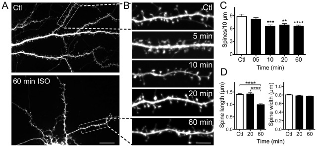
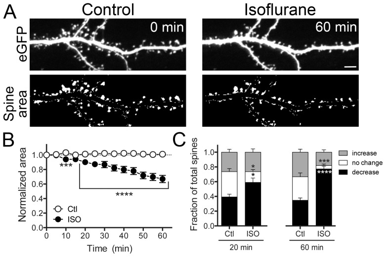
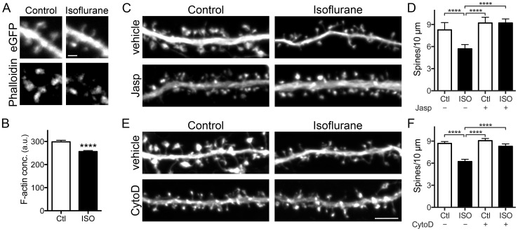
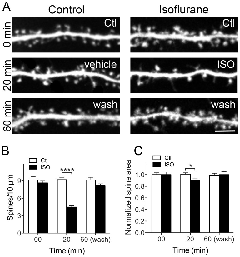
General anesthetics produce a reversible coma-like state through modulation of excitatory and inhibitory synaptic transmission. Recent evidence suggests that anesthetic exposure can also lead to sustained cognitive dysfunction. However, the subcellular effects of anesthetics on the structure of established synapses are not known. We investigated effects of the widely used volatile anesthetic isoflurane on the structural stability of hippocampal dendritic spines, a postsynaptic structure critical to excitatory synaptic transmission in learning and memory. Exposure to clinical concentrations of isoflurane induced rapid and non-uniform shrinkage and loss of dendritic spines in mature cultured rat hippocampal neurons. Spine shrinkage was associated with a reduction in spine F-actin concentration. Spine loss was prevented by either jasplakinolide or cytochalasin D, drugs that prevent F-actin disassembly. Isoflurane-induced spine shrinkage and loss were reversible upon isoflurane elimination. Thus, isoflurane destabilizes spine F-actin, resulting in changes to dendritic spine morphology and number. These findings support an actin-based mechanism for isoflurane-induced alterations of synaptic structure in the hippocampus. These reversible alterations in dendritic spine structure have important implications for acute anesthetic effects on excitatory synaptic transmission and synaptic stability in the hippocampus, a locus for anesthetic-induced amnesia, and have important implications for anesthetic effects on synaptic plasticity.


Conflict of interest statement

Competing Interests: The authors have declared that no competing interests exist.

Figures

Figure 1
Figure 1. Isoflurane reduces dendritic spine density and alters spine morphology.
Hippocampal neuron cultures transfected with eGFP as a cell volume indicator were exposed to 95% air/5% CO2 (Ctl) or 2 vol% isoflurane in 95% air/5% CO2 for 60 min at 37°C. A-representative full image showing the dendritic arbor of a control neuron and of a neuron treated with isoflurane for 60 min; B-representative images of single ROIs at various times of exposure to isoflurane. Spine number was counted manually at the indicated time points. Isoflurane significantly reduced spine density (C) by one-way ANOVA with Dunnett's post hoc test (**p<0.01; ***p<0.001; ****p<0.0001). Exposure to isoflurane reduced mean spine length (D-left panel) with no significant change in spine width (D-right panel) by one-way ANOVA with Holm-Sidak post hoc test (****p<0.0001). Data are mean ± SEM; n = 30 to 40 dendritic ROIs per experimental group; n = 600 to 1260 dendritic spines for spine density, spine length and spine head width per experimental group. Scale bar in 1A = 10 µm, in 1B = 5 µm.
Figure 2
Figure 2. Isoflurane reduces dendritic spine area.
Hippocampal neuron cultures transfected with eGFP were exposed to 95% air/5% CO2 (Ctl) or 2 vol% isoflurane in 95% air/5% CO2 for 60 min at 37°C. Representative images show eGFP fluorescence (A-top panels) and thresholded overall spine area after manual dendrite subtraction (A-bottom panels). A time-dependent decrease in total spine area was observed (B) by two-way ANOVA with Sidak post hoc test (***p<0.001). Changes in individual spine area of >10% (increase) or <10% (decrease), or no change in area were evident at 20 and 60 min (C) by two-way ANOVA with Sidak post hoc test (*p<0.05; ***p<0.001; ****p<0.0001). Data are mean ± SEM; n = 1500 to 1800 dendritic spines per experimental group. Scale bar  = 5 µm.
Figure 3
Figure 3. Isoflurane reduces F-actin concentration in spines, and actin targeted drugs prevent isoflurane-induced spine loss.
Hippocampal neuron cultures transfected with eGFP were exposed to 95% air/5% CO2 (Ctl) or 2 vol% isoflurane in 95% air/5% CO2 at 37°C for 20 min and stained with phalloidin-Alexa 568 (A-representative images). Quantification of phalloidin fluorescence intensity shows that isoflurane reduced F-actin concentration (B) by Student unpaired t-test (****p<0.0001). Pretreatment with 0.1 µM jasplakinolide (C-representative images) or 0.1 µM cytochalasin D (E-representative images) for 2 min before exposure to 95% air/5% CO2 (Ctl) or 2 vol% isoflurane in 95% air/5% CO2 at 37°C for 20 min prevented isoflurane-induced spine loss (D,F) by one-way ANOVA with Tukey's post hoc test (****p<0.0001). Data are mean ± SEM; n = 30 to 40 dendritic ROIs (600–1260 spines) per experimental group for phalloidin staining and actin stabilizing drug treatments. Scale bar  = 1 µm for 3A and 5 µm for 3C,E.
Figure 4
Figure 4. Isoflurane-induced spine loss is reversible.
Hippocampal neuron cultures transfected with eGFP were exposed to 95% air/5% CO2 (Ctl) or 2 vol% isoflurane in 95% air/5% CO2 for 20 min at 37°C. At 20 min, isoflurane exposure was terminated and spine density and area was quantified at different time points (A, only 40 min wash shown). Changes in spine number and area observed after 20 min of isoflurane exposure recovered at 40 min of wash-out (B, C) by two-way ANOVA with Sidak post hoc test (*p<0.05; ****p<0.0001). Data are mean ± SEM; n = 30 to 40 dendritic ROIs per experimental group for spine density; n = 600 to 1800 dendritic spines per experimental group for spine area. Scale bar  = 5 µm.

References

    1. Hemmings HC Jr, Yan W, Westphalen RI, Ryan TA (2005) The general anesthetic isoflurane depresses synaptic vesicle exocytosis. Mol Pharmacol 67: 1591–1599. - PubMed
    1. Eger EI 2nd, Raines DE, Shafer SL, Hemmings HC Jr, Sonner JM (2008) Is a new paradigm needed to explain how inhaled anesthetics produce immobility? Anesth Analg 107: 832–848. - PMC - PubMed
    1. Jevtovic-Todorovic V, Absalom AR, Blomgren K, Brambrink A, Crosby G, et al. (2013) Anaesthetic neurotoxicity and neuroplasticity: an expert group report and statement based on the BJA Salzburg Seminar. Br J Anaesth 111: 143–51. - PMC - PubMed
    1. Sanders RD, Hassell J, Davidson AJ, Robertson NJ, Ma D (2013) Impact of anaesthetics and surgery on neurodevelopment: an update. Br J Anaesth 110 Suppl 1i53–i72. - PMC - PubMed
    1. Mintz CD, Barrett KM, Smith SC, Benson DL, Harrison NL (2013) Anesthetics interfere with axon guidance in developing mouse neocortical neurons in vitro via a g-aminobutyric acid type A receptor mechanism. Anesthesiology 118: 825–833. - PMC - PubMed

Publication types