. 2014 Jul 28:13:179.
doi: 10.1186/1476-4598-13-179.

Effective inhibition of melanoma tumorigenesis and growth via a new complex vaccine based on NY-ESO-1-alum-polysaccharide-HH2

Affiliations

Effective inhibition of melanoma tumorigenesis and growth via a new complex vaccine based on NY-ESO-1-alum-polysaccharide-HH2

Meng Li et al. Mol Cancer. .

Abstract

Background: A safe and effective adjuvant plays an important role in the development of a vaccine. However, adjuvants licensed for administration in humans remain limited. Here, for the first time, we developed a novel combination adjuvant alum-polysaccharide-HH2 (APH) with potent immunomodulating activities, consisting of alum, polysaccharide of Escherichia coli and the synthetic cationic innate defense regulator peptide HH2.

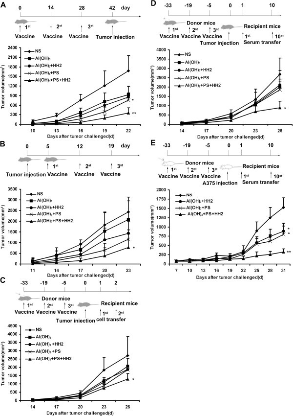
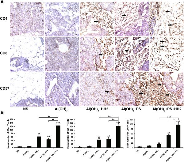
Methods: The adjuvant effects of APH were examined using NY-ESO-1 protein-based vaccines in prophylactic and therapeutic models. We further determined the immunogenicity and anti-tumor effect of NY-ESO-1-APH (NAPH) vaccine using adoptive cellular/serum therapy in C57/B6 and nude mice. Cell-mediated and antibody-mediated immune responses were evaluated.

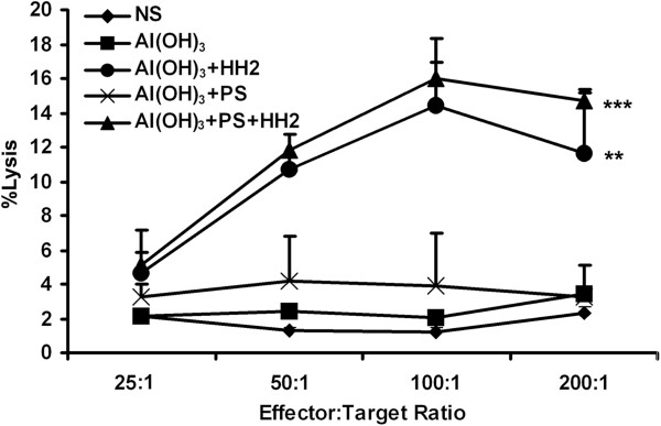
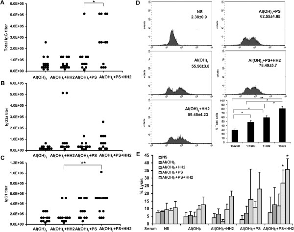
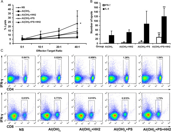
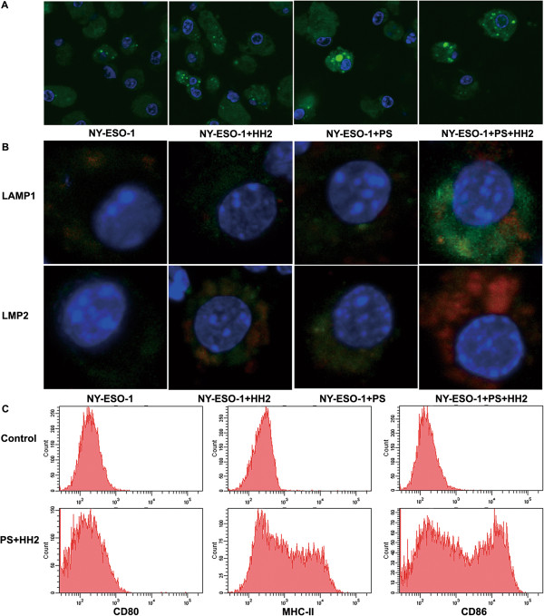
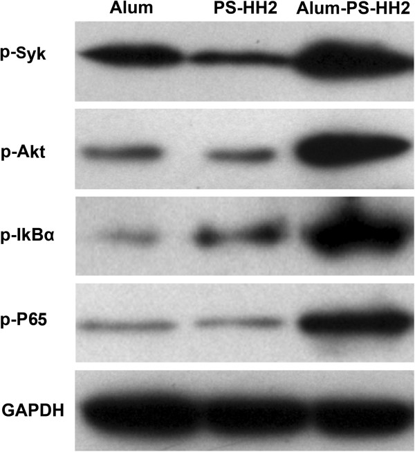
Results: The APH complex significantly promoted antigen uptake, maturation and cross-presentation of dendritic cells and enhanced the secretion of TNF-α, MCP-1 and IFN-γ by human peripheral blood mononuclear cells compared with individual components. Vaccination of NAPH resulted in significant tumor regression or delayed tumor progression in prophylactic and therapeutic models. In addition, passive serum/cellular therapy potently inhibited tumor growth of NY-ESO-1-B16. Mice treated with NAPH vaccine produced higher antibody titers and greater antibody-dependent/independent cellular cytotoxicity. Therefore, NAPH vaccination effectively stimulated innate immunity, and boosted both arms of the adaptive humoral and cellular immune responses to suppress tumorigenesis and growth of melanoma.

Conclusions: Our study revealed the potential application of APH complex as a novel immunomodulatory agent for vaccines against tumor refractory and growth.


Figures

Figure 1
Figure 1
NAPH vaccine inhibits the melanoma tumorigenesis and growth. A, C57/B6 mice (n = 12) were treated with different vaccines (on Days 0, 14 and 28) and challenged with 2 × 105 NY-ESO-1-B16 cells (on Day 42) in a prophylactic model. B, NY-ESO-1-B16-bearing mice (n = 12) were treated with vaccines (on Days 5, 12 and 19) in a therapeutic model. C, D, Adoptive transfer of cells or serum from NAPH-treated mice induced tumor regression in recipient mice. Donor splenocytes (2 × 107) were injected i.v. into recipient mice for 2 consecutive days (on Days 1 and 2). In serum adoptive therapy, tumor-bearing mice were injected i.v. with 0.1 ml of the sera from Days 1 to 10. E, Adoptive transfer of serum from immunized-Balb/c mice inhibited A375 melanoma growth in a nude mouse model. * P < 0.05; ** P < 0.005.
Figure 2
Figure 2
NAPH vaccine stimulates the innate immune response. 48 hours after NAPH injection, the mice were sacrificed. A standard 4 h NK assay against YAC-1 targets revealed that the NK cytotoxicity in the NAH group was higher than that in the NS, NA and NAP groups; in particular, NAPH-treated mice had the greatest cytotoxic activity against YAC-1 cells among all groups (* P < 0.05; ** P < 0.005).
Figure 3
Figure 3
NAPH increases NY-ESO-1-specific antibody immune response. A, The titers of anti-NY-ESO-1 total IgG were measured using ELISA. Dot, titer of each mouse. B, C, The titers of anti-NY-ESO-1 IgG2a/IgG1 titers in the treated groups were measured. D, FACS analysis using NY-ESO-1-B16 cells to detect NY-ESO-1-specific IgG antibodies (n = 3 mice, pooled composite results are shown). E, Antibody-dependent cellular cytotoxicity was determined using the 51Cr-release assay. NY-ESO-1-B16 cells were labeled with 51Cr and coated with sera from control or treated mice. Splenocytes from untreated or treated mice were used as effectors. The assay was run in three replicates.
Figure 4
Figure 4
NAPH vaccination elicits robust NY-ESO-1-specific cellular response in vitro. A, The cytotoxicity of splenocytes against NY-ESO-1-B16 cells was examined in a 4-h 51Cr-release assay. Points, mean of triplicate samples; bars, SD. B, IL-4/IFN-γ ELISPOT responses were performed at one week post-immunization (n = 3, three replicates). C, Intracellular staining of IFN-γ in CD4+ and CD8+ T cells. Similar results were obtained from three independent experiments (*P < 0.05; ** P < 0.005).
Figure 5
Figure 5
Immunohistochemistry analyses were performed to study infiltrating lymphocytes (TILs) in tumors. A. Tumor tissue sections were separately incubated with anti-CD4, anti-CD8 and anti-CD57 mAbs overnight at 4°C. Sections were then incubated with biotinylated anti-mouse antibody and streptavidin-biotinylated peroxidase complex. B. Intensity of lympocyte infiltration in the tumors was determined by the mean positive cell counts in the dermis around the B16-NY-ESO-1 tumor per field (10 randomly selected high power fields/slide). For each site, 3 pathologists performed a blind read of the glass slides.
Figure 6
Figure 6
PS-HH2 complex adjuvant promotes the antigen uptake and cross -presentation and maturation of DCs. A, DC were incubated with NHS-conjugated native NY-ESO-1 or protein adjuvant complex for 3 h, fixed, stained with DAPI and analyzed via confocal laser microscopy. B, DCs were incubated with Alexa Fluor-labeled NY-ESO-1 or NY-ESO-1/complex adjuvant for 3 h, fixed, permeabilized, blocked and stained with mouse FITC-anti-LAMP1 or FITC-anti-LMP2 antibody, respectively. DNA was stained with DAPI and analyzed via confocal laser microscopy. C, Analysis of surface maturation markers of DCs stimulated with PS-HH2 for 24 h.
Figure 7
Figure 7
Alum and PS-HH2 synergistically triggers activation of the NF-κB signal pathway. Immature DCs derived from bone marrow were incubated and harvested on Day 5 for Western blotting. After treatment with alum (125 μg/ml), PS-HH2 (PS: 20 μg/ml and HH2: 40 μg/ml), or alum-PS-HH2 complex for 0, 5, 10 and 30 min, respectively. DCs were washed, lysed and used for Western blot analysis using antibodies to phospho-Syk, phospho-Akt, phospho-IkBα and phospho-NF-κB, respectively. GAPDH was detected on the same membrane and used as the loading control.

References

    1. Valmori D, Souleimanian NE, Tosello V, Bhardwaj N, Adams S, O’Neill D, Pavlick A, Escalon JB, Cruz CM, Angiulli A, Angiulli F, Mears G, Vogel SM, Pan L, Jungbluth AA, Hoffmann EW, Venhaus R, Ritter G, Old LJ, Ayyoub M. Vaccination with NY-ESO-1 protein and CpG in Montanide induces integrated antibody/Th1 responses and CD8 T cells through cross-priming. Proc Natl Acad Sci U S A. 2007;104:8947–8952. doi: 10.1073/pnas.0703395104. - DOI - PMC - PubMed
    1. Sugita Y, Wada H, Fujita S, Nakata T, Sato S, Noguchi Y, Jungbluth AA, Yamaguchi M, Chen YT, Stockert E, Gnjatic S, Williamson B, Scanlan MJ, Ono T, Sakita I, Yasui M, Miyoshi Y, Tamaki Y, Matsuura N, Noguchi S, Old LJ, Nakayama E, Monden M. NY-ESO-1 expression and immunogenicity in malignant and benign breast tumors. Cancer Res. 2004;64:2199–2204. doi: 10.1158/0008-5472.CAN-03-3070. - DOI - PubMed
    1. Caballero OL, Chen YT. Cancer/testis (CT) antigens: potential targets for immunotherapy. Cancer Sci. 2009;100:2014–2021. doi: 10.1111/j.1349-7006.2009.01303.x. - DOI - PMC - PubMed
    1. Jager E, Jager D, Karbach J, Chen YT, Ritter G, Nagata Y, Gnjatic S, Stockert E, Arand M, Old LJ, Knuth A. Identification of NY-ESO-1 epitopes presented by human histocompatibility antigen (HLA)-DRB4*0101-0103 and recognized by CD4 (+) T lymphocytes of patients with NY-ESO-1-expressing melanoma. J Exp Med. 2000;191:625–630. doi: 10.1084/jem.191.4.625. - DOI - PMC - PubMed
    1. Purbhoo MA, Sutton DH, Brewer JE, Mullings RE, Hill ME, Mahon TM, Karbach J, Jager E, Cameron BJ, Lissin N, Vyas P, Chen JL, Cerundolo V, Jakobsen BK. Quantifying and imaging NY-ESO-1/LAGE-1-derived epitopes on tumor cells using high affinity T cell receptors. J Immunol. 2006;176:7308–7316. doi: 10.4049/jimmunol.176.12.7308. - DOI - PubMed

Publication types

MeSH terms