. 2014 Sep 1;193(5):2394-404.
doi: 10.4049/jimmunol.1302718. Epub 2014 Jul 28.

The DNA sensor, cyclic GMP-AMP synthase, is essential for induction of IFN-β during Chlamydia trachomatis infection

Affiliations

The DNA sensor, cyclic GMP-AMP synthase, is essential for induction of IFN-β during Chlamydia trachomatis infection

Yugen Zhang et al. J Immunol. .

Abstract

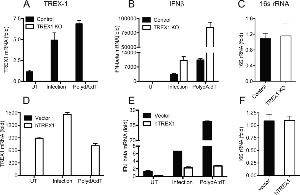
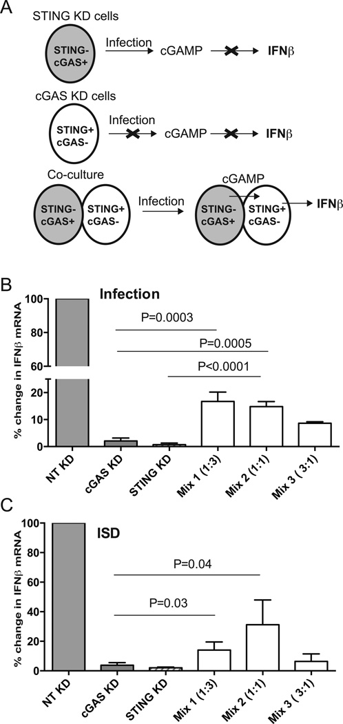
IFN-β has been implicated as an effector of oviduct pathology resulting from genital chlamydial infection in the mouse model. In this study, we investigated the role of cytosolic DNA and engagement of DNA sensors in IFN-β expression during chlamydial infection. We determined that three-prime repair exonuclease-1, a host 3' to 5' exonuclease, reduced IFN-β expression significantly during chlamydial infection using small interfering RNA and gene knockout fibroblasts, implicating cytosolic DNA as a ligand for this response. The DNA sensor cyclic GMP-AMP synthase (cGAS) has been shown to bind cytosolic DNA to generate cyclic GMP-AMP, which binds to the signaling adaptor stimulator of IFN genes (STING) to induce IFN-β expression. We determined that cGAS is required for IFN-β expression during chlamydial infection in multiple cell types. Interestingly, although infected cells deficient for STING or cGAS alone failed to induce IFN-β, coculture of cells depleted for either STING or cGAS rescued IFN-β expression. These data demonstrate that cyclic GMP-AMP produced in infected cGAS(+)STING(-) cells can migrate into adjacent cells via gap junctions to function in trans in cGAS(-)STING(+) cells. Furthermore, we observed cGAS localized in punctate regions on the cytosolic side of the chlamydial inclusion membrane in association with STING, indicating that chlamydial DNA is most likely recognized outside the inclusion as infection progresses. These novel findings provide evidence that cGAS-mediated DNA sensing directs IFN-β expression during Chlamydia trachomatis infection and suggest that effectors from infected cells can directly upregulate IFN-β expression in adjacent uninfected cells during in vivo infection, contributing to pathogenesis.


Figures

Figure 1
Figure 1. IFNβ expression during chlamydial infection is elevated in TREX1 KO MEFs and reduced by TREX-1 over-expression
(A–C) Control MEFs and TREX1 KO MEFs were infected with C. muridarum at 1 MOI or transfected with poly dA:dT for 6 h, before harvest. Infected cells were harvested at 24 h p.i. TREX1 mRNA (A), IFNβ mRNA (B) and chlamydial 16S rRNA (C) levels were measured by qRT-PCR. (D–F) TREX1 KO cells were transfected with human TREX1 or vector control. Twenty four hour post transfection, cells were infected with C.muridarum or transfected with poly dA:dT. Relative expression levels of TREX1 (D), IFNβ (E) and chlamydial 16S rRNA (F) normalized to GAPDH are shown. Panels are representative of three independent experiments and error bars represent the range in technical replicates. Statistical significance for qPCR data between multiple experiments was determined by using paired T tests on percent change in IFNβ levels between WT and TREX1 KO infected cells (P=0.01). In experiment involving transfection of TREX KO cells, percent change in IFNβ between vector and TREX-1 cDNA from multiple experiments were used to calculate significance (Infection; P=0.001, poly dA:dT; P=0.009). UT = Untreated.
Figure 2
Figure 2. siRNA knockdown of TREX-1 in epithelial cells increases IFNβ expression, during chlamydial infection
(A–C) Mouse BM1.11 were transfected with TREX1 siRNA or non-targeting (NT) siRNA (A) and infected with C. muridarum at 1 MOI, 72 h post transfection. TREX-1 (A) and IFNβ (B) mRNA were measured at 24 h p.i. In parallel, cells were transfected with dsDNA analog poly dA:dT, 6 h before harvest. Chlamydial replication was monitored by comparing 16S rRNA levels (C) in infected cells between treatments. (D–F) HeLa cells were transfected with siRNA for human TREX1 or NT siRNA. Cells were infected with C.muridarum at 1 MOI for 24 h or transfected 6 h before harvest with ISD or RNA analog poly I:C. TREX1 (D), IFN beta (E) and chlamydial 16S rRNA levels (F) were measured by qRT-PCR. Panels are representative of three independent experiments and error bars represent the mean ± error of technical replicates. Statistical significance for qPCR data between multiple experiments was determined by using paired T tests on percent change in IFNβ levels between TREX1 siRNA relative to NT siRNA in each experiment. For BM1.11 cells, infection; P=0.014 and poly dA-dT; P=0.047. For HeLa, infection; P=0.04, ISD; P=0.04, poly IC; P=NS. UT=Un-treated.
Figure 3
Figure 3. cGAS is required for IFNβ expression in HeLa cells infected with C. muridarum or C. trachomatis (D and L2)
HeLa cells were transfected with non-targeting siRNA (NT), 2 different siRNA for cGAS (cGAS1 and cGAS2), or a siRNA for STING as described in Methods. Significant knock down of STING (99%) and cGAS (90%) mRNA was achieved. Cells were infected with 1 MOI of C. muridarum (C.M) or 5 MOI of C.trachomatis -serovar D (C.T-D) or C. trachomatis L2 (C.T-L2). Cells were harvested at 24 h p.i and analyzed for expression of IFNβ (A), IFN λ (D), IL-8 (G), and chlamydial 16S rRNA (H). In parallel, cells were transfected with ISD (positive control) or poly IC-LyoVec (negative control) and harvested at 6 h post transfection and analyzed for expression of IFNβ (B, C), IFNλ (E, F). Culture supernatants from infected or transfected cells were collected at 24 h and CXCL10 protein levels assayed by ELISA (I). Mean ± SD of samples from 3 independent experiments are shown for ELISA. A representative western blot showing the levels of STING and cGAS following siRNA knock down in uninfected HeLa cells (J). A representative of five independent experiments is presented in A–H for qRT-PCR data and error bars represent range in technical replicates. Statistical significance for qPCR data between multiple experiments was determined by one way ANOVA with multiple comparison tests on the percent decrease in IFNβ levels for the siRNA used relative to NT siRNA in each experiment (K). UT=Un-treated.
Figure 4
Figure 4. cGAS is required for IFNβ expression during Chlamydia spp. infection of mouse oviduct epithelial cells (BM1.11 cells) and human oviduct epithelial cells (OE-E6/E7)
siRNA knock down in mouse BM1.11 cells were carried out using accell™ NT, mouse cGAS or STING siRNA pools. Seventy-two hours after transfection, cells were infected with C. muridarum or transfected with ISD (DNA) 6 h before harvest, and analyzed simultaneously at 24 h p.i for expression of mouse IFNβ (A), cGAS (B) and STING (C). Human oviduct epithelial cells (OE-E6/E7) were transfected with non-targeting (NT), human cGAS (cGAS siRNA 1 and 2) or human STING siRNA. Seventy-two hours after transfection, cells were infected with C. trachomatis (serovar D) at 5 MOI or transfected with ISD (DNA) 6 h before harvest and analyzed concurrently at 24 h p.i for expression of human IFNβ (E), cGAS (F) and STING (G). A representative of three experiments for BM1.11 cells and OE cells is presented for qRT-PCR data. Statistical significance for qPCR data between multiple experiments was determined by one way ANOVA with multiple comparison tests on the percent decrease in IFNβ levels for the siRNA used relative to NT siRNA in each experiment (D and H).
Figure 5
Figure 5. cGAS localizes in punctate regions around the chlamydial inclusion membrane
HeLa cells infected with C. muridarum (A) or C. trachomatis, serovar D (B) were fixed and stained for endogenous cGAS (red) and Chlamydia (green). Cells were fixed with Prolong gold™ with DAPI (blue) and analyzed by confocal microscopy. Uninfected HeLa cells (C) and cells infected with C. muridarum (D) for 18 h were fixed and stained for endogenous cGAS (red) and STING (green). In an independent experiment HeLa cells were transfected with FLAG-cGAS (E) and infected with C. muridarum 24 h later. Infected cells were fixed at 18 h p.i and stained using mouse monoclonal Ab for FLAG. Chlamydial inclusions and cell nucleus are marked with an “I” and “N” respectively on the DAPI staining.
Figure 6
Figure 6. Evidence of cGAMP transfer from infected cells to adjacent cells
Schematic representation of the experimental plan (A). STINGcGAS+ can produce cGAMP but cannot induce IFNβ since they lack STING, while STING+cGAS cells cannot produce cGAMP upon infection. Co-culture can rescue IFNβ expression during infection if cGAMP from cGAS+ cells can migrate into STING+ cells. HeLa cells knocked down for STING (>99% KD) or cGAS (>95% KD) were cultured individually or co-cultured at different ratios 18 h before infection in 24 well dishes (2 × 10⁵ cells/well). The mix numbers (1, 2, and 3) represent cGAS KD:STING KD cells in the indicated ratios. Cells were infected with C. muridarum at 3 MOI and IFNβ mRNA measured at 24 h post infection (B). A parallel set of cells were transfected with ISD as a positive control, 6 h before harvesting all the cells for RNA (C). Data is represented as mean of percent decrease relative to NT control from three experiments with SD. Significance determined by one way ANOVA with multiple comparison tests and indicated. Differences between Mix 1:3 and 1:1 co-culture were not significant.
Figure 7
Figure 7. Both cGAS and STING are required for maximal IFNβ expression during chlamydial infection
Protein levels of cGAS and STING in HeLa, HEK293T, and HEK293 cell lysates (A). Actin blots were carried out using 1/ 10th of cell lysates used for cGAS and STING western blots. HEK293 cells were permeabilized with cGAMP and IFNβ expression measured 6 h post treatment (B). HEK293 cells were infected with C. muridarum or transfected with ISD and IFNβ expression measured 24 h post infection or 6 h post treatment, respectively (C). HEK293T cells were transiently transfected with pcDNA3.1 (vector), STING, or cGAS expression constructs. Twenty-four hours post transfection, cells were trypsinized and plated individually or mixed at indicated ratio. Six hours after plating, individual cells were transfected with ISD or permeabilized with cGAMP (D). Individual or mixed cells were transfected with ISD or infected with C. muridarum (E). Expression of IFNβ measured at 24 h p.i or 6 h after ISD/cGAMP transfection. Data represents average of three experiments and error bars indicate SD. Significance was determined by one way ANOVA with multiple comparison test. For (D), p=0.004 for STING transfected-untreated (UT) vs cGAMP treated cells.
Figure 8
Figure 8. A schematic model of cGAS recognizing DNA during Chlamydia infection, in human epithelial cells
Left panel shows an electron micrograph of an inclusion, where chlamydial RBs are in close contact with the inclusion membrane and rough ER (arrows). Right panel shows a hypothetical model of DNA recognition by cGAS leading to cGAMP generation, STING activation and IRF3 phosphorylation, resulting in IFNβ expression during infection. Contribution of TREX-1 in diminishing this response is also shown.

References

    1. Xia M, Bumgarner RE, Lampe MF, Stamm WE. Chlamydia trachomatis infection alters host cell transcription in diverse cellular pathways. J Infect Dis. 2003;187:424–434. - PubMed
    1. Rasmussen SJ, Eckmann L, Quayle AJ, Shen L, Zhang YX, Anderson DJ, Fierer J, Stephens RS, Kagnoff MF. Secretion of proinflammatory cytokines by epithelial cells in response to Chlamydia infection suggests a central role for epithelial cells in chlamydial pathogenesis. J Clin Invest. 1997;99:77–87. - PMC - PubMed
    1. Lad SP, Fukuda EY, Li J, de la Maza LM, Li E. Up-regulation of the JAK/STAT1 signal pathway during Chlamydia trachomatis infection. J Immunol. 2005;174:7186–7193. - PubMed
    1. Nagarajan UM, Prantner D, Sikes JD, Andrews CW, Jr, Goodwin AM, Nagarajan S, Darville T. Type I interferon signaling exacerbates Chlamydia muridarum genital infection in a murine model. Infect Immun. 2008;76:4642–4648. - PMC - PubMed
    1. Qiu H, Fan Y, Joyee AG, Wang S, Han X, Bai H, Jiao L, Rooijen NVan, Yang X. Type I IFNs enhance susceptibility to Chlamydia muridarum lung infection by enhancing apoptosis of local macrophages. J Immunol. 2008;181:2092–2102. - PubMed

Publication types

MeSH terms