. 2014 Sep 12;289(37):25957-75.
doi: 10.1074/jbc.M114.592873. Epub 2014 Jul 28.

Multiple regulatory mechanisms control the expression of the Geobacillus stearothermophilus gene for extracellular xylanase

Affiliations

Multiple regulatory mechanisms control the expression of the Geobacillus stearothermophilus gene for extracellular xylanase

Smadar Shulami et al. J Biol Chem. .

Abstract

Geobacillus stearothermophilus T-6 produces a single extracellular xylanase (Xyn10A) capable of producing short, decorated xylo-oligosaccharides from the naturally branched polysaccharide, xylan. Gel retardation assays indicated that the master negative regulator, XylR, binds specifically to xylR operators in the promoters of xylose and xylan-utilization genes. This binding is efficiently prevented in vitro by xylose, the most likely molecular inducer. Expression of the extracellular xylanase is repressed in medium containing either glucose or casamino acids, suggesting that carbon catabolite repression plays a role in regulating xynA. The global transcriptional regulator CodY was shown to bind specifically to the xynA promoter region in vitro, suggesting that CodY is a repressor of xynA. The xynA gene is located next to an uncharacterized gene, xynX, that has similarity to the NIF3 (Ngg1p interacting factor 3)-like protein family. XynX binds specifically to a 72-bp fragment in the promoter region of xynA, and the expression of xynA in a xynX null mutant appeared to be higher, indicating that XynX regulates xynA. The specific activity of the extracellular xylanase increases over 50-fold during early exponential growth, suggesting cell density regulation (quorum sensing). Addition of conditioned medium to fresh and low cell density cultures resulted in high expression of xynA, indicating that a diffusible extracellular xynA density factor is present in the medium. The xynA density factor is heat-stable, sensitive to proteases, and was partially purified using reverse phase liquid chromatography. Taken together, these results suggest that xynA is regulated by quorum-sensing at low cell densities.

Keywords: Gene Regulation; Glycoside Hydrolase; Gram-positive Bacteria; Nif3-like Family; Plant Cell Wall; Quorum Sensing; Thermophile; Transcription Repressor; Transformation; Xylanase.


Figures

FIGURE 1.
FIGURE 1.
Schematic view of the xylanolytic system from G. stearothermophilus T-6. The utilization of xylan by G. stearothermophilus is initiated by residual levels of xylose or xylo-oligosaccharides present in the environment. These sugars interact with the extracellular domain of the class I histidine kinase sensor protein XynD, which triggers phosphorylation of the response regulator XynC (21). The phosphorylated XynC activates the expression of a dedicated ABC xylo-oligosaccharides transporter, XynEFG, which facilitated the entrance of xylosaccharides into the cell. Inside the cell, xylose serves as the molecular inducer and interacts with the master repressor XylR, which negatively regulates five transcriptional units. The expression of the extracellular xylanase provides the cell with ample amounts of decorated xylo-oligosaccharides that enter the cell via two dedicated ABC sugar transporters, XynEFG for xylooligosaccharides and AguEFG for aldotetrauronic acid. The decorated xylooligomers are hydrolyzed to their corresponding monomers by intracellular side chain-cleaving enzymes, including α-glucuronidase (Agu67A) (35, 39), two α-l-arabinofuranosidases (Abf51A and Abf51B) (36), two xylan acetylesterases (CE4) (31, 42), an intracellular xylanase (Xyn10A) (43), and three β-xylosidases (Xyn39B, Xyn52B2, and Xyn43B3) (33, 34, 37, 38, 41). The extracellular xylanase gene (xynA) is subjected to carbon catabolite repression mediated by the global negative repressor CcpA and most likely by CodY. Furthermore, xynA is also regulated by XynX and cell density by uncharacterized mechanisms.
FIGURE 2.
FIGURE 2.
Transcriptional analyses of the xynA and xynX-axe2-xynB3 transcripts. A, mapping the 5′ termini of the xynA transcript by primer extension analysis. Extension products resulting from RNA obtained from cultures grown on 0.5% glucose (lane 1) or with 0.5% xylose (lane 2) as the carbon source are shown. Dideoxynucleotide sequencing reactions were carried out with the same primer used for the reverse transcriptase reactions. The position of the transcriptional start point is indicated with an asterisk on the inferred nontemplate strand sequence. B, sequence data for the xynA and xynX regulatory regions. The 5′ end of the xynX gene was determined by 5′-RACE analysis. Total RNA was isolated from mid-exponential phase cultures of G. stearothermophilus T-6 grown on minimal media supplemented with 0.5% xylose as a sole carbon source. Vertical arrow indicates the transcriptional start point (+1). The potential inverted repeats binding site for the xylose repressor XylR is indicated by horizontal arrows. The −35 and −10 regions, the proposed ribosome binding site (RBS), the initiating methionine codon, and the potential catabolite-responsive elements (CRE) are in boldface. The proposed catabolite-responsive element sequence is TG(T/A)AANC|GNTN(A/T) CA, where underlined letters represent the most critical bases; N is any base, and the vertical line catabolite-responsive element denotes an axis of symmetry (68). A putative CodY-binding site is underlined and bold letters are identical to the canonical 15-bp motif (AATTTTCWGAAAATT) (61). P indicates the proposed promoter regions; Ω indicates Rho-independent terminator-like transcription terminator.
FIGURE 3.
FIGURE 3.
XylR binds to the promoter regions of xylose and xylan-utilization genes in gel retardation assays. All lanes contained about 0.15 ng of radioactively labeled DNA fragment. A, XylR binds to the xynA promoter region. Lanes 1–5 contained different amounts (in micrograms) of crude extracts of E. coli cells producing XylR. Lane 6 contained an unrelated 32-bp fragment containing a 14-bp inverted repeat of the GlcUA operator. Lane 7 contained micrograms of crude extract from cells carrying only the vector (pET11d). The shifted bands are indicated by arrows: B, higher mobility band; a, lower mobility band. XylR binds to the promoter regions of xynX (B), xylA (C), and xylR (D). Lanes 1–4 contained different amounts (in micrograms) of crude extracts of E. coli cells producing XylR. E, binding of XylR to the xynA promoter in the presence of xylose, glucose, and xylotetraose (Xyl4). All lanes contained 0.12 ng of labeled xynA promoter DNA and crude extract of E. coli cells producing XylR (100 ng/μl) together with different concentrations (in millimolar) of xylose (lanes 1 and 2), glucose (lanes 3–5), and Xyl4 (lanes 6 and 7). F, alignment of XylR-binding sites in the xylose and xylan-utilization genes promoter regions.
FIGURE 4.
FIGURE 4.
Extracellular xylanase activity is repressed during growth with glucose or in nutrient-rich medium. A, effect of glucose or arabinose on xylanase activity in a Chemostat operating under nitrogen limitation. The dilution rate was 0.3 h−1, and the medium contained 0.2 g/liter ammonium sulfate (the limiting nutrient) and 0.4% xylose as sole carbon source. Glucose or arabinose (0.2%) was added directly to the fermenter (indicated by arrows), and the effect on xylanase-specific activity was determined. B, repression of xylanase activity at different nutritional conditions. Cells were grown in minimal medium supplemented with xylose (mBSMX), xylose and glucose (mBSMXG), xylose and casamino acids (mBSMXCA), and on rich medium with xylose (mLBX). When reaching mid-exponential phase (A600 ranging from 0.7–0.8 to 2 for mLBX media), the specific activity of the extracellular xylanase was measured. Briefly, 1 ml of culture was clarified by centrifugation, and 80 μl of the supernatant fluid was mixed with 80 μl of 50 mm phosphate buffer and 80 μl of chromogenic substrate, p-nitrophenol-cellobioside. The increase in absorbance at 420 nm was measured at 60 °C in a plate reader. The specific activity was calculated dividing A420 min−1 per cell turbidity (A600).
FIGURE 5.
FIGURE 5.
Gel retardation assay demonstrating the binding of CodY to the xynA promoter region. A, CodY and a biotin-labeled 82-bp DNA fragment corresponding to positions −96 to −14 with respect to the xynA transcriptional start site (shown at the bottom) were incubated with increasing concentrations of His6-CodY and 2 mm GTP and 10 mm of each of the three branched-chain amino acids (isoleucine, leucine, and valine) (lanes 1–5). Lane 6 contained a 79-bp DNA fragment corresponding to positions −172 to −93 with respect to the xynA transcriptional start site, which lacks potential binding sites for CodY. The shifted bands are indicated by arrows: B, CodY-DNA complex; a, unbound DNA. B, motif logo for the putative CodY-binding site identified in hemicellulolytic genes from G. stearothermophilus T-6. Searching for sequences containing the CodY-binding motif (AATTTTCWGAAAATT) was done using the program Gene Runner version 3.05 allowing five mismatches. The logo was generated by the MEME function of the Galaxy site (96).
FIGURE 6.
FIGURE 6.
XynX binds to the xynA promoter region in gel retardation assay. A, His6-XynX and a biotin-labeled 212-bp DNA fragment containing the xynA promoter were incubated as described under “Materials and Methods.” Lane 1 contained no protein. Lanes 2–4 contained increasing concentrations of purified XynX. XynX and a biotin-labeled 72-bp DNA fragment positioned (−102 to −30) (B) or with 70-bp DNA fragment (−49 to +21) with respect to the xynA transcriptional start site (C) were incubated with increasing concentrations of His6-XynX. D, schematic map summarizing the gel mobility experiments. The regulatory region that binds to XynX is shown by gray box, and black horizontal bars indicate the DNA fragments (a, b, c, and d) tested by gel mobility analysis. The asterisks indicate fragments retarded by XynX. The −35 is underlined, and the inverted repeat sequence is indicated by horizontal arrowheads.
FIGURE 7.
FIGURE 7.
Confirmation of xynX deletion by genomic PCR. Deletion of the entire xynX coding sequence (264 amino acid residues) was confirmed by PCR using genomic DNA and primers 1 and 2 (Table 2). In the wild type strain the PCR fragment is indicates by dashed line and is expected to be 1502 bp (lane 1) and in xynX-null mutant it is 710 bp.
FIGURE 8.
FIGURE 8.
Extracellular xylanase activity in xynX null mutant. G. stearothermophilus T-6 and its xynX null mutant were grown in a 0.5-liter fermenter (Multifors, INFORS HT, Basel, Switzerland) in mBSM supplemented with xylose (mBSMX) (A) or with xylose and glucose (mBSMXG) (B). Samples were taken from exponentially growing cells at the indicated time points, and xylanase extracellular activity was measured. Briefly, 1 ml of culture was clarified by centrifugation, and 80 μl of the supernatant fluid was mixed with 80 μl of 50 mm phosphate buffer, and 80 μl of substrate, p-nitrophenol-cellobioside. The increase in absorbance at 420 nm was measured at 50 °C using a plate reader. Specific activity was calculated dividing A420 min−1 per cell turbidity (A600). Error bars present S.D. of the means of three independent experiments.
FIGURE 9.
FIGURE 9.
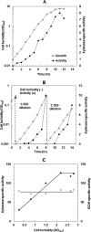
Cell density affects extracellular xylanase-specific activity. A, effect of cell turbidity on xylanase T-6 production in batch growth. Growth was carried out in a well aerated 10-liter fermenter in mBSM with 1% xylose. B, xylanase activity in cycled batch culture of G. stearothermophilus T-6. Strain T-6 culture was grown in mBSM with 1% xylose, and the culture was repeatedly diluted 500-fold into fresh medium. C, effect of cell turbidity on xylanase-specific activity in a carbon-limited Chemostat. Growth was carried out on defined medium at a constant dilution rate (D = 0.2 h−1). The specific activity of xylanase (●) and the intracellular enzyme isocitrate dehydrogenase (○) were determined at various culture densities at steady state. Xylanase-specific activity was determined by hydrolytic activity on the synthetic substrate pNPX2 at 50 °C, measured by absorbance at A420 nm (OD420). One unit of activity was defined as (A420/min/ml of culture per A600) × 100 (Miller units).
FIGURE 10.
FIGURE 10.
Expression of extracellular xylanase gene xynA and xynX, axe2, and xynB3 genes is increased by cell density. Strain T-6 cultures were grown in mBSM supplemented with 1% xylose and amino acids in a 10-liter fermenter. Samples (up to 100 ml) were collected at various cell densities along the logarithmic phase, and total RNA was extracted. Relative mRNA levels of the indicated genes at different cell densities were measured by quantitative real time RT PCR using the isocitrate dehydrogenase gene for normalization. A, xynA, xynA2, xynB1 and xynB2; B, xynX, axe2, xynB3.
FIGURE 11.
FIGURE 11.
Quantitative xynA-mRNA levels at different cell densities from strain T-6 cultures grown on medium containing xylose or glucose. A, strain T-6 cultures were grown in a 10-liter fermenter, and samples (up to 200 ml) were drawn at various cell densities along the logarithmic growth phase for total RNA isolation. RNase protection assays were performed with a 467-nt 32P-labeled antisense RNA probe. The probe was hybridized with 4 μg of total RNA. The protected RNAs were loaded on a 5% polyacrylamide gel, which was further visualized using a phosphorimager system. Mr is the labeled DNA fragments of pUC19 digested with Sau3A. Lane 1, probe hybridized with 10 μg of nonspecific yeast RNA incubated without RNase; lane 2, same as lane 1 but with RNase digestion; lanes 3–8, RNA from cultures grown on xylose, lanes 9–13, RNA from cultures grown on glucose. Cell turbidities at A600 (OD600): 3–0.02; 4–0.03; 5–0.05; 6–0.1; 7–0.23; 8–0.47; 9–0.008; 10–0.02; 11–0.04; 12–0.23; 13–0.65. B, xynA mRNA molecules per cell as function of cell density. The number of xylanase transcripts per cell was calculated based on the following: total RNA per cell is 1.78 × 10⁻⁷ μg, 1 A600 culture contains 2.6 × 10⁸ cells/ml, and probe molecular weight is 75,240. Xylanase mRNA levels at different cell densities from three independent experiments. (○ indicates glucose; ●, ■, ▴ xylose).
FIGURE 12.
FIGURE 12.
Partial characterization and purification of XDF. A, activation of xylanase using conditioned medium before and after absorption to XAD-2 matrix. 100 ml of conditioned medium was concentrated 2-fold using freeze dryer and then boiled for 10 min. Cells from early logarithmic growth phase were diluted 200-fold (∼0.001 A600 (OD600)) in fresh mBSM, conditioned medium (10–30 μl), and pNPX2 and placed in a 96-well plate. The plate is then incubated with shaking at 60 °C and monitored simultaneously for both cell growth (A600) and p-nitrophenol release (A420). Specific activity = A420/(A600 × min). The remaining medium was loaded on XAD-2 column and the flow-through was used to activate xylanase production at low cell-density. B, effect of proteinase K treatment of conditioned medium on xylanase cell density activation. Conditioned medium was incubated with proteinase K-beaded agarose (Sigma) for 1 h at 37 °C, subsequent by bead removal by centrifugation. The treated conditioned medium was used to assay xylanase activation at low cell density. C, elution pattern of the active XDF fraction by reverse phase HPLC on C18 column. 2.5 liters of conditioned medium were concentrated 500-fold by absorption on XAD-2 column. Aliquots of 0.25 ml of concentrated active sample were fractionated by reverse-phase chromatography on C18 column with a linear gradient of ACN in 0.1% aqueous TFA at 0.5% min−1. Fraction containing XDF activity was eluted with 20–25% ACN, and it is marked with dashed circle. mAU, milliabsorbance units.
FIGURE 13.
FIGURE 13.
Alignment of regulatory regions upstream to xylanases genes from Geobacillus spp. The sequences upstream the putative −10 regions of several xylanases genes from Geobacillus strains: G. stearothermophilus T-6, G. stearothermophilus T-1, Geobacillus sp. MAS1, Geobacillus sp. Y412MC6, Geobacillus sp. C56-T3, Geobacillus sp. GHH01 and G. thermodenitrificans NG80–2. The first sequence aligned is the sequence upstream of the xynX gene from G. stearothermophilus T-6. Conserved nucleotides are shown in bold, and the inverted repeat sequence, the putative XynX-binding site, is indicated by arrowheads.
FIGURE 14.
FIGURE 14.
Schematic representation of multiple regulatory mechanisms controlling the expression of xynA. Xylose and short xylo-oligomers are sensed by the two-component system XynDC, which in turn activates the ABC xylo-oligosaccharide transporter, XynEFG, facilitating the entrance of xylose and xylosaccharides into the cell. Inside the cell, xylose serves as the molecular inducer and interacts with the master repressor XylR, which negatively regulates the transcription of the xynA gene. XylR can also interact with glucose 6-phosphate, which exerts inducer prevention. Expression of the xynA gene is also repressed by glucose via carbon catabolite repression, most likely mediated by CcpA. In rapidly growing cells in high nutrition availability, xynA is repressed by CodY, which is subjected to regulation by GTP and branched-chain amino acids. Additionally, xynA is regulated by two yet uncharacterized mechanisms, a NIF3-like protein, XynX, and by cell density. The xynA gene is induced via quorum sensing regulation at relatively low cell density. The XDFs are most likely short modified peptides that can either bind to a specific sensor component of a two-component signal-transduction system or can be imported into the cell by a dedicated peptide transporter. The DNA sequence corresponds to position −131 bp with respect to the transcriptional start point (+1). The proposed catabolite-responsive element (cre) is presented in red letters. The potential inverted repeat binding site for the xylose repressor, XylR, and the putative XynX-binding site are indicated by arrows. Two of the proposed CodY-binding site sequences are shown in green letters. The table below summarizes the proposed regulatory proteins and their effectors involved in the regulation of the xynA gene.

References

    1. Shallom D., Shoham Y. (2003) Microbial hemicellulases. Curr. Opin. Microbiol. 6, 219–228 - PubMed
    1. Yu H., Zeng G., Huang H., Xi X., Wang R., Huang D., Huang G., Li J. (2007) Microbial community succession and lignocellulose degradation during agricultural waste composting. Biodegradation 18, 793–802 - PubMed
    1. Taylor L. E., 2nd, Henrissat B., Coutinho P. M., Ekborg N. A., Hutcheson S. W., Weiner R. M. (2006) Complete cellulase system in the marine bacterium Saccharophagus degradans strain 2-40T. J. Bacteriol. 188, 3849–3861 - PMC - PubMed
    1. Ouyang J., Yan M., Kong D., Xu L. (2006) A complete protein pattern of cellulase and hemicellulase genes in the filamentous fungus Trichoderma reesei. Biotechnol. J. 1, 1266–1274 - PubMed
    1. Gilbert H. J., Stålbrand H., Brumer H. (2008) How the walls come crumbling down: recent structural biochemistry of plant polysaccharide degradation. Curr. Opin. Plant Biol. 11, 338–348 - PubMed

Publication types

MeSH terms

Associated data

LinkOut - more resources