

Ablation of EIF5A2 induces tumor vasculature remodeling and improves tumor response to chemotherapy via regulation of matrix metalloproteinase 2 expression
- PMID: 25071013
- PMCID: PMC4196158
- DOI: 10.18632/oncotarget.2236
Ablation of EIF5A2 induces tumor vasculature remodeling and improves tumor response to chemotherapy via regulation of matrix metalloproteinase 2 expression
Abstract
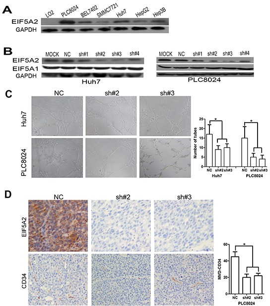
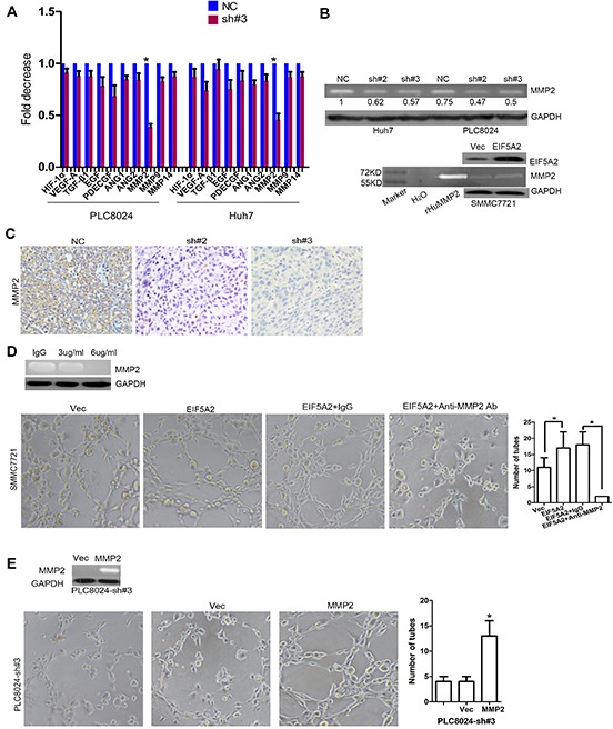
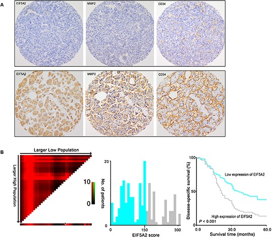
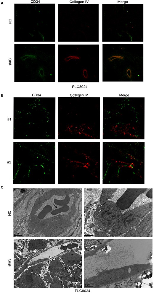
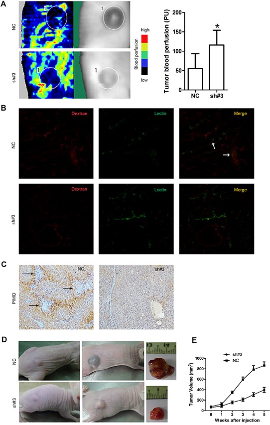
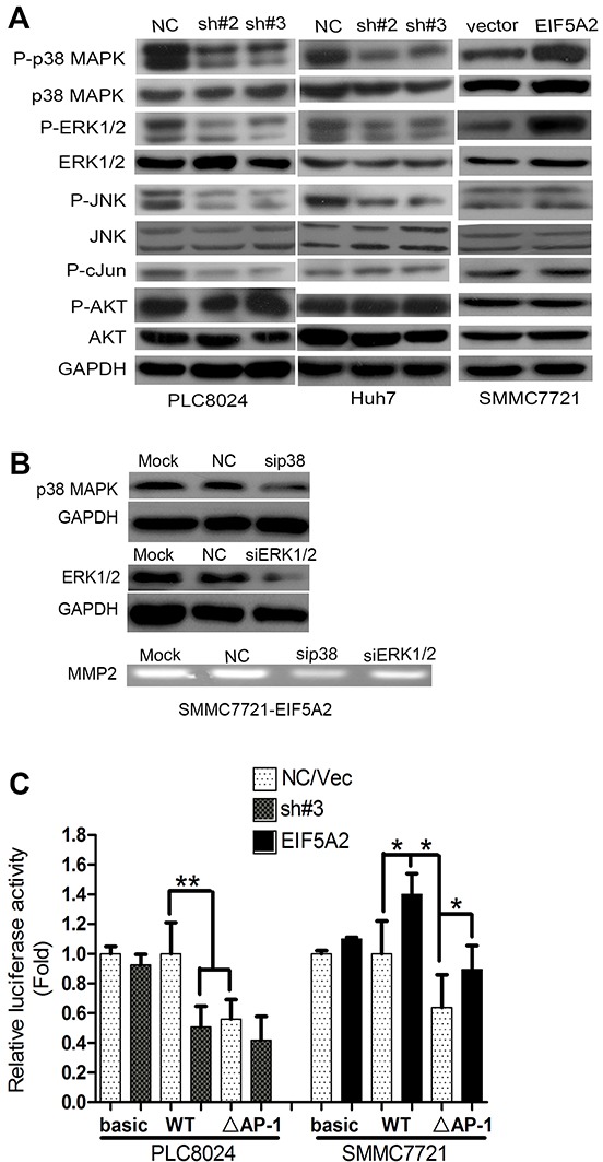
Hepatocellular carcinoma (HCC) is a highly vascularized tumor with poor clinical outcome. Our previous work has shown that eukaryotic initiation factor 5A2 (EIF5A2) over-expression enhances HCC cell metastasis. In this study, EIF5A2 was identified to be an independent risk factor for poor disease-specific survival among HCC patients. Both in vitro and in vivo assays indicated that ablation of endogenous EIF5A2 inhibited tumor angiogenesis by reducing matrix metalloproteinase 2 (MMP-2) expression. Given that MMP-2 degrades collagen IV, a main component of the vascular basement membrane (BM), we subsequently investigated the effect of EIF5A2 on tumor vasculature remodeling using complementary approaches, including fluorescent immunostaining, transmission electron microscopy, tumor perfusion assays and tumor hypoxia assays. Taken together, our results indicate that EIF5A2 silencing increases tumor vessel wall continuity, increases blood perfusion and improves tumor oxygenation. Additionally, we found that ablation of EIF5A2 enhanced the chemosensitivity of HCC cells to 5-Fluorouracil (5-FU). Finally, we demonstrated that EIF5A2 might exert these functions by enhancing MMP-2 activity via activation of p38 MAPK and JNK/c-Jun pathways.
Conclusion: This study highlights an important role of EIF5A2 in HCC tumor vessel remodeling and indicates that EIF5A2 represents a potential therapeutic target in the treatment of HCC.
Conflict of interest statement
The authors declare no conflict of interest.
Figures
Similar articles
-
Knockdown of eukaryotic translation initiation factor 5A2 enhances the therapeutic efficiency of doxorubicin in hepatocellular carcinoma cells by triggering lethal autophagy.Int J Oncol. 2020 Dec;57(6):1368-1380. doi: 10.3892/ijo.2020.5143. Epub 2020 Nov 2. Int J Oncol. 2020. PMID: 33174013 Free PMC article.
-
Eukaryotic Initiation Factor 5A2 Contributes to the Maintenance of CD133(+) Hepatocellular Carcinoma Cells via the c-Myc/microRNA-29b Axis.Stem Cells. 2018 Feb;36(2):180-191. doi: 10.1002/stem.2734. Epub 2017 Nov 20. Stem Cells. 2018. PMID: 29119708
-
MicroRNA-383 inhibits doxorubicin resistance in hepatocellular carcinoma by targeting eukaryotic translation initiation factor 5A2.J Cell Mol Med. 2019 Nov;23(11):7190-7199. doi: 10.1111/jcmm.14197. Epub 2019 Feb 23. J Cell Mol Med. 2019. PMID: 30801960 Free PMC article.
-
Eukaryotic translation initiation factor 5A2 promotes metabolic reprogramming in hepatocellular carcinoma cells.Carcinogenesis. 2017 Jan;38(1):94-104. doi: 10.1093/carcin/bgw119. Epub 2016 Nov 22. Carcinogenesis. 2017. PMID: 27879277
-
Unveiling EIF5A2: A multifaceted player in cellular regulation, tumorigenesis and drug resistance.Eur J Pharmacol. 2025 Jun 15;997:177596. doi: 10.1016/j.ejphar.2025.177596. Epub 2025 Apr 5. Eur J Pharmacol. 2025. PMID: 40194645 Review.
Cited by
-
Chemolipiodolization with or without embolization in transcatheter arterial chemoembolization combined with radiofrequency ablation for hepatocellular carcinoma-propensity score matching analysis.Oncotarget. 2016 May 24;7(21):31311-21. doi: 10.18632/oncotarget.8897. Oncotarget. 2016. PMID: 27121318 Free PMC article.
-
Targeting the MAPK signaling pathway: implications and prospects of flavonoids in 3P medicine as modulators of cancer cell plasticity and therapeutic resistance in breast cancer patients.EPMA J. 2025 Apr 10;16(2):437-463. doi: 10.1007/s13167-025-00407-6. eCollection 2025 Jun. EPMA J. 2025. PMID: 40438489 Free PMC article.
-
Comparison of the therapeutic effectiveness of human CD34+ and rat bone marrow mesenchymal stem cells on improvement of experimental liver fibrosis in Wistar rats.Int J Physiol Pathophysiol Pharmacol. 2016 Sep 30;8(3):128-139. eCollection 2016. Int J Physiol Pathophysiol Pharmacol. 2016. PMID: 27785340 Free PMC article.
-
Eukaryotic translation initiation factor 5A in the pathogenesis of cancers.Oncol Lett. 2020 Oct;20(4):81. doi: 10.3892/ol.2020.11942. Epub 2020 Aug 3. Oncol Lett. 2020. PMID: 32863914 Free PMC article. Review.
-
Expression of EIF5A2 associates with poor survival of nasopharyngeal carcinoma patients treated with induction chemotherapy.BMC Cancer. 2016 Aug 22;16(1):669. doi: 10.1186/s12885-016-2714-2. BMC Cancer. 2016. PMID: 27549330 Free PMC article.
References
-
- El-Serag HB, Rudolph KL. Hepatocellular carcinoma: epidemiology and molecular carcinogenesis. Gastroenterology. 2007;132:2557–2576. - PubMed
-
- Llovet JM, Ricci S, Mazzaferro V, Hilgard P, Gane E, Blanc JF, de Oliveira AC, Santoro A, Raoul JL, Forner A, Schwartz M, Porta C, Zeuzem S, et al. Sorafenib in advanced hepatocellular carcinoma. N Engl J Med. 2008;359:378–390. - PubMed
-
- Guan XY, Sham JS, Tang TC, Fang Y, Huo KK, Yang JM. Isolation of a novel candidate oncogene within a frequently amplified region at 3q26 in ovarian cancer. Cancer Res. 2001;61:3806–3809. - PubMed
-
- Guan XY, Fung JM, Ma NF, Lau SH, Tai LS, Xie D, Zhang Y, Hu L, Wu QL, Fang Y, Sham JS. Oncogenic role of eIF-5A2 in the development of ovarian cancer. Cancer Res. 2004;64:4197–4200. - PubMed
Publication types
MeSH terms
Substances
LinkOut - more resources
Full Text Sources
Other Literature Sources
Medical
Research Materials
Miscellaneous
