. 2015 Jan;64(1):23-35.
doi: 10.2337/db13-1070. Epub 2014 Jul 28.

Compartmentalized acyl-CoA metabolism in skeletal muscle regulates systemic glucose homeostasis

Affiliations

Compartmentalized acyl-CoA metabolism in skeletal muscle regulates systemic glucose homeostasis

Lei O Li et al. Diabetes. 2015 Jan.

Abstract

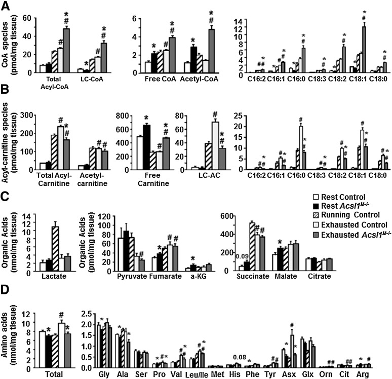
The impaired capacity of skeletal muscle to switch between the oxidation of fatty acid (FA) and glucose is linked to disordered metabolic homeostasis. To understand how muscle FA oxidation affects systemic glucose, we studied mice with a skeletal muscle-specific deficiency of long-chain acyl-CoA synthetase (ACSL)1. ACSL1 deficiency caused a 91% loss of ACSL-specific activity and a 60-85% decrease in muscle FA oxidation. Acsl1(M-/-) mice were more insulin sensitive, and, during an overnight fast, their respiratory exchange ratio was higher, indicating greater glucose use. During endurance exercise, Acsl1(M-/-) mice ran only 48% as far as controls. At the time that Acsl1(M-/-) mice were exhausted but control mice continued to run, liver and muscle glycogen and triacylglycerol stores were similar in both genotypes; however, plasma glucose concentrations in Acsl1(M-/-) mice were ∼40 mg/dL, whereas glucose concentrations in controls were ∼90 mg/dL. Excess use of glucose and the likely use of amino acids for fuel within muscle depleted glucose reserves and diminished substrate availability for hepatic gluconeogenesis. Surprisingly, the content of muscle acyl-CoA at exhaustion was markedly elevated, indicating that acyl-CoAs synthesized by other ACSL isoforms were not available for β-oxidation. This compartmentalization of acyl-CoAs resulted in both an excessive glucose requirement and severely compromised systemic glucose homeostasis.


Figures

Figure 1
Figure 1
ACSL1 deficiency decreased ACSL-specific activity and FA oxidation in skeletal muscle. A: ACSL1 protein in the gastrocnemius muscle and heart in Acsl1M−/− and control mice. B: mRNA expression of Acsl1 isoforms in gastrocnemius relative to tubulin (males; n = 8). C: ACSL-specific activity in homogenates from the gastrocnemius muscle and heart (n = 4). Histology (×20) of control (D) and Acsl1M−/− (E) gastrocnemius. Thin arrows, central nuclei; thick arrow, macrophage infiltration. FA oxidation in homogenates from gastrocnemius and soleus muscle from [1-14C]palmitate (F) or [14C]palmitoyl-CoA (G) (n = 3–4). Oxidation (H) or incorporation (I) in isolated soleus or extensor digitorum longus with [14C]16:0, or with [14C]glucose (J) (n = 5). *P < 0.05, compared with control littermates. EDL, extensor digitorum longus; Gastroc, gastrocnemius.
Figure 2
Figure 2
Acsl1M−/− mice were more vulnerable to hypoglycemia and hepatic lipid accumulation after an overnight fast and required less insulin to maintain euglycemia. Plasma glucose (A), nonesterified FAs (NEFAs) (B), TAG (C), and β-hydroxybutyrate (D) in female Acsl1M−/− after food removal for 4 h (n = 9–11) or fasting overnight (O/N) (n = 7–17). E: Histology of representative liver slides from female control and Acsl1M−/− mice after an overnight fast (hematoxylin-eosin staining). Measurement after an overnight fast of liver TAG (F) and glycogen (G) content (n = 4–8). H: Gene expression of glucose-6-phosphatase (G6Pase), phosphoenolpyruvate (PEPCK), and hydroxymethylglutaryl-CoA synthase-2 (HMGS2) in male mice (n = 6–10). I: Glycerol tolerance test in male mice after an overnight fast (n = 4). J: GLUT1 protein in gastrocnemius muscle (n = 4–5). In control and Acsl1M−/− female mice fed a high-fat diet (HFD) or matched standard diet (SD): glucose tolerance (K); blood glucose and insulin levels before and 15 min after intraperitoneal administration of insulin (L); and insulin tolerance tests and areas above the curve (AAC) after food removal for 4 h (M). N: pAkt (T308) and total Akt in female liver and gastrocnemius muscle 10 min after intraperitoneal PBS or insulin. C, control mice; K, knockout mice. #P < 0.05, vs. 4-h state (same genotype); *P < 0.05, vs. control (same treatment).
Figure 3
Figure 3
Acsl1M−/− mice used more glucose as fuel source. A: Male mice (13 wk) were placed in metabolic chambers at 1:00 p.m. and measurements were taken for 2 days before overnight (O/N; 5:00 p.m. to 10:00 a.m.) fasting. Mice were then refed with chow for 24 h (n = 4); RER (VCO2/VO2) was monitored by indirect calorimetry during O/N fasting. Average RER (B), food intake (C), and heat production (D) during day and night cycles when the mice were fed, fasted, or refed. *P < 0.05, compared with control.
Figure 4
Figure 4
Muscle ACSL1 deficiency impaired endurance capacity. A: Total distance run during treadmill endurance exercise for Acsl1M−/− mice (16 weeks of age, female). Blood lactate (B) and glucose (C) levels during endurance exercise. *P < 0.05, glucose from exhausted Acsl1M−/− mice compared with control mice that had run same distance (Run). D: mRNA expression of glucose-6-phosphatase (G6Pase), phosphoenolpyruvate carboxykinase (PEPCK), FGF21, hydroxymethylglutaryl-CoA synthase-2 (HMGS2), and liver CPT1 (LCPT1) (n = 5–13). *P < 0.05, compared with exhausted controls. Run, data from control mice collected at the time their Acsl1M−/− littermates were exhausted.
Figure 5
Figure 5
Metabolite changes in Acsl1M−/− gastrocnemius muscle during endurance exercise (16–26 weeks of age, female). A: Muscle free- and acyl-CoAs. B: Muscle free- and acyl-carnitines. C: Muscle Krebs cycle intermediates. D: Muscle amino acids (n = 5–9). #P < 0.05, vs. 4-h state (same genotype); *P < 0.05, vs. control (same treatment). Running, data from control mice collected when their Acsl1M−/− littermates were exhausted. a-KG, α-ketoglutarate; LC-AC, long-chain acyl-carnitine; LC-CoA, long-chain acyl-CoA; C, carbon number.
Figure 6
Figure 6
Muscle ACSL1 regulates fuel use and whole-body metabolism. A: mRNA expression of Acsl isoforms in gastrocnemius muscle relative to tubulin (female, n = 7–9). B: ACSL-specific activity within gastrocnemius homogenates (n = 4). C: Intramuscular TAG in gastrocnemius muscle after endurance exercise (n = 6–8). #P < 0.05, vs. 4-h state (same genotype); *P < 0.05, vs. control (same treatment). Exh, exhausted; Rest, resting.
Figure 7
Figure 7
Skeletal muscle metabolism in control and Acsl1M−/− mice. Exercising control muscle. Initial fuels include glucose hydrolyzed from hepatic and muscle glycogen and intramuscular TAG. Activation of AMPK suppresses TAG synthesis and accelerates mitochondrial FA oxidation, and autophagy releases amino acids, which are exported to provide a substrate for hepatic gluconeogenesis (37). As exercise continues, Acsl1 mRNA increases, and epinephrine increases the supply of adipocyte-derived long-chain (LC)-FA, which are activated by ACSL1 and converted to LC-acyl-carnitines (ACs) by CPT1 and, within the mitochondria, back to LC-CoA by CPT2. Mitochondrial oxidation produces shorter-chain acyl-CoAs and their AC counterparts. Some amino acids that arise from protein hydrolysis contribute to TCA cycle intermediates and to acetyl-CoA for energy production. Hepatic glucose, now derived via amino acid–fueled gluconeogenesis, continues to enter the glycolytic pathway, but the use of LC-FA spares muscle demand for glucose. In addition to ACs of different chain lengths that move out of the mitochondria into the cytosol, ACs derived from hepatic FA oxidation are released into the blood and equilibrate across the muscle plasma membrane. Exercising Acsl1M−/− muscle. When ACSL1 is absent, other ACSL isoforms (ACSL?) activate the large amount of entering LC-FA. Most, but perhaps not all, of the LC-CoA produced by these ACSL? lack ready access to CPT1 and cannot be converted to LC-AC and enter the mitochondria. (For simplicity, we have not shown a theoretical pathway by which some FAs might be activated by ACSL? and oxidized in the absence of ACSL1 [see Fig. 1D]; these pathways 1) might provide some LC-CoA at the mitochondrial surface with access to CPT1 or 2) might provide medium-chain (MC)-CoA in Acsl1M−/− muscle [Supplementary Fig. 4A], which can enter the mitochondria without CPT1.) The cell content of LC-CoAs increases because of their diminished entry into both oxidative and synthetic pathways. Failure to switch to the use of LC-FA for oxidation enhances glucose entry and oxidation and increases the use of muscle protein–derived amino acid that is converted to acetyl-CoA and TCA cycle intermediates. Fewer amino acids are released by muscle to supply the substrate for hepatic gluconeogenesis. As a consequence, systemic hypoglycemia ensues and the capacity for endurance exercise is impaired.

References

    1. Kelley DE, Mandarino LJ. Fuel selection in human skeletal muscle in insulin resistance: a reexamination. Diabetes 2000;49:677–683 - PubMed
    1. Thyfault JP, Rector RS, Noland RC. Metabolic inflexibility in skeletal muscle: a prelude to the cardiometabolic syndrome? J Cardiometab Syndr 2006;1:184–189 - PubMed
    1. Watt MJ, Hoy AJ. Lipid metabolism in skeletal muscle: generation of adaptive and maladaptive intracellular signals for cellular function. Am J Physiol Endocrinol Metab 2012;302:E1315–E1328 - PubMed
    1. Zhang D, Liu ZX, Choi CS, et al. . Mitochondrial dysfunction due to long-chain Acyl-CoA dehydrogenase deficiency causes hepatic steatosis and hepatic insulin resistance. Proc Natl Acad Sci USA 2007;104:17075–17080 - PMC - PubMed
    1. Ukropcova B, Sereda O, de Jonge L, et al. . Family history of diabetes links impaired substrate switching and reduced mitochondrial content in skeletal muscle. Diabetes 2007;56:720–727 - PubMed

Publication types

MeSH terms