

Rebound burst firing in the reticular thalamus is not essential for pharmacological absence seizures in mice
- PMID: 25071191
- PMCID: PMC4136605
- DOI: 10.1073/pnas.1408609111
Rebound burst firing in the reticular thalamus is not essential for pharmacological absence seizures in mice
Abstract
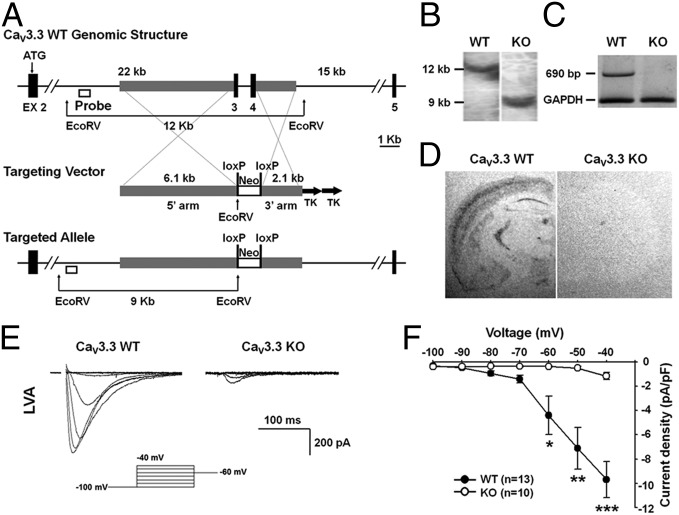
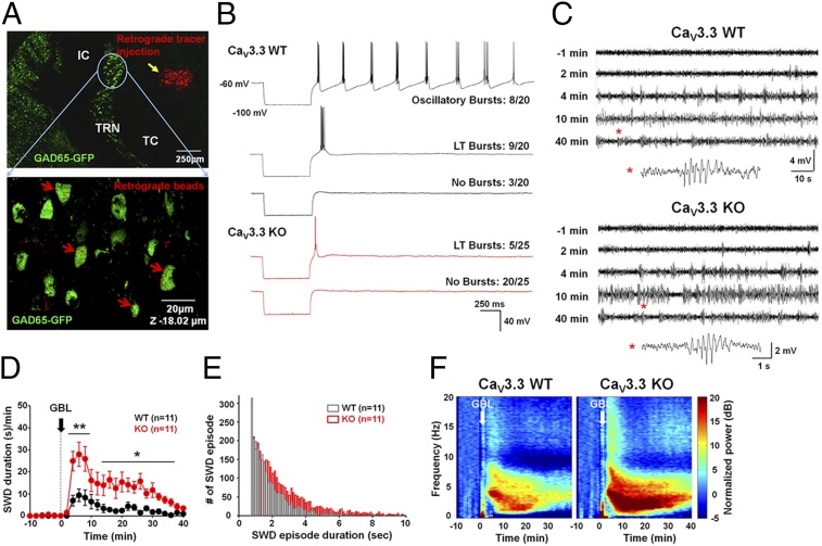
Intrinsic burst and rhythmic burst discharges (RBDs) are elicited by activation of T-type Ca(2+) channels in the thalamic reticular nucleus (TRN). TRN bursts are believed to be critical for generation and maintenance of thalamocortical oscillations, leading to the spike-and-wave discharges (SWDs), which are the hallmarks of absence seizures. We observed that the RBDs were completely abolished, whereas tonic firing was significantly increased, in TRN neurons from mice in which the gene for the T-type Ca(2+) channel, CaV3.3, was deleted (CaV3.3(-/-)). Contrary to expectations, there was an increased susceptibility to drug-induced SWDs both in CaV3.3(-/-) mice and in mice in which the CaV3.3 gene was silenced predominantly in the TRN. CaV3.3(-/-) mice also showed enhanced inhibitory synaptic drive onto TC neurons. Finally, a double knockout of both CaV3.3 and CaV3.2, which showed complete elimination of burst firing and RBDs in TRN neurons, also displayed enhanced drug-induced SWDs and absence seizures. On the other hand, tonic firing in the TRN was increased in these mice, suggesting that increased tonic firing in the TRN may be sufficient for drug-induced SWD generation in the absence of burst firing. These results call into question the role of burst firing in TRN neurons in the genesis of SWDs, calling for a rethinking of the mechanism for absence seizure induction.
Conflict of interest statement
The authors declare no conflict of interest.
Figures


Similar articles
-
T-type Ca2+ channels in absence epilepsy.Pflugers Arch. 2014 Apr;466(4):719-34. doi: 10.1007/s00424-014-1461-y. Epub 2014 Feb 13. Pflugers Arch. 2014. PMID: 24519464 Review.
-
Lack of the burst firing of thalamocortical relay neurons and resistance to absence seizures in mice lacking alpha(1G) T-type Ca(2+) channels.Neuron. 2001 Jul 19;31(1):35-45. doi: 10.1016/s0896-6273(01)00343-9. Neuron. 2001. PMID: 11498049
-
CaV 3.2 drives sustained burst-firing, which is critical for absence seizure propagation in reticular thalamic neurons.Epilepsia. 2018 Apr;59(4):778-791. doi: 10.1111/epi.14018. Epub 2018 Feb 21. Epilepsia. 2018. PMID: 29468672 Free PMC article.
-
T-type Ca²⁺ channels in absence epilepsy.Biochim Biophys Acta. 2013 Jul;1828(7):1560-71. doi: 10.1016/j.bbamem.2013.02.002. Epub 2013 Feb 13. Biochim Biophys Acta. 2013. PMID: 23416255 Review.
-
Adult loss of Cacna1a in mice recapitulates childhood absence epilepsy by distinct thalamic bursting mechanisms.Brain. 2020 Jan 1;143(1):161-174. doi: 10.1093/brain/awz365. Brain. 2020. PMID: 31800012 Free PMC article.
Cited by
-
Effects of a patient-derived de novo coding alteration of CACNA1I in mice connect a schizophrenia risk gene with sleep spindle deficits.Transl Psychiatry. 2020 Jan 23;10(1):29. doi: 10.1038/s41398-020-0685-1. Transl Psychiatry. 2020. PMID: 32066662 Free PMC article.
-
Unbalanced Peptidergic Inhibition in Superficial Neocortex Underlies Spike and Wave Seizure Activity.J Neurosci. 2015 Jun 24;35(25):9302-14. doi: 10.1523/JNEUROSCI.4245-14.2015. J Neurosci. 2015. PMID: 26109655 Free PMC article.
-
µ-Theraphotoxin Pn3a inhibition of CaV3.3 channels reveals a novel isoform-selective drug binding site.Elife. 2022 Jul 20;11:e74040. doi: 10.7554/eLife.74040. Elife. 2022. PMID: 35858123 Free PMC article.
-
Neuronal Cav3 channelopathies: recent progress and perspectives.Pflugers Arch. 2020 Jul;472(7):831-844. doi: 10.1007/s00424-020-02429-7. Epub 2020 Jul 7. Pflugers Arch. 2020. PMID: 32638069 Free PMC article. Review.
-
Heantos-4, a natural plant extract used in the treatment of drug addiction, modulates T-type calcium channels and thalamocortical burst-firing.Mol Brain. 2016 Dec 5;9(1):94. doi: 10.1186/s13041-016-0274-7. Mol Brain. 2016. PMID: 27919294 Free PMC article.
References
-
- Crunelli V, Leresche N. Childhood absence epilepsy: Genes, channels, neurons and networks. Nat Rev Neurosci. 2002;3(5):371–382. - PubMed
-
- Steriade M, Deschênes M, Domich L, Mulle C. Abolition of spindle oscillations in thalamic neurons disconnected from nucleus reticularis thalami. J Neurophysiol. 1985;54(6):1473–1497. - PubMed
-
- Steriade M, McCormick DA, Sejnowski TJ. Thalamocortical oscillations in the sleeping and aroused brain. Science. 1993;262(5134):679–685. - PubMed
-
- Huntsman MM, Porcello DM, Homanics GE, DeLorey TM, Huguenard JR. Reciprocal inhibitory connections and network synchrony in the mammalian thalamus. Science. 1999;283(5401):541–543. - PubMed
Publication types
MeSH terms
Substances
LinkOut - more resources
Full Text Sources
Other Literature Sources
Molecular Biology Databases
Miscellaneous
