Review
. 2014 Jul 8:7:63.
doi: 10.3389/fnmol.2014.00063. eCollection 2014.

Ubiquitin pathways in neurodegenerative disease

Affiliations
Review

Ubiquitin pathways in neurodegenerative disease

Graham Atkin et al. Front Mol Neurosci. .

Abstract

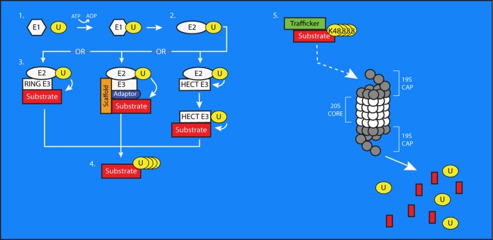
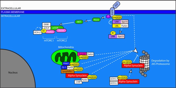
Control of proper protein synthesis, function, and turnover is essential for the health of all cells. In neurons these demands take on the additional importance of supporting and regulating the highly dynamic connections between neurons that are necessary for cognitive function, learning, and memory. Regulating multiple unique synaptic protein environments within a single neuron while maintaining cell health requires the highly regulated processes of ubiquitination and degradation of ubiquitinated proteins through the proteasome. In this review, we examine the effects of dysregulated ubiquitination and protein clearance on the handling of disease-associated proteins and neuronal health in the most common neurodegenerative diseases.

Keywords: Alzheimer's disease; Amyotrophic Lateral Sclerosis; Huntington's disease; Parkinson disease; neurodegenerative diseases; proteasome; protein quality control; ubiquitin.


Figures

Figure 1
Figure 1
The process of ubiquitin conjugation. 1. Ubiquitin (U) is bound via a thioester bond to the active-site cysteine of an E1 Ubiquitin-Activating enzyme, through a process requiring ATP. 2. The ubiquitin molecule is then passed to an E2 Ubiquitin-Conjugating Enzyme through trans(thio)esterification. 3. An E3 ubiquitin ligase brings the E2 into sufficiently close proximity and correct alignment with a substrate protein to facilitate the transfer of ubiquitin to a target residue. In the case of HECT-type E3s, the ubiquitin is first transferred to an active site cysteine on the E3 before being conjugated to the substrate. E3s can also exist as multi-subunit complexes including scaffolding and adaptor proteins that confer substrate specificity to the process of ubiquitin transfer. 4. Additional ubiquitin molecules can be added onto the first to create polyubiquitin chains on substrate proteins. 5. The 26S proteasome is composed of a cylindrical, proteolytic 20S core which is capped at both ends by a 19S regulatory cap. Polyubiquitinated substrate proteins, typically bearing K48-linked chains, are targeted to the proteasome by trafficking proteins. The proteasome digests these substrates into smaller peptides and free ubiquitin molecules, which then can be used to modify further substrates.
Figure 2
Figure 2
Regulation of protein trafficking, receptor signaling, and protein clearance by ubiquitination in Alzheimer's disease. Improper processing of APP and Tau contribute to the pathology of AD. Excessive or improperly folded APP is cleared from the ER by HDR1 and Fbxo2 through the addition of K-48 linked chains. Ubiquitin-mediated processes are indicated by dashed lines. Maturation of APP is arrested in the early secretory pathway by non-degradative, K-63 linked ubiquitination that is stimulated by Ubiquilin 1. Surface APP is endocytosed to the late Golgi, where it is cleaved by secretases including BACE-1 and PS-1, whose levels are regulated by the E3 ligases Fbxo2 and FBXW-7, respectively. The cleavage of APP results in the production of Amyloid-Beta, which can be targeted for degradation by CHIP. Uncleared Amyloid-Beta is exocytosed to the extracellular space, where it aggregates to form plaques. Amyloid-Beta can influence NMDA receptor signaling. NMDA receptor activation stimulates the kinase Cdk5, which results in the downstream inhibition of the E3 ligase APC and blocks the degradation of cyclin B1. NMDA receptor signaling also increases the activation of GSK3B, which phosphorylates Tau. GSK3B is targeted for degradation by NUB1, which also blocks the interaction between GSK3B and Tau. GSK3B activity is increased by complexing with p53, whose levels are regulated by the E3 ligase MDM2. Under conditions of cellular stress, MDM2 auto-ubiquitinates and targets itself for degradation. The E3 ligase CHIP targets Tau, but can have divergent effects on its handling. When working in combination with Hsp70, CHIP targets Tau for degradation. However, when Hsp90 is involved, CHIP facilitates an alternative ubiquitination of unknown linkage type, resulting in the accumulation of phosphorylated tau. Through an unknown mechanism, this accumulated Tau then forms insoluble protein aggregates.
Figure 3
Figure 3
Ubiquitination in pro-survival pathways, mitochondria stability, and protein clearance in Parkinson's disease. Dysregulation of Parkin and UCHL1, both of which are associated with PD, has widespread effects on neuronal health. The Epidermal Growth Factor (EGF) binds to EGFR and initiates pro-survival signaling through the Akt and mTOR pathways. Ligand-binding to EGFR stimulates its ubiquitination, which allows for its UIM-dependent recognition by Eps15. Eps15 internalizes EGFR, allowing it to be trafficked to the proteasome for degradation. The EGFR-Eps15 interaction is blocked by the interaction of the UBL of the E3 ligase Parkin with the UIM of Eps15; this interaction increases Parkin's ligase activity, causing Eps15 to become ubiquitinated and dissociated from its UIM-dependent interactors. Downstream of EGFR signaling, mTOR's participation in the protein complex mTORC1 requires the ubiquitination of Raptor by DDB1 and Cul4. This ubiquitination is undone by the de-ubiquitinating enzyme UCHL1. Parkin also plays a role in the degradation of mitochondrial outer membrane proteins including Tom20, and through its involvement with PINK1 and Dj-1, facilitates the degradation of Synphilin-1 and itself. Synphilin interacts with alpha-synuclein, which can be degraded by Parkin in association with CHIP. Dimerized UCHL1 may also modify alpha-synuclein, though the result is non-degradative, K-63 linked ubiquitination which promotes its accumulation. Accumulated alpha-synuclein binds to the 20S core of the proteasome and inhibits proteasome function.
Figure 4
Figure 4
A role for ubiquitination in the pathogenesis of Amyotrophic Lateral Sclerosis. Impaired turnover of SOD1 and TPD-43 are implicated in the etiology of ALS. Mutated or excess SOD1 in the ER is targeted for degradation by the E3 ligase NEDL1 via the translocon protein TRAP. SOD1 in the cytosol is targeted by the canonical HECT-type E3 ligase, E6-AP, and the RING-type E3 Dorfin. TDP-43 can be ubiquitinated by Parkin in association with HDAC6. This modification can include both K48 and K63-linked chains and does not lead to the degradation of TDP-43, instead increasing the proportion of TDP-43 in the cytosol. In the nucleus, TDP-43 regulates the transcription of genes including Parkin. In the cytosol, accumulated TDP-43 can inhibit the proteasome and form protein aggregates. In cases associated with mutations in the C9ORF72 gene, aggregates of DPR proteins are present with ubiquitin- and p62-positive immunoreactivity, but do no colocalize with TPD-43.
Figure 5
Figure 5
Ubiquitin-mediated handling of the pathologic Huntingtin protein in Huntington's disease. The Huntingtin protein is improperly cleared from neurons in HD. Mutant Huntingtin can be targeted for degradation in the ER by HRD1, and in the cytosol by NUB1. However, this ubiquitination can be edited by UCHL1, inhibiting the degradation of Huntingtin and promoting its aggregation in the cytosol. Traf6 facilitates the non-canonical ubiquitination of Huntingtin through the formation of K6, K27, and K29-linked polyubiquitin chains, and these modifications selectively promote the aggregation of mutant Huntingtin. Aggregates of mutant Huntingtin include components of the UPS including proteasomes. Rhes promotes the sumoylation of Huntingtin, which reduces its aggregation, but causes sumoylated, mutant Huntingtin to interfere with other cellular processes including gene transcription in the nucleus.

References

    1. Abou-Sleiman P. M., Healy D. G., Quinn N., Lees A. J., Wood N. W. (2003). The role of pathogenic DJ-1 mutations in Parkinson's disease. Ann. Neurol. 54, 283–286 10.1002/ana.10675 - DOI - PubMed
    1. Adachi H., Waza M., Tokui K., Katsuno M., Minamiyama M., Tanaka F., et al. (2007). CHIP overexpression reduces mutant androgen receptor protein and ameliorates phenotypes of the spinal and bulbar muscular atrophy transgenic mouse model. J. Neurosci. 27, 5115–5126 10.1523/JNEUROSCI.1242-07.2007 - DOI - PMC - PubMed
    1. Alves-Rodrigues A., Gregori L., Figueiredo-Pereira M. E. (1998). Ubiquitin, cellular inclusions and their role in neurodegeneration. Trends Neurosci. 21, 516–520 10.1016/S0166-2236(98)01276-4 - DOI - PubMed
    1. Andersen P. M., Al-Chalabi A. (2011). Clinical genetics of amyotrophic lateral sclerosis: what do we really know? Nat. Rev. Neurol. 7, 603–615 10.1038/nrneurol.2011.150 - DOI - PubMed
    1. Ardley H. C., Robinson P. A. (2005). E3 ubiquitin ligases. Essays Biochem. 41, 15–30 10.1042/EB0410015 - DOI - PubMed

LinkOut - more resources