. 2014 Jul 4:8:59.
doi: 10.3389/fnana.2014.00059. eCollection 2014.

Combinatorial expression of Lef1, Lhx2, Lhx5, Lhx9, Lmo3, Lmo4, and Prox1 helps to identify comparable subdivisions in the developing hippocampal formation of mouse and chicken

Affiliations

Combinatorial expression of Lef1, Lhx2, Lhx5, Lhx9, Lmo3, Lmo4, and Prox1 helps to identify comparable subdivisions in the developing hippocampal formation of mouse and chicken

Antonio Abellán et al. Front Neuroanat. .

Abstract

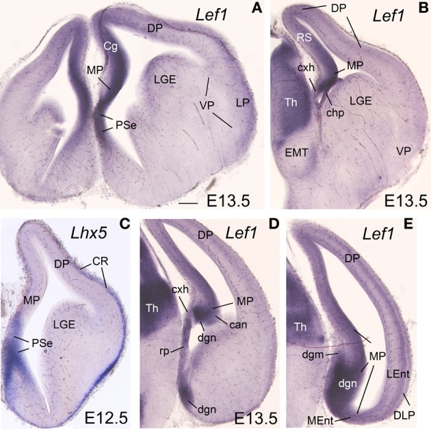
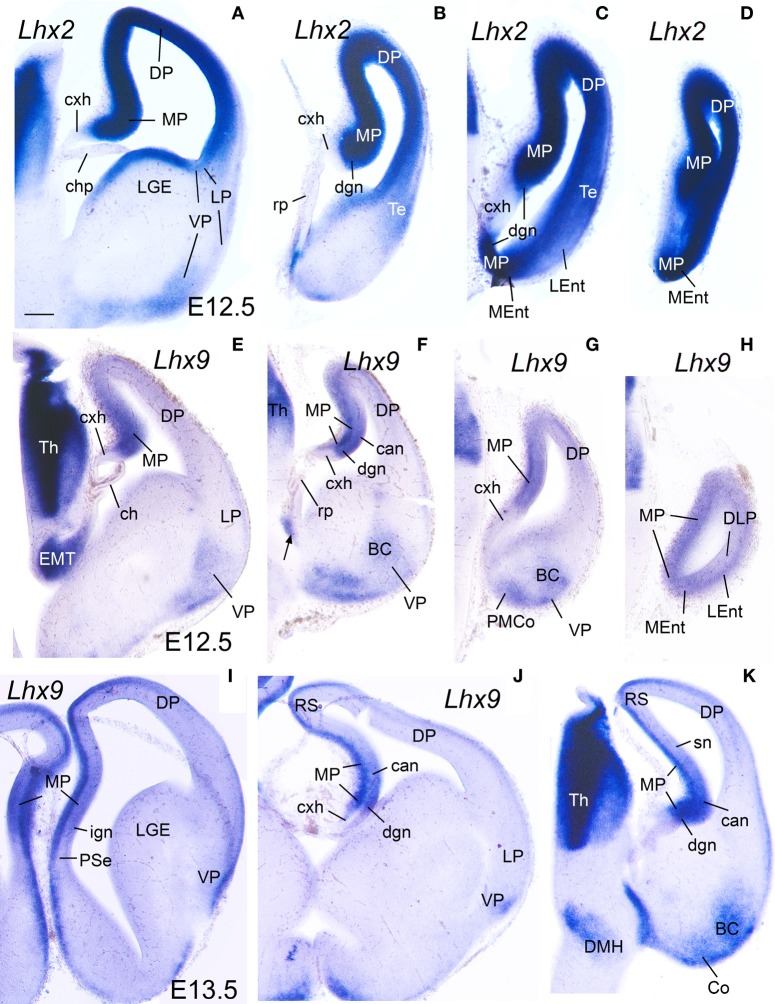
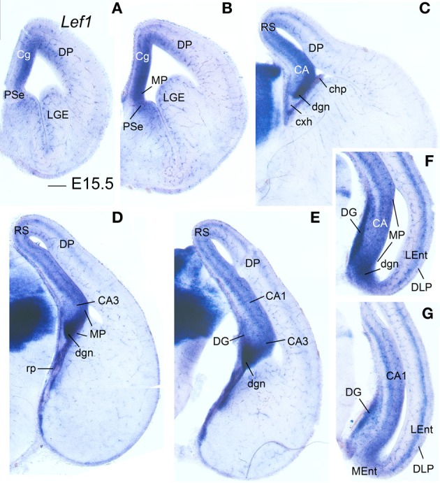
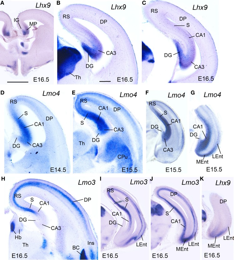
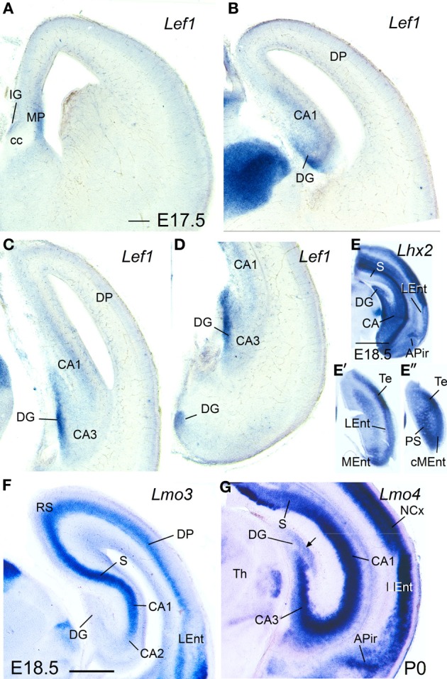
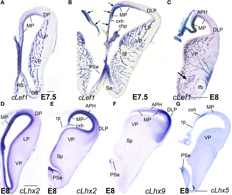
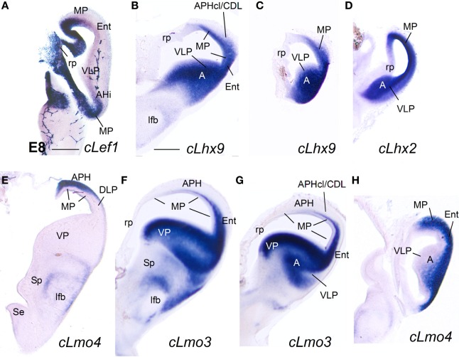
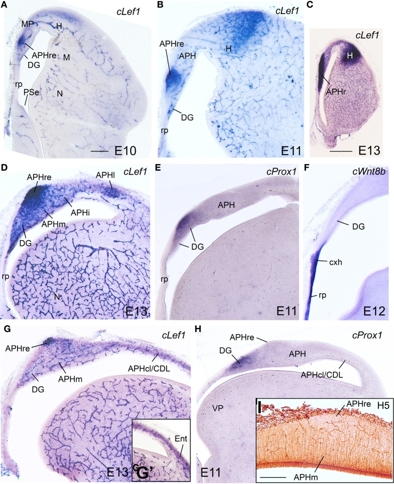
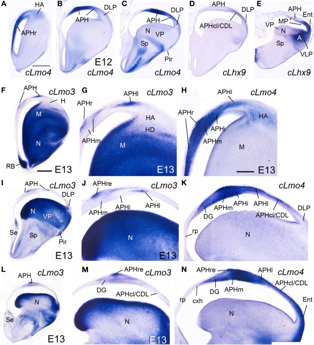
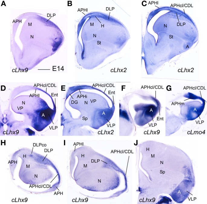
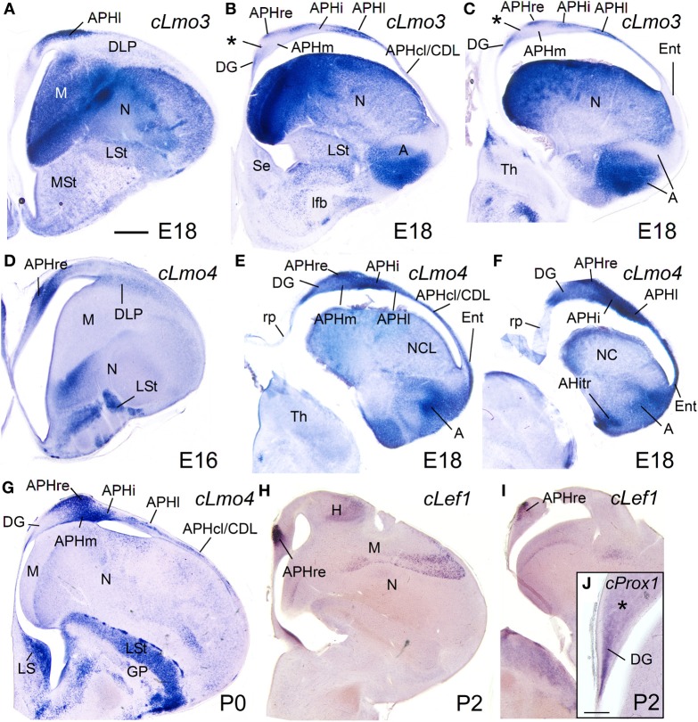
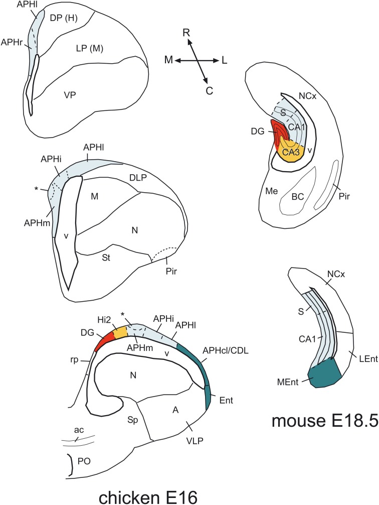
We carried out a study of the expression patterns of seven developmental regulatory genes (Lef1, Lhx2, Lhx9, Lhx5, Lmo3, Lmo4, and Prox1), in combination with topological position, to identify the medial pallial derivatives, define its major subdivisions, and compare them between mouse and chicken. In both species, the medial pallium is defined as a pallial sector adjacent to the cortical hem and roof plate/choroid tela, showing moderate to strong ventricular zone expression of Lef1, Lhx2, and Lhx9, but not Lhx5. Based on this, the hippocampal formation (indusium griseum, dentate gyrus, Ammon's horn fields, and subiculum), the medial entorhinal cortex, and part of the amygdalo-hippocampal transition area of mouse appeared to derive from the medial pallium. In the chicken, based on the same position and gene expression profile, we propose that the hippocampus (including the V-shaped area), the parahippocampal area (including its caudolateral part), the entorhinal cortex, and the amygdalo-hippocampal transition area are medial pallial derivatives. Moreover, the combinatorial expression of Lef1, Prox1, Lmo4, and Lmo3 allowed the identification of dentate gyrus/CA3-like, CA1/subicular-like, and medial entorhinal-like comparable sectors in mouse and chicken, and point to the existence of mostly conserved molecular networks involved in hippocampal complex development. Notably, while the mouse medial entorhinal cortex derives from the medial pallium (similarly to the hippocampal formation, both being involved in spatial navigation and spatial memory), the lateral entorhinal cortex (involved in processing non-spatial, contextual information) appears to derive from a distinct dorsolateral caudal pallial sector.

Keywords: Ammon's horn fields; dentate gyrus; dorsolateral caudal pallium; entorhinal cortex; evolution; hippocampus; medial pallium.


Figures

Figure 1
Figure 1
Expression of Lef1 in mouse embryonic telencephalon at E13.5. Digital images of coronal sections of mouse embryonic telencephalon (E13.5), from rostral (A) to caudal (E) levels, hybridized for Lef1. Note the strong expression in the ventricular zone of the medial pallium and pallial septum. For abbreviations see list. Scale bar: (A) = 200 μm (applies to all).
Figure 2
Figure 2
Expression of Lhx2 and Lhx9 in mouse embryonic telencephalon at early stages. Digital images of coronal sections of mouse embryonic telencephalon (A–H: E12.5; I–K: E13.5), from intermediate (left panels) to caudal (right panels) levels, hybridized for Lhx2 (A–D) or Lhx9 (I–K). Note the strong expression in the ventricular zone of the medial pallium. As noted previously, Lhx9 is also distinctly expressed in ventral pallial (VP) derivatives, such as part of the basal amygdalar complex (BC) and cortical amygdalar areas (Co, PMCo). Although weak transient expression is also present in part of the dorsal pallium (DP; Rétaux et al., 1999), this pallial sector is clearly distinguished from MP and VP based on its distinct position and combinatorial genetic profile (Puelles et al., ; Abellán et al., 2009). For abbreviations see list. Scale bar: (A) = 200 μm (applies to all).
Figure 3
Figure 3
Expression of Lef1 in mouse embryonic telencephalon at E15.5. Digital images of coronal sections of mouse embryonic telencephalon (E15.5), from rostral (A) to caudal (G) levels, hybridized for Lef1. The medial pallial (MP) vz and derivatives show expression of Lef1. Note the lack of Lef1 expression in the vz of the dorsolateral caudal pallium (DLP), giving rise to LEnt. For abbreviations see list. Scale bar: (A) = 200 μm (applies to all).
Figure 4
Figure 4
Expression of Lhx9, Lmo3, and Lmo4 in mouse embryonic telencephalon at intermediate stages. Digital images of coronal sections of mouse embryonic telencephalon (E15.5 or E16.5), at intermediate (A–E,H) or caudal (F,G,I–K) levels, hybridized for Lhx9, Lmo3, or Lmo4. For abbreviations see list. Scale bar: (A) = 1 mm (applies to A,F,G,I–K); (B) = 300 μm (applies to B–E,H).
Figure 5
Figure 5
Expression of Lef1, Lhx2, Lmo3, and Lmo4 in mouse embryonic telencephalon at prenatal or neonatal ages. Digital images of coronal sections of prenatal or neonatal mouse telencephalon (E17.5, E18.5, or P0), at intermediate (A,B,F) or caudal (C–E″,G) levels, hybridized for Lef1, Lhx2, Lmo3, or Lmo4. For abbreviations see list. Scale bar: (A) = 200 μm (applies to A–D,F,G). (E) = 1 mm (applies to E–E″).
Figure 6
Figure 6
Expression of cLef1, cLhx2, cLhx9, and cLhx5 in chicken embryonic telencephalon at early stages. Digital images of coronal sections of chicken embryonic telencephalon (E7.5 or E8), at rostral (A,D) or intermediate (B,C,E–G) levels, hybridized for cLef1, cLhx2, cLhx9, or cLhx5. Note the moderate to strong expression of cLef1, cLhx2, and cLhx9 in the ventricular zone of the medial pallium (MP) and pallial septum (PSe). The pallial septum also expresses cLhx5. From E8, the dorsolateral pallium (DLP) can be distinguished from MP because it does not express cLef1, but shows moderate to strong expression of cLhx9 in the mantle. Note the expression of cLef1 in the meninges (pia mater; arrows in B), in forebrain blood vessels, and in some cell aggregates around the lateral forebrain bundle (arrow in C). For abbreviations see list. Scale bar: (A) = 200 μm (applies to A,B); (C) = 400 μm; (D) = 500 μm (applies to D–F); (G) = 500 μm.
Figure 7
Figure 7
Expression of cLef1, cLhx2, cLhx9, cLmo3, and cLmo4 in the chicken embryonic telencephalon at early stages. Digital images of coronal sections of chicken embryonic telencephalon (E8), at intermediate (E,F) or caudal (A–D,G,H) levels, hybridized for cLef1, cLhx2, cLhx9, cLmo3, or cLmo4. The medial pallium is characterized by strong expression of cLef1, cLhx2, and cLhx9 in the ventricular zone, and strong expression of cLhx2 and cLmo4 in the mantle. For abbreviations see list. Scale bar: (A) = 400 μm; (B) = 500 μm (applies to B–H).
Figure 8
Figure 8
Expression of several genes and radial glial fibers in the chicken embryonic medial pallium at intermediate stages. (A–H) Digital images of coronal sections of chicken embryonic telencephalon (E10–E13), at rostral (A–C), intermediate (D–F), or caudal (G–I) levels, hybridized for cLef1, cProx1, or cWnt8b (the latter is used to distinguish the roof plate and cortical hem) Note the strong expression of cLef1 in the rostral APH, which extends caudally to a small area that occupies a superficial position above APHm. cProx1 allows distinction of the dentate gyrus (DG). I: Detail of radial glial fibers in the APH (immunohistochemical staining using H5 antibody). Note that the caudal extension of APHr (ectopic APHr or APHre in G–I) is avoided by fibers. For abbreviations see list. Scale bar: (A) = 200 μm (applies to A,B,D–H); (C) = 1 mm, (I) = 200 μm.
Figure 9
Figure 9
Expression of several genes in the chicken embryonic telencephalon at intermediate stages. Digital images of coronal sections of chicken embryonic telencephalon (E12, E13), at rostral (A,F–H), intermediate (B–D, I–K) or caudal (E, L–N) levels, hybridized for cLhx9, cLmo3, or cLmo4. Note the strong expression of cLmo4 in medial pallial derivatives, which is remarkable in APHm and APHi. cLhx9 is also expressed in the vz of the medial pallium, and at caudal levels the expression becomes stronger and is additionally present in the mantle. Moreover, cLhx9 is expressed in derivatives of the ventral pallium (VP; in particular, the caudal nidopallium, N), and in both vz and derivatives of the ventrolateral caudal pallium (VLP, which gives rise to the arcopallium). For abbreviations see list. Scale bar: (A) = 1 mm (applies to A–E); (F) = 1 mm (applies to F,I,L); (H) = 500 μm (applies to G,H,J,K,M,N).
Figure 10
Figure 10
Expression of several genes in the chicken embryonic telencephalon at intermediate stages. (A–G) Digital images of coronal sections of chicken embryonic telencephalon (E14), at intermediate (A–C) or caudal (D–G) levels, hybridized for cLhx2, cLhx9, or cLmo4. (H–J) Digital images of horizontal sections of chicken embryonic telencephalon (E14), from top (H) to bottom (J), hybridized for cLhx9. Note the distinct genetic profile of DLP and VLP. For abbreviations see list. Scale bar: (A) = 1 mm (applies to all).
Figure 11
Figure 11
Expression of several genes in the chicken embryonic telencephalon at prehatching and early posthatching stages. Digital images of coronal sections of chicken telencephalon (E16, E18, P0, or P2), at intermediate (A,D,G,H) or caudal (B,C,E,F,I,J) levels, hybridized for cLmo3, cLmo4, cLef1, or cProx1. Note the expression of cProx1 in the dentate gyrus, which includes a large part of V-shaped area, but not its dorsal part (asterisk in J). This dorsal part of V-shaped area (hippocampal sector 2) is also free of cLmo3 (asterisk in B,C) and may be comparable to CA3 of mammals. For abbreviations see list. Scale bar: (A) = 1 mm (applies to A–I); (J) = 200 μm.
Figure 12
Figure 12
Comparison of chicken and mouse medial pallial subdivisions. Schematic drawings of frontal sections through the telencephalon of a chicken (at E16) and a mouse (at E18.5), at rostral intermediate, or caudal levels, showing the major subdivisions of the medial pallium. A color code is used to compare these subdivisions between species. In these schemes, dorsal is to the top and medial is to the left. In the chicken, the rostralmost part is represented by the APHr. The asterisk points to an ectopic part of chicken APHr (possibly a group tangentially migrated cells), observed at the surface of APHm at intermediate and caudal levels of the medial pallium. The rostralmost part of mouse is not represented here, but appears to include the indusium griseum. For abbreviations see list. See text for more details.

References

    1. Abellán A., Desfilis E., Medina L. (2013). The olfactory amygdala in amniotes: an evo-devo approach. Anat. Rec. (Hoboken) 296, 1317–1332 10.1002/ar.22744 - DOI - PubMed
    1. Abellán A., Legaz I., Vernier B., Rétaux S., Medina L. (2009). Olfactory and amygdalar structures of the chicken ventral pallium based on the combinatorial expression patterns of LIM and other developmental regulatory genes. J. Comp. Neurol. 516, 166–186 10.1002/cne.22102 - DOI - PubMed
    1. Abellán A., Medina L. (2009). Subdivisions and derivatives of the chicken subpallium based on expression of LIM and other regulatory genes and markers of neuron subpopulations during development. J. Comp. Neurol. 515, 465–501 10.1002/cne.22083 - DOI - PubMed
    1. Abellán A., Vernier B., Rétaux S., Medina L. (2010). Similarities and differences in the forebrain expression of Lhx1 and Lhx5 between chicken and mouse: insights for understanding telencephalic development and evolution. J. Comp. Neurol. 518, 3512–3528 10.1002/cne.22410 - DOI - PubMed
    1. Aboitiz F., Zamorano F. (2013). Neural progenitors, patterning and ecology in neocortical origins. Front. Neuroanat. 7:38 10.3389/fnana.2013.00038 - DOI - PMC - PubMed

LinkOut - more resources