. 2014 Jul 2:8:117.
doi: 10.3389/fnsys.2014.00117. eCollection 2014.

Comparative organization of the claustrum: what does structure tell us about function?

Affiliations

Comparative organization of the claustrum: what does structure tell us about function?

Joan S Baizer et al. Front Syst Neurosci. .

Abstract

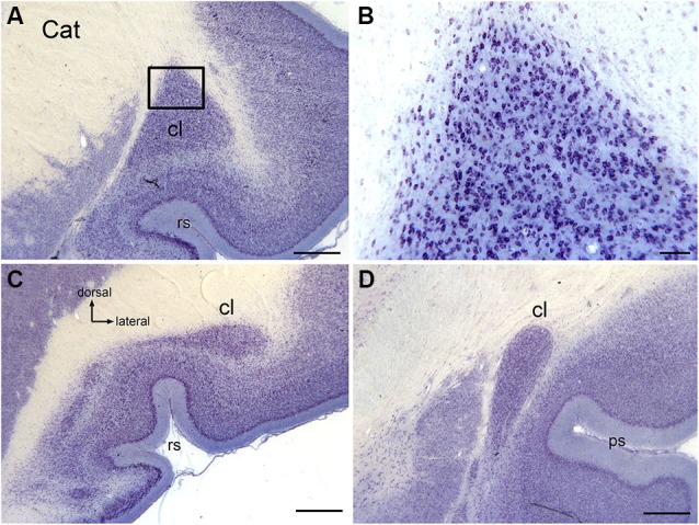
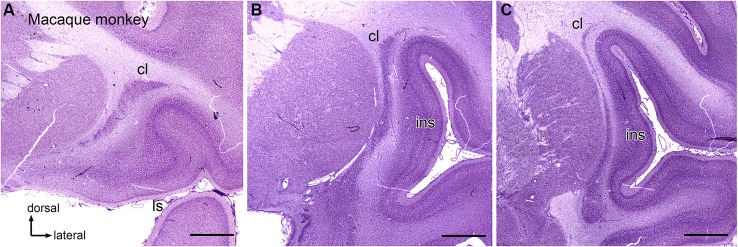
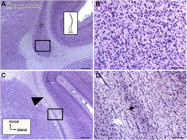
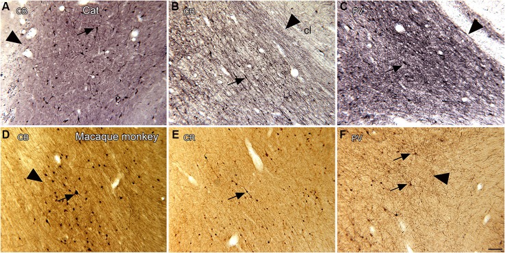
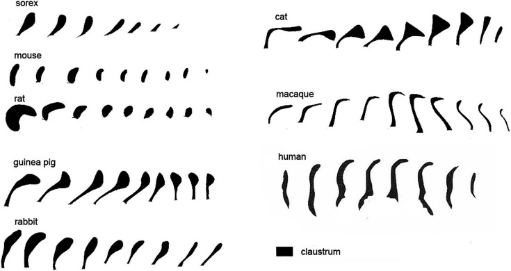
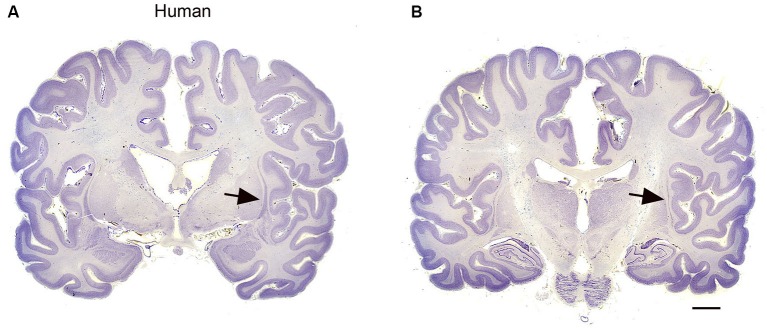
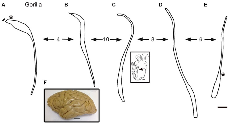
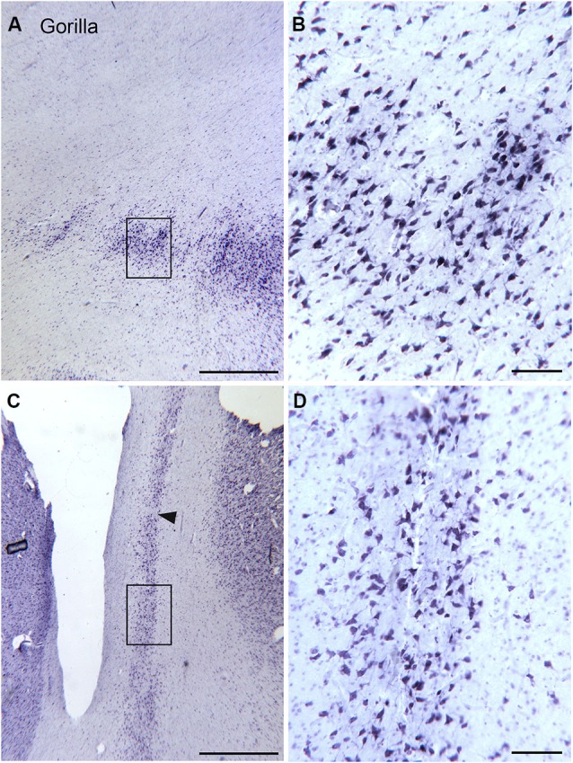
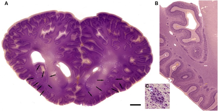
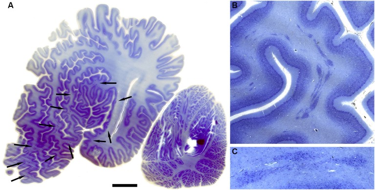
The claustrum is a subcortical nucleus present in all placental mammals. Many anatomical studies have shown that its inputs are predominantly from the cerebral cortex and its outputs are back to the cortex. This connectivity thus suggests that the claustrum serves to amplify or facilitate information processing in the cerebral cortex. The size and the complexity of the cerebral cortex varies dramatically across species. Some species have lissencephalic brains, with few cortical areas, while others have a greatly expanded cortex and many cortical areas. This evolutionary diversity in the cerebral cortex raises several questions about the claustrum. Does its volume expand in coordination with the expansion of cortex and does it acquire new functions related to the new cortical functions? Here we survey the organization of the claustrum in animals with large brains, including great apes and cetaceans. Our data suggest that the claustrum is not always a continuous structure. In monkeys and gorillas there are a few isolated islands of cells near the main body of the nucleus. In cetaceans, however, there are many isolated cell islands. These data suggest constraints on the possible function of the claustrum. Some authors propose that the claustrum has a more global role in perception or consciousness that requires intraclaustral integration of information. These theories postulate mechanisms like gap junctions between claustral cells or a "syncytium" to mediate intraclaustral processing. The presence of discontinuities in the structure of the claustrum, present but minimal in some primates, but dramatically clear in cetaceans, argues against the proposed mechanisms of intraclaustral processing of information. The best interpretation of function, then, is that each functional subdivision of the claustrum simply contributes to the function of its cortical partner.

Keywords: calcium-binding proteins; dolphin; gorilla; visual cortex; whale.


Figures

Figure 1
Figure 1
The claustrum in the cat shown on cresyl violet-stained celloidin-embedded sections. (A) The claustrum at about its rostro-caudal center. The rectangle shows the location of the image in (B). (B) Cells in the claustrum. (C) The claustrum at its rostral limit. (D) The claustrum at its caudal limit. Scale bars: A, C, D = 1 mm; B = 100 μm . Abbreviations: cl, claustrum; ps, pseudosylvian sulcus; rs, rhinal sulcus.
Figure 2
Figure 2
The claustrum in the macaque monkey. (A) Rostral claustrum. (B) The claustrum at about its rostro-caudal center. (C) The caudal claustrum. Note the long, thin vertical stem and the ventral enlargement in (B), (C). Scale bars: A, B, C = 2 mm. Abbreviations: cl, claustrum; ls, lateral sulcus; ins, insula.
Figure 3
Figure 3
Neuronal organization of the claustrum in the macaque monkey shown on higher magnification images. (A) The ventral expansion of the claustrum in the macaque monkey. The rectangle shows the location of the image in (B). The inset shows an outline drawing of the entire claustrum. (B) Neurons in the ventral enlargement of the monkey have round or polygonal somata. (C) Cells in the long ascending stem of the claustrum. The arrowhead indicates a cell-sparse region. The rectangle shows the location of the image in (D). (D) Many neurons in the thin ascending stem have somata that are elongated parallel to the borders of the structure (arrow). Scale bars: A, C = 500 μm; A, inset = 2 mm; B, D = 100 μm.
Figure 4
Figure 4
Spacing and density of neurons in the claustrum of the cat (A, B, C) and macaque monkey (D, E, F) claustrum that are immunoreactive for the calcium-binding proteins. (A) CB, cat. The arrowhead indicates the medial border of the claustrum; the arrow indicates a labeled cell. (B) CR, cat. The arrowhead shows the border of the claustrum and the arrow a cell with a fusiform soma. (C) PV, cat. The claustrum is darkly stained; the border (arrowhead) is very clear. The arrow shows a PV-ir neuron. A, B, C glucose oxidase modification of DAB for visualization of immunoreactivity. (D) CB, monkey. The arrowhead shows the border of the claustrum, the arrow a labeled neuron. (E) CR, monkey. The arrow shows a neuron with an elongated soma. (F) PV, monkey. The arrowhead shows the edge of the claustrum. The arrows show two large neurons and their dendrites. D, E, F, standard DAB visualization of immunoreactivity. Scale bar: F = 100 μm; same magnification for all other panels. Abbreviations: CB, calbindin; CR, calretinin; PV, parvalbumin.
Figure 5
Figure 5
Variations in the shape of the dorsal claustrum in eight different species. Modified from Figure 7 in Kowiański et al. (1999). The different drawings are not to scale. Note the presence of a dorsal enlargement and thin stem in sorex, cat and guinea pig compared to the dorsal-ventral symmetry seen in the mouse and the rat and, at some levels, human. In all species the images are arranged with the most rostral on the left.
Figure 6
Figure 6
(A, B) The claustrum in the human brain. The claustrum is shown on two coronal sections through the human brain about 17 mm apart. A is the more rostral. The arrows show the claustrum, which is quite small relative to the total size of the section. The images were downloaded from http://www.brains.rad.msu.edu and, http://brainmuseum.org sites supported by the US National Science Foundation, and used with permission. The brain had been embedded in celloidin and sectioned at 35 μm. Scale bar = 10 mm.
Figure 7
Figure 7
(A–E) Outline drawings of the claustrum of the gorilla from five coronal cresyl-violet stained sections. A is the most rostral. The spacing of the sections (mm) is indicated by the numbers between the arrows. (A) The asterisk shows the approximate location of the image in Figures 8A,C. The inset shows an outline drawing of the left half of the cerebral cortex on the section for the level of the claustrum shown; the arrow indicates the location of the claustrum on this section. (E) The asterisk shows the approximate location of the image in Figure 8C. (F) Lateral view of the left hemisphere of a gorilla showing the sulci and gyri. Caudal is to the right. Scale bars: E = 2 mm, same scale for A–D; C, inset = 5 mm; F = 2 cm.
Figure 8
Figure 8
Size shape and density of neurons in the claustrum of the gorilla at different rostro-caudal and dorso-ventral levels. (A) Cell islands in the dorsal claustrum (section outlined on Figure 7A). The rectangle shows the location of the image in (B). (B) Stained somata in the claustrum; many somata are multipolar. Scale bar = 100 μm. (C) Image of the more ventral and caudal claustrum; the section was outlined in Figure 7E. The rectangle shows the location of the image in (D). The arrowhead indicates a cell-sparse region. (D) Somata in the ventral and caudal claustrum; many are multipolar. Scale bars: A, C = 1 mm; B, D = 100 μm.
Figure 9
Figure 9
Morphology of the claustrum in the bottlenose dolphin (Tursiops truncatus). (A) Coronal section through the brain of a 4 year old Tursiops. The arrows point to the location of the claustrum around the anterior portion of the insular pocket and to the highly unusual distribution of many claustral island along cortical gyri in the prefrontal cortex, shown at higher magnification in (B). (C) Cellular details of one island of claustral neurons in the anterior portion of the ectosylvian gyrus. Scale bars = 1 cm (A), 4 mm (B), and 100 μm (C).
Figure 10
Figure 10
Morphology of the claustrum in the humpback whale (Megaptera novaeangliae). (A) Parasagittal section through one hemisphere of an adult humpback whale. There are a very large number of claustral islands dispersed in the white matter underlying the perisylvian, ectosylvian, and suparsylvian cortex, as well as in the frontal pole (arrows). (B) Higher magnification image of a large cluster of these claustral neurons in the white matter of the suprasylvian gyrus. (C) Shows cellular details. Note that as in the bottlenose dolphin, these claustral island are completely separate from the neocortex. Scale bars = 2 cm (A), 6 mm (B), and 1.5 mm (C).

References

    1. Agulhon C., Charnay Y., Vallet P., Abitbol M., Kobetz A., Bertrand D., et al. (1998). Distribution of mRNA for the alpha4 subunit of the nicotinic acetylcholine receptor in the human fetal brain. Mol. Brain Res. 58, 123–131 10.1016/s0169-328x(98)00113-2 - DOI - PubMed
    1. Amaral D. G., Cowan W. M. (1980). Subcortical afferents to the hippocampal formation in the monkey. J. Comp. Neurol. 189, 573–591 10.1002/cne.901890402 - DOI - PubMed
    1. Arikuni T., Kubota K. (1985). Claustral and amygdaloid afferents to the head of the caudate nucleus in macaque monkeys. Neurosci. Res. 2, 239–254 10.1016/0168-0102(85)90003-3 - DOI - PubMed
    1. Baizer J. S. (2014). “The neurochemical organization of the claustrum,” in The Claustrum. Structural, Functional and Clinical Neuroscience, eds Smythies J. R., Edelstein L. R., Ramachandran V. S. (Amsterdam: Elsevier; ), 85–118
    1. Baizer J. S. (2001). Serotonergic innervation of the primate claustrum. Brain Res. Bull. 55, 431–434 10.1016/s0361-9230(01)00535-4 - DOI - PubMed

LinkOut - more resources