

Frequency-specific corticofugal modulation of the dorsal cochlear nucleus in mice
- PMID: 25071477
- PMCID: PMC4076887
- DOI: 10.3389/fnsys.2014.00125
Frequency-specific corticofugal modulation of the dorsal cochlear nucleus in mice
Abstract
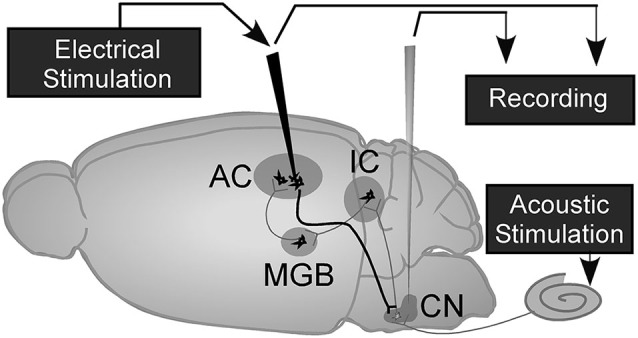
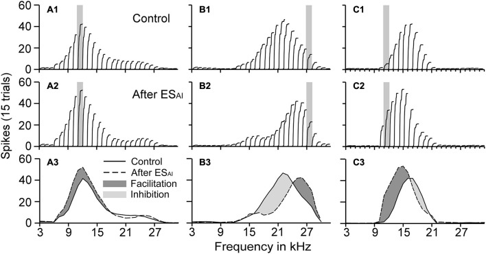
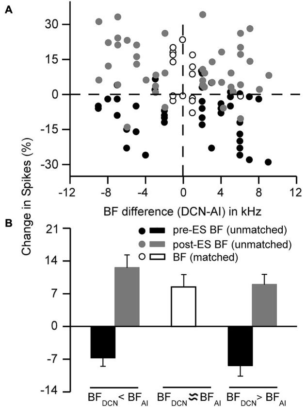
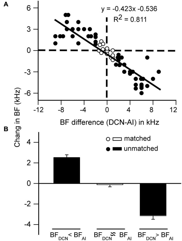
The primary auditory cortex (AI) modulates the sound information processing in the lemniscal subcortical nuclei, including the anteroventral cochlear nucleus (AVCN), in a frequency-specific manner. The dorsal cochlear nucleus (DCN) is a non-lemniscal subcortical nucleus but it is tonotopically organized like the AVCN. However, it remains unclear how the AI modulates the sound information processing in the DCN. This study examined the impact of focal electrical stimulation of AI on the auditory responses of the DCN neurons in mice. We found that the electrical stimulation induced significant changes in the best frequency (BF) of DCN neurons. The changes in the BFs were highly specific to the BF differences between the stimulated AI neurons and the recorded DCN neurons. The DCN BFs shifted higher when the AI BFs were higher than the DCN BFs and the DCN BFs shifted lower when the AI BFs were lower than the DCN BFs. The DCN BFs showed no change when the AI and DCN BFs were similar. Moreover, the BF shifts were linearly correlated to the BF differences. Thus, our data suggest that corticofugal modulation of the DCN is also highly specific to frequency information, similar to the corticofugal modulation of the AVCN. The frequency-specificity of corticofugal modulation does not appear limited to the lemniscal ascending pathway.
Keywords: corticofugal modulation; dorsal cochlear nucleus; frequency-specific modulation; lemniscal; neural plasticity; non-lemniscal; primary auditory cortex.
Figures
Similar articles
-
The onset and post-onset auditory responses of cochlear nucleus neurons are modulated differently by cortical activation.Hear Res. 2019 Mar 1;373:96-102. doi: 10.1016/j.heares.2018.12.013. Epub 2018 Dec 31. Hear Res. 2019. PMID: 30640070
-
Corticofugal modulation of initial neural processing of sound information from the ipsilateral ear in the mouse.PLoS One. 2010 Nov 16;5(11):e14038. doi: 10.1371/journal.pone.0014038. PLoS One. 2010. PMID: 21124980 Free PMC article.
-
Modulation of responses and frequency tuning of thalamic and collicular neurons by cortical activation in mustached bats.J Neurophysiol. 2000 Jul;84(1):325-33. doi: 10.1152/jn.2000.84.1.325. J Neurophysiol. 2000. PMID: 10899207
-
Tuning shifts of the auditory system by corticocortical and corticofugal projections and conditioning.Neurosci Biobehav Rev. 2012 Feb;36(2):969-88. doi: 10.1016/j.neubiorev.2011.11.006. Epub 2011 Dec 2. Neurosci Biobehav Rev. 2012. PMID: 22155273 Free PMC article. Review.
-
Plasticity of the adult auditory system based on corticocortical and corticofugal modulations.Neurosci Biobehav Rev. 2020 Jun;113:461-478. doi: 10.1016/j.neubiorev.2020.03.021. Epub 2020 Mar 21. Neurosci Biobehav Rev. 2020. PMID: 32209362 Review.
Cited by
-
Characterization of three cholinergic inputs to the cochlear nucleus.J Chem Neuroanat. 2023 Sep;131:102284. doi: 10.1016/j.jchemneu.2023.102284. Epub 2023 May 8. J Chem Neuroanat. 2023. PMID: 37164181 Free PMC article.
-
Recurrent Circuits Amplify Corticofugal Signals and Drive Feedforward Inhibition in the Inferior Colliculus.J Neurosci. 2023 Aug 2;43(31):5642-5655. doi: 10.1523/JNEUROSCI.0626-23.2023. Epub 2023 Jun 12. J Neurosci. 2023. PMID: 37308295 Free PMC article.
-
The Corticofugal Effects of Auditory Cortex Microstimulation on Auditory Nerve and Superior Olivary Complex Responses Are Mediated via Alpha-9 Nicotinic Receptor Subunit.PLoS One. 2016 May 19;11(5):e0155991. doi: 10.1371/journal.pone.0155991. eCollection 2016. PLoS One. 2016. PMID: 27195498 Free PMC article.
-
Top-Down Inference in the Auditory System: Potential Roles for Corticofugal Projections.Front Neural Circuits. 2021 Jan 22;14:615259. doi: 10.3389/fncir.2020.615259. eCollection 2020. Front Neural Circuits. 2021. PMID: 33551756 Free PMC article. Review.
-
Imbalance of excitation and inhibition at threshold level in the auditory cortex.Front Neural Circuits. 2015 Mar 18;9:11. doi: 10.3389/fncir.2015.00011. eCollection 2015. Front Neural Circuits. 2015. PMID: 25852485 Free PMC article.
References
LinkOut - more resources
Full Text Sources
Other Literature Sources
Miscellaneous