Review
. 2014 Jul 4:5:240.
doi: 10.3389/fphys.2014.00240. eCollection 2014.

Kallikrein-related peptidase-4 (KLK4): role in enamel formation and revelations from ablated mice

Affiliations
Review

Kallikrein-related peptidase-4 (KLK4): role in enamel formation and revelations from ablated mice

John D Bartlett et al. Front Physiol. .

Abstract

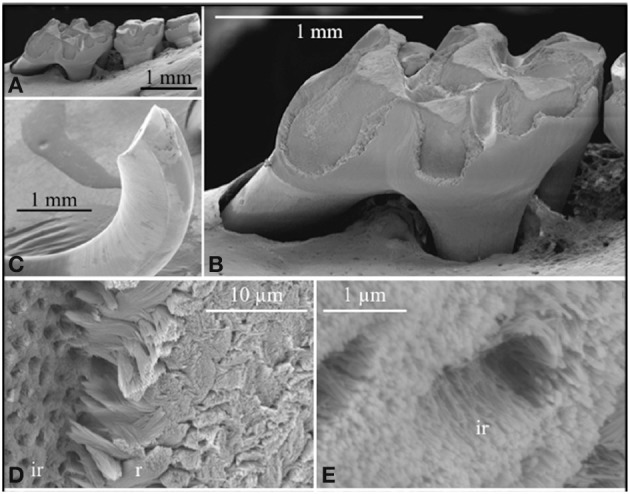
Enamel development occurs in stages. During the secretory stage, a soft protein rich enamel layer is produced that expands to reach its final thickness. During the maturation stage, proteins are removed and the enamel matures into the hardest substance in the body. KLK4 is expressed during the transition from secretory to the maturation stage and its expression continues throughout maturation. KLK4 is a glycosylated chymotrypsin-like serine protease that cleaves enamel matrix proteins prior to their export out of the hardening enamel layer. Mutations in KLK4 can cause autosomal recessive, non-syndromic enamel malformations in humans and mice. Klk4 ablated mice initially have normal-looking teeth with enamel of full thickness. However, the enamel is soft and protein-rich. Three findings are notable from Klk4 ablated mice: first, enamel rods fall from the interrod enamel leaving behind empty holes where the enamel fractures near the underlying dentin surface. Second, the ~10,000 crystallites that normally fuse to form a solid enamel rod fail to grow together in the ablated mice and can fall out of the rods. Third, and most striking, the crystallites grow substantially in width and thickness (a- and b-axis) in the ablated mice until they almost interlock. The crystallites grow in defined enamel rods, but interlocking is prevented presumably because too much protein remains. Conventional thought holds that enamel proteins bind specifically to the sides of enamel crystals to inhibit growth in width and thickness so that the thin, ribbon-like enamel crystallites grow predominantly in length. Results from Klk4 ablated mice demonstrate that this convention requires updating. An alternative mechanism is proposed whereby enamel proteins serve to form a mold or support structure that shapes and orients the mineral ribbons as they grow in length. The remnants of this support structure must be removed by KLK4 so that the crystallites can interlock to form fully hardened enamel.

Keywords: MMP20; ameloblastin; amelogenin; enamel crystallites; enamel development; enamel rods; enamelin.


Figures

Figure 1
Figure 1
Histochemical detection of β-galactosidase activity in maxillary first molars of wild-type (Wt) heterozygous (Klk4+/lacZ) and homozygous (Klk4lacZ/lacZ) mice at postnatal days 5, 8, 11, and 14. LacZ histochemistry shows nuclear localized β-galactosidase activity where KLK4 is normally expressed. At the 5-h incubation used, no endogenous (lysosomal) β-gal activity was observed and the Wt mice were negative. In mouse molars, a positive signal was only observed in transition and maturation ameloblasts. No expression was observed in odontoblasts. B, Bone; Od, odontoblasts; D, dentin; E, enamel; Am, ameloblasts. Scale bars = 100 μm. This figure was reprinted with permission (S. Karger AG, Basel) from Simmer et al. (2011a).
Figure 2
Figure 2
Scanning electron micropscopy of the mandibular molars (A,B) and mandibular incisor (C–E) of a Klk4 null mouse at 7 weeks. The enamel of all molars showed a significant loss of enamel from all working surfaces (buccal cusps, occlusal surface, and marginal ridges) (A,B). Similarly, the enamel layer was abraded at the working (buccal) surface of the mandibular incisor at its tip (C). Higher magnification of the chipped area near the tip of the incisor showed the break was in the enamel layer, close to, but not at the DEJ. The broken surface appears to be composed of interrod (ir) enamel with holes where enamel rods (r) had pulled out and separated (D) from the initial deposit of interrod enamel near the DEJ. The holes are too numerous to be made by odontoblastic processes penetrating the enamel (enamel spindles). The orientation of the crystallites on the walls of the holes is parallel to the direction of the tubular holes and to the crystallites between the holes (E). This figure was originally published by the American Society for Biochemistry and Molecular Biology in Simmer et al. (2009).
Figure 3
Figure 3
Comparison of enamel rods from (A) wild-type and (B) Klk4 null mice. Enamel rods in wild-type mice have tightly packed crystallites that lose some aspect of their individuality. Enamel rods in the Klk4 null mice are composed of distinctly individual crystallites resembling angel hair spaghetti. Holes or vacancies in some rods give the impression that smaller bundles of crystallites broke at a slightly deeper level and slid out of the rod. This figure was originally published by the American Society for Biochemistry and Molecular Biology in Simmer et al. (2009).

References

    1. Bartlett J. D. (2013). Dental enamel development: proteinases and their enamel matrix substrates. ISRN Dent. 2013, 684607 10.1155/2013/684607 - DOI - PMC - PubMed
    1. Bartlett J. D., Simmer J. P. (1999). Proteinases in developing dental enamel. Crit. Rev. Oral Biol. Med. 10, 425–441 10.1177/10454411990100040101 - DOI - PubMed
    1. Bartlett J. D., Simmer J. P., Xue J., Margolis H. C., Moreno E. C. (1996). Molecular cloning and mRNA tissue distribution of a novel matrix metalloproteinase isolated from porcine enamel organ. Gene 183, 123–128 10.1016/S0378-1119(96)00525-2 - DOI - PubMed
    1. Begue-Kirn C., Krebsbach P. H., Bartlett J. D., Butler W. T. (1998). Dentin sialoprotein, dentin phosphoprotein, enamelysin and ameloblastin: tooth-specific molecules that are distinctively expressed during murine dental differentiation. Eur. J. Oral Sci. 106, 963–970 10.1046/j.0909-8836.1998.eos106510.x - DOI - PubMed
    1. Beniash E., Metzler R. A., Lam R. S., Gilbert P. U. (2009). Transient amorphous calcium phosphate in forming enamel. J. Struct. Biol. 166, 133–143 10.1016/j.jsb.2009.02.001 - DOI - PMC - PubMed

LinkOut - more resources