Review
. 2014 Jul 2:5:244.
doi: 10.3389/fphys.2014.00244. eCollection 2014.

Vitamin D in inflammatory diseases

Affiliations
Review

Vitamin D in inflammatory diseases

Thea K Wöbke et al. Front Physiol. .

Abstract

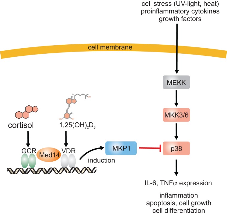
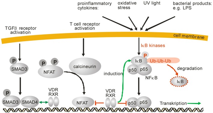
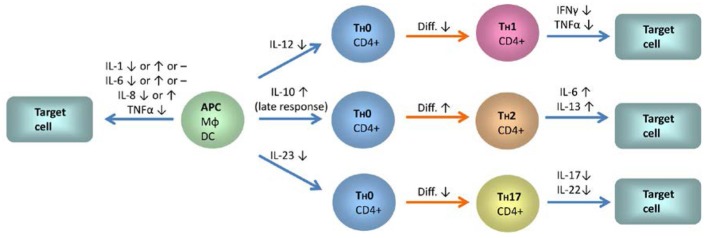
Changes in vitamin D serum levels have been associated with inflammatory diseases, such as inflammatory bowel disease (IBD), rheumatoid arthritis, systemic lupus erythematosus, multiple sclerosis (MS), atherosclerosis, or asthma. Genome- and transcriptome-wide studies indicate that vitamin D signaling modulates many inflammatory responses on several levels. This includes (i) the regulation of the expression of genes which generate pro-inflammatory mediators, such as cyclooxygenases or 5-lipoxygenase, (ii) the interference with transcription factors, such as NF-κB, which regulate the expression of inflammatory genes and (iii) the activation of signaling cascades, such as MAP kinases which mediate inflammatory responses. Vitamin D targets various tissues and cell types, a number of which belong to the immune system, such as monocytes/macrophages, dendritic cells (DCs) as well as B- and T cells, leading to individual responses of each cell type. One hallmark of these specific vitamin D effects is the cell-type specific regulation of genes involved in the regulation of inflammatory processes and the interplay between vitamin D signaling and other signaling cascades involved in inflammation. An important task in the near future will be the elucidation of the regulatory mechanisms that are involved in the regulation of inflammatory responses by vitamin D on the molecular level by the use of techniques such as chromatin immunoprecipitation (ChIP), ChIP-seq, and FAIRE-seq.

Keywords: 1α,25(OH)2D3; MKP1; NFAT; NFκB; VDR; cyclooxygenase; innate immune system; interleukins.


Figures

Figure 1
Figure 1
Inhibition of the p38 MAP kinase pathway by 1α,25(OH)2D3 and a mechanism for the synergistic anti-inflammatory effects of 1α,25(OH)2D3 and glucocorticoids. Proinflammatory stimuli lead to p38 MAP kinase phosphorylation and activation which subsequently induces expression of many proinflammatory proteins such as IL-6 and TNFα. 1α,25(OH)2D3 induces MKP1 expression which dephosphorylates and inactivates p38 MAP kinase. 1α,25(OH)2D3 stimulates glucocorticoid-induced MKP1 expression via enhanced expression of Med14.
Figure 2
Figure 2
SMAD, NFAT and NFκB signaling and modulation of these signaling pathways by 1α,25(OH)2D3, respective VDR/RXR. IκB phosphorylation after various cell stress signals leads to its ubiquitinylation and subsequent proteosomal degradation. After IκB degradation, NFκB is released and translocates into the nucleus where it binds to DNA and modulates gene expression. Activation of NFAT is mediated by the protein phosphatase calcineurin which dephosphorylates NFAT. After dephosphorylation, NFAT translocates into the nucleus, interacts with a variety of other transcription factors and modulates gene expression. Activation of TGFβ receptors leads to phosphorylation of SMAD2 and SMAD3 as well as subsequent translocation into the nucleus. SMAD3 forms a complex with SMAD4 and modulates gene expression of its target genes. After activation by 1α,25(OH)2D3 the VDR/RXR heterodimer can inhibit NFκB signaling either by induction of IκB or by interference with NFκB DNA binding. Also, inhibition of NFAT signaling was reported by prevention of NFAT binding to its response elements.
Figure 3
Figure 3
The influence of 1α,25(OH)2D3 on the expression of interleukins, TNFα and IFNγ in monocytes, dendritic cells, and different T-cell subsets. Blue arrows indicate IL signaling between the different cell types and red arrows indicate differentiation processes. IL-12 and IL-23 expression is downregulated in monocytes and dendritic cells by 1α,25(OH)2D3. In contrast, IL-10 expression is enhanced. A shift from a Th1 profile toward the Th2 type and a decrease in Th17 responses is to be anticipated from these changes. Inhibition of T-cell autoregulation by 1α,25(OH)2D3-mediated suppression of IL-2 expression is not shown. Abbreviations and symbols: APC, antigen presenting cell; MΦ, macrophage; DC, dendritic cell; ↑, upregulation; ↓, downregulation; -, no changes.

References

    1. Abe J., Moriya Y., Saito M., Sugawara Y., Suda T., Nishii Y. (1986). Modulation of cell growth, differentiation, and production of interleukin-3 by 1 alpha,25-dihydroxyvitamin D3 in the murine myelomonocytic leukemia cell line WEHI-3. Cancer Res. 46, 6316–6321 - PubMed
    1. Adorini L., Penna G. (2008). Control of autoimmune diseases by the vitamin D endocrine system. Nat. Clin. Pract. Rheumatol. 4, 404–412 10.1038/ncprheum0855 - DOI - PubMed
    1. Alroy I., Towers T. L., Freedman L. P. (1995). Transcriptional repression of the interleukin-2 gene by vitamin D3: direct inhibition of NFATp/AP-1 complex formation by a nuclear hormone receptor. Mol. Cell. Biol. 15, 5789–5799 - PMC - PubMed
    1. Ananthakrishnan A. N. (2013). Environmental triggers for inflammatory bowel disease. Curr. Gastroenterol. Rep. 15, 302 10.1007/s11894-012-0302-4 - DOI - PMC - PubMed
    1. Barnes P. J. (1998). Anti-inflammatory actions of glucocorticoids: molecular mechanisms. Clin. Sci. (Lond.) 94, 557–572 - PubMed