. 2014 Jul 8:5:353.
doi: 10.3389/fmicb.2014.00353. eCollection 2014.

Biofilm formation of mucosa-associated methanoarchaeal strains

Affiliations

Biofilm formation of mucosa-associated methanoarchaeal strains

Corinna Bang et al. Front Microbiol. .

Abstract

Although in nature most microorganisms are known to occur predominantly in consortia or biofilms, data on archaeal biofilm formation are in general scarce. Here, the ability of three methanoarchaeal strains, Methanobrevibacter smithii and Methanosphaera stadtmanae, which form part of the human gut microbiota, and the Methanosarcina mazei strain Gö1 to grow on different surfaces and form biofilms was investigated. All three strains adhered to the substrate mica and grew predominantly as bilayers on its surface as demonstrated by confocal laser scanning microscopy analyses, though the formation of multi-layered biofilms of Methanosphaera stadtmanae and Methanobrevibacter smithii was observed as well. Stable biofilm formation was further confirmed by scanning electron microscopy analysis. Methanosarcina mazei and Methanobrevibacter smithii also formed multi-layered biofilms in uncoated plastic μ-dishes(TM), which were very similar in morphology and reached a height of up to 40 μm. In contrast, biofilms formed by Methanosphaera stadtmanae reached only a height of 2 μm. Staining with the two lectins ConA and IB4 indicated that all three strains produced relatively low amounts of extracellular polysaccharides most likely containing glucose, mannose, and galactose. Taken together, this study provides the first evidence that methanoarchaea can develop and form biofilms on different substrates and thus, will contribute to our knowledge on the appearance and physiological role of Methanobrevibacter smithii and Methanosphaera stadtmanae in the human intestine.

Keywords: biofilms; human gut; methanoarchaea; microbiota.


Figures

FIGURE 1
FIGURE 1
Growth of different methanoarchaea on mica. Methanosarcina mazei, Methanobrevibacter smithii, and Methanosphaera stadtmanae were grown in 3 ml standard medium under an N2/CO2 atmosphere for Methanosarcina mazei or an H2/CO2 gas phase for Methanobrevibacter smithii and Methanosphaera stadtmanae; the cultures were supplemented with 1–2 pieces of mica. Growth on mica of all three strains was monitored by phase-contrast microscopy at defined time points of 48, 72, and 96 h.
FIGURE 2
FIGURE 2
Growth of methanogens on mica examined by confocal laser scanning microscopy. Methanosarcina mazei, Methanobrevibacter smithii, and Methanosphaera stadtmanae were grown on mica in hungate tubes with 3 ml of the respective medium. After 48 h of growth, cells were fixed to mica by 2% glutaraldehyde. The autofluorescence of glutaraldehyde was used for CLSM pictures at a wavelength of 520 nm. The scale bar is 50 μm.
FIGURE 3
FIGURE 3
Growth of methanogens on mica examined by SEM. Methanosarcina mazei, Methanobrevibacter smithii, and Methanosphaera stadtmanae were grown on mica in hungate tubes with 3 ml of the respective medium. After 48 h (Methanosarcina mazei and Methanosphaera stadtmanae) and 72 h (Methanobrevibacter smithii) of growth, cells were fixed to mica by 2% glutaraldehyde. Images are representative for the respective sample.
FIGURE 4
FIGURE 4
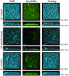
Structures of static biofilms formed by Methanosarcina mazei, Methanosphaera stadtmanae, and Methanobrevibacter smithii. Cells were grown in 4 ml standard medium in μ-dishesTM under the respective gas atmosphere. After 72 h of growth, the biofilms were treated with DAPI (blue channel), ConA (green channel) and IB4 (yellow channel) and visualized by CLSM; single channels and overlays of the images are displayed. Both top view (upper lane) and side view (lower lane) of the biofilms are shown. The scale bar is 20 μm.
FIGURE 5
FIGURE 5
Analysis of the surface coverage of biofilms formed by Methanosarcina mazei, Methanosphaera stadtmanae, and Methanobrevibacter smithii. Differential interference contrast (DIC) pictures (A, left panel) were taken from the bottom layer of static biofilms and converted into black/white (B/W; A, right panel) to calculate the surface coverage. The ratio of B/W pixels was determined and used to obtain the surface coverage (B). The mean and standard deviations of three biological replicates are shown.

References

    1. Abreu M. T., Fukata M., Arditi M. (2005). TLR signaling in the gut in health and disease. J. Immunol. 174 4453–4460 10.4049/jimmunol.174.8.4453 - DOI - PubMed
    1. An D., Parsek M. R. (2007). The promise and peril of transcriptional profiling in biofilm communities. Curr. Opin. Microbiol. 10 292–296 10.1016/j.mib.2007.05.011 - DOI - PubMed
    1. Artis D. (2008). Epithelial-cell recognition of commensal bacteria and maintenance of immune homeostasis in the gut. Nat. Rev. Immunol. 8 411–420 10.1038/nri2316 - DOI - PubMed
    1. Ashok N., Warad S., Singh V. P., Chaudhari H., Narayanan A., Rodrigues J. (2013). Prevalence of archaea in chronic periodontitis patients in an Indian population. Ind. J. Dent. Res. 24 289–293 10.4103/0970-9290.117988 - DOI - PubMed
    1. Backhed F., Ley R. E., Sonnenburg J. L., Peterson D. A., Gordon J. I. (2005). Host-bacterial mutualism in the human intestine. Science 307 1915–1920 10.1126/science.1104816 - DOI - PubMed