

Clinical significance of computed tomography assessment for third molar surgery
- PMID: 25071882
- PMCID: PMC4109093
- DOI: 10.4329/wjr.v6.i7.417
Clinical significance of computed tomography assessment for third molar surgery
Abstract
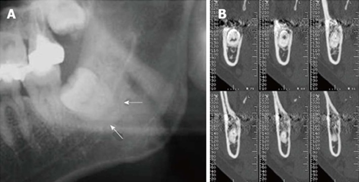
Surgical extraction of the third molar is the most commonly performed surgical procedure in the clinical practice of oral surgery. Third molar surgery is warranted when there is inadequate space for eruption, malpositioning, or risk for cyst or odontogenic tumor formation. Preoperative assessment should include a detailed morphologic analysis of the third molar and its relationship to adjacent structures and surrounding tissues. Due to developments in medical engineering technology, computed tomography (CT) now plays a critical role in providing the clear images required for adequate assessment prior to third molar surgery. Removal of the maxillary third molar is associated with a risk for maxillary sinus perforation, whereas removal of the mandibular third molar can put patients at risk for a neurosensory deficit from damage to the lingual nerve or inferior alveolar nerve. Multiple factors, including demographic, anatomic, and treatment-related factors, influence the incidence of nerve injury during or following removal of the third molar. CT assessment of the third molar prior to surgery can identify some of these risk factors, such as the absence of cortication between the mandibular third molar and the inferior alveolar canal, prior to surgery to reduce the risk for nerve damage. This topic highlight presents an overview of the clinical significance of CT assessment in third molar surgery.
Keywords: Assessment; Computed tomography; Extraction; Oral surgery; Third molar.
Figures


References
-
- Susarla SM, Dodson TB. Preoperative computed tomography imaging in the management of impacted mandibular third molars. J Oral Maxillofac Surg. 2007;65:83–88. - PubMed
-
- Palma-Carrió C, García-Mira B, Larrazabal-Morón C, Peñarrocha-Diago M. Radiographic signs associated with inferior alveolar nerve damage following lower third molar extraction. Med Oral Patol Oral Cir Bucal. 2010;15:e886–e890. - PubMed
-
- Monaco G, Montevecchi M, Bonetti GA, Gatto MR, Checchi L. Reliability of panoramic radiography in evaluating the topographic relationship between the mandibular canal and impacted third molars. J Am Dent Assoc. 2004;135:312–318. - PubMed
-
- Leone SA, Edenfield MJ, Cohen ME. Correlation of acute pericoronitis and the position of the mandibular third molar. Oral Surg Oral Med Oral Pathol. 1986;62:245–250. - PubMed
Publication types
LinkOut - more resources
Full Text Sources
Other Literature Sources
