Clinical Trial
. 2015 Jan 1;24(1):132-43.
doi: 10.1089/scd.2014.0234.

Immune regulatory properties of CD117(pos) amniotic fluid stem cells vary according to gestational age

Affiliations
Clinical Trial

Immune regulatory properties of CD117(pos) amniotic fluid stem cells vary according to gestational age

Mariano Di Trapani et al. Stem Cells Dev. .

Abstract

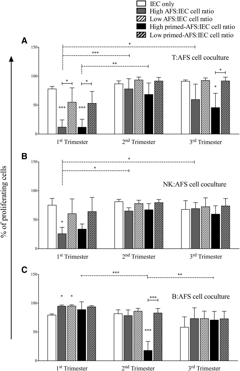
Amniotic Fluid Stem (AFS) cells are broadly multipotent fetal stem cells derived from the positive selection and ex vivo expansion of amniotic fluid CD117/c-kit(pos) cells. Considering the differentiation potential in vitro toward cell lineages belonging to the three germ layers, AFS cells have raised great interest as a new therapeutic tool, but their immune properties still need to be assessed. We analyzed the in vitro immunological properties of AFS cells from different gestational age in coculture with T, B, and natural killer (NK) cells. Nonactivated (resting) first trimester-AFS cells showed lower expression of HLA class-I molecules and NK-activating ligands than second and third trimester-AFS cells, whose features were associated with lower sensitivity to NK cell-mediated lysis. Nevertheless, inflammatory priming with interferon gamma (IFN-γ) and tumor necrosis factor alpha (TNF-α) enhanced resistance of all AFS cell types to NK cytotoxicity. AFS cells modulated lymphocyte proliferation in a different manner according to gestational age: first trimester-AFS cells significantly inhibited T and NK cell proliferation, while second and third trimester-AFS cells were less efficient. In addition, only inflammatory-primed second trimester-AFS cells could suppress B cell proliferation, which was not affected by the first and third trimester-AFS cells. Indolamine 2,3 dioxygenase pathway was significantly involved only in T cell suppression mediated by second and third trimester-AFS cells. Overall, this study shows a number of significant quantitative differences among AFS cells of different gestational age that have to be considered in view of their clinical application.


Figures

<b>FIG. 1.</b>
FIG. 1.
Heterogeneity of amniotic fluid stem (AFS) cells isolated at different gestational age. (A) Microscopy images showing morphologic features of the AFS cell types (the scale bar indicates 100 μm). Arrowheads show oval-round shaped cells. Images were obtained by Axiovert ObserverZ1, Zeiss. (B) Fluorescence-activated cell sorting (FACS) analysis of AFS cells showing the expression profile of hematopoietic and mesenchymal stromal cell markers. The histograms display the isotopic controls (open curve) and specific markers (filled curve) in four (first and second trimester-AFS cells) and five (third trimester-AFS cells) different donors. The relative mean fluorescence intensity (rMFI)±SEM are reported in Table 1.
<b>FIG. 2.</b>
FIG. 2.
Expression of specific markers involved in immunomodulation. AFS cells were cultured with or without inflammatory priming; after 48 h, the expression of CD40, CD80, CD86, HLA-ABC, HLA-DR, CD274, CD54, and CD106 was evaluated by FACS analysis. The results are expressed as rMFI±SEM of four (first and second trimester-AFS cells) or five (third trimester-AFS cells) independent experiments. *P<0.05, **P<0.01, ***P<0.001.
<b>FIG. 3.</b>
FIG. 3.
Immunomodulatory effect of different AFS cells on immune effector cell (IEC) proliferation. Carboxyfluorescein succinimidyl ester (CFSE)-labeled IECs, activated by specific stimuli, were cultured alone or in presence of resting and primed AFS cells. All experiments were carried out at high and low IEC:AFS cell ratios, that is 10:1 (corresponding to 2×10⁵ IECs and 2×10⁴ AFS cells) and 100:1 (corresponding to 2×10⁵ T cells and 2×10³ AFS cells) for T cells (A), and 1:1 (corresponding to 2×10⁴ IECs and 2×10⁴ AFS cells) and 10:1 (corresponding to 2×10⁴ IECs and 2×10³ AFS cells) for natural killer (NK) (B) and B cells (C). Data are represented as IEC proliferation percentage, corresponding to the mean±SD of CD45pos TOPRO-3neg cell proliferation derived from four independent experiments. *P<0.05, **P<0.01, ***P<0.001.
<b>FIG. 4.</b>
FIG. 4.
Role of indoleamine 2,3-dioxygenase (IDO) on T cell inhibition. CFSE-labeled T cells, stimulated with anti-CD3 and anti-CD28 monoclonal antibodies, were cultured alone or in presence of resting or primed AFS cells. L-1-methyltryptophan (L-1MT), the specific inhibitor of IDO was added to cocultures to evaluate the involvement of IDO on AFS-mediated immunosuppression. At the end of coculture (6 days), cell proliferation was analyzed by FACS. Graphs represent the dot plots of CFSE fluorescence (log scale) and proliferation was evaluated as percentage of T cells undergoing at least one cell division. Each box represents the mean±SD of proliferating CD45pos TOPRO-3neg cells derived from four independent experiments. *P<0.05, **P<0.01.
<b>FIG. 5.</b>
FIG. 5.
Inflammatory microenvironment reduces AFS sensitivity to NK cell-mediated lysis. (A) Interleukin-2 (IL-2)-activated NK cells were cocultured in the presence of bis-acetoxymethyl terpyridine dicarboxylate (BATDA)-labeled AFS cells at different ratios. The cytotoxic effect on resting and inflammatory-primed AFS cells was expressed as release of fluorescence by lysed cells and quantified by time-resolved fluorimeter (Victor×4 Multilabel Plate Reader; PerkinElmer). Data are showed as percentage of fluorescence release. Error bars represent the mean±SD of four (first trimester-AFS cells) or five (second and third trimester-AFS cells) experiments. As internal control, we measured spontaneous and maximum release of BATDA and then normalized the values. (B) AFS cells were cultured in presence or not of inflammatory cytokines. At day 2 the expression of CD112, CD155, MHC class I-related A and B molecule (MICA/B), UL16-binding proteins (ULBPs) was analyzed by FACS. The results are expressed as rMFI±SEM of four (first and second trimester-AFS cells) or five (third trimester-AFS cells) different experiments. *P<0.05, **P<0.01, ***P<0.001.
<b>FIG. 6.</b>
FIG. 6.
Antiapoptotic effect of AFS cells on resting IECs. Resting and inflammatory-primed AFS cells were cultured in presence of unstimulated IECs at different ratios, that is 10:1 (corresponding to 2×10⁵ IECs and 2×10⁴ AFS cells) and 100:1 (corresponding to 2×10⁵ T cells and 2×10³ AFS cells) for T cells (A), and 1:1 (corresponding to 2×10⁴ IECs and 2×10⁴ AFS cells) and 10:1 (corresponding to 2×10⁴ IECs and 2×10³ AFS cells) for NK (B) and B cells (C). At the end of coculture (4 days for NK and B cells, and 6 days for T cells) cells were harvested and labeled with anti-CD45 and anti-cytoplasmic active-caspase-3 antibodies to evaluate the percentage of apoptotic IECs by FACS analysis. Data are expressed as percentage of caspase-3neg CD45pos cells. Error bars represent the mean±SD of four different experiments for each (first, second and third trimester) AFS cell types. *P<0.05, **P<0.01, ***P<0.001.

References

    1. Prusa AR, Marton E, Rosner M, Bernaschek G. and Hengstschlager M. (2003). Oct-4-expressing cells in human amniotic fluid: a new source for stem cell research?. Hum Reprod 18:1489–1493 - PubMed
    1. Karlmark KR, Freilinger A, Marton E, Rosner M, Lubec G. and Hengstschlager M. (2005). Activation of ectopic Oct-4 and Rex-1 promoters in human amniotic fluid cells. Int J Mol Med 16:987–992 - PubMed
    1. Tsai MS, Hwang SM, Tsai YL, Cheng FC, Lee JL. and Chang YJ. (2006). Clonal amniotic fluid-derived stem cells express characteristics of both mesenchymal and neural stem cells. Biol Reprod 74:545–551 - PubMed
    1. De Coppi P, Bartsch G, Jr., Siddiqui MM, Xu T, Santos CC, Perin L, Mostoslavsky G, Serre AC, Snyder EY, et al. (2007). Isolation of amniotic stem cell lines with potential for therapy. Nat Biotechnol 25:100–106 - PubMed
    1. Pozzobon M, Piccoli M, Schiavo AA, Atala A. and De Coppi P. (2013). Isolation of c-Kit+ human amniotic fluid stem cells from second trimester. Methods Mol Biol 1035:191–198 - PubMed

Publication types

Substances