. 2014 Jul 29:5:4552.
doi: 10.1038/ncomms5552.

Myocardium-derived angiopoietin-1 is essential for coronary vein formation in the developing heart

Affiliations

Myocardium-derived angiopoietin-1 is essential for coronary vein formation in the developing heart

Yoh Arita et al. Nat Commun. .

Abstract

The origin and developmental mechanisms underlying coronary vessels are not fully elucidated. Here we show that myocardium-derived angiopoietin-1 (Ang1) is essential for coronary vein formation in the developing heart. Cardiomyocyte-specific Ang1 deletion results in defective formation of the subepicardial coronary veins, but had no significant effect on the formation of intramyocardial coronary arteries. The endothelial cells (ECs) of the sinus venosus (SV) are heterogeneous population, composed of APJ-positive and APJ-negative ECs. Among these, the APJ-negative ECs migrate from the SV into the atrial and ventricular myocardium in Ang1-dependent manner. In addition, Ang1 may positively regulate venous differentiation of the subepicardial APJ-negative ECs in the heart. Consistently, in vitro experiments show that Ang1 indeed promotes venous differentiation of the immature ECs. Collectively, our results indicate that myocardial Ang1 positively regulates coronary vein formation presumably by promoting the proliferation, migration and differentiation of immature ECs derived from the SV.


Figures

Figure 1
Figure 1. Myocardial Ang1 is crucial for subepicardial coronary vessel formation.
(ad) Whole-mount immunostaining of embryonic hearts at E14.0 with anti-CD31 antibody. Myocardial Ang1 was required for subepicardial CD31-positive vessel remodelling (a,b: arrowheads in magnified image of inset). The CD31-positive vessel formation was impaired in the ventricles of Ang1CKO embryos (c,d: arrowheads in magnified image of inset). (e,f) Sectioned analysis of the whole-mount immunostained embryonic heart. Subepicardial CD31-positive vessels were detected uniformly in all the sections from control embryo ventricles (e), whereas the density of subepicardial CD31-positive vessels decreased gradually from the dorsal (area 1) to the ventral side (area 3) in Ang1CKO embryos (f). Arrows and arrowheads indicate the intramyocardial CD31-positive vessels and subepicardial CD31-positive vessels, respectively. Area 1, dorsal side; area 2, lateral side; area 3, ventral side of the ventricles. (g,h) Quantification of the number of subepicardial and intramyocardial CD31-positive vessels in the transverse section including inflow-tract of ventricle from E14.0 (n=3). Scale bars, 400 μm in a,c; 100 μm in b,d; 200 μm in e,f; and 50 μm in magnified images of insets 1–3. LV, left ventricle; RV, right ventricle. Values are shown as means±s.e.m. for three separate experiments. Student’s t-test was used to analyse differences. **P<0.01 compared with control. NS, not significant.
Figure 2
Figure 2. Myocardial Ang1 is essential for coronary vein formation.
(ad) Whole-mount immunostaining of embryonic hearts with anti-APJ antibody. APJ-positive coronary veins were observed on the surfaces of both the RA (a, arrowheads) and RV (c, arrowheads) of control embryos, but not on the RA (b) or RV (d) of Ang1CKO embryos. (eh) Sectioned analyses of the whole-mount immunostained embryonic hearts revealed subepicardial APJ-positive coronary veins with vessel-like structures in the RA (e, arrowheads) and RV (g, arrowheads) of control, but not Ang1CKO embryos (f,h). (il; Note: for the experiment presented in il, both ‘control’ and ‘Ang1CKO’ mice contained the EphB4 tau-lacZ knockin allele; see Results.) EphB4-lacZ-positive signals in the hearts of control and Ang1CKO embryos at E13.0. EphB4-lacZ-positive subepicardial coronary veins were observed in control (i,j, arrowhead), but not Ang1CKO embryos (k,l, arrowhead). EphB4-lacZ-positive signals were also detected in the endocardial endothelium in both control and Ang1CKO embryos. (mp) Expression patterns of COUP-TFII in the hearts at E13.5. COUP-TFII-positive subepicardial coronary veins were observed in control (m,n, arrowhead), but not in Ang1CKO embryos (o,p, arrowhead). (qs) Quantification of the number of subepicardial APJ (q, E13.5), EphB4-lacZ (r, E13.0) and COUP-TFII (s, E13.5) -positive vessels in the transverse section including inflow-tract of ventricle (n=3). (r) The number of the EphB4-positive vessels especially with vessel-like structures or with red blood cells were quantified. Scale bars, 100 μm in ah; 300 μm in i,k,m,o; 25 μm in j,l; and 75 μm in n,p. RA, right atrium; RV, right ventricle. Values are shown as means±s.e.m. for three separate experiments. Student’s t-test was used to analyse differences. **P<0.01 compared with control.
Figure 3
Figure 3. Myocardial Ang1 is dispensable for coronary artery formation.
(ad) Coronary angiography by ink injection into the hearts of control (a,b) and Ang1CKO embryos (c,d) at E13.5. (eh; Note: for the experiment presented in eh, both ‘control’ and ‘Ang1CKO’ mice contained the EphrinB2 tau-lacZ knockin allele; see Results.) EphrinB2-lacZ-positive signals in the hearts of control and Ang1CKO embryos at E13.5. EphrinB2-lacZ-positive intramyocardial coronary arteries (containing red blood cells) were similarly observed in control (e,f, arrowhead) and Ang1CKO embryos (g,h, arrowhead). The arrow indicates impaired formation of the interventricular septum in Ang1CKO embryos. (i) Quantification of the number of RV+LV free wall EphrinB2-lacZ-positive vessels in the transverse section including inflow-tract of ventricle from E13.5 (n=3). Scale bars, 100 μm in ad; 300 μm in e,g; and 50 μm in f,h. Ao, aorta; LCA, left coronary artery; RCA, right coronary artery. Values are shown as means±s.e.m. for three separate experiments. Student’s t-test was used to analyse differences. NS, not significant.
Figure 4
Figure 4. The expression levels of venous marker genes are significantly lower in the hearts of Ang1CKO embryos than in those of control.
(ai) Quantitative expression analysis of venous and arterial marker mRNAs in the ventricles at E12.5-E13.0 (normalized to GAPDH mRNA; n=3). The expression levels of venous marker genes such as APJ (a), Ephb4 (b), COUP-TFII (c), were significantly reduced in Ang1CKO embryos compared with control. However, the expression levels of arterial marker genes such as EfnbB2 (d), Nrp1 (e), Dll4 (f), Hes1 (g), Acvrl1 (h) and Notch1 (i) were not significantly affected in Ang1CKO embryos compared with control. Values are shown as means±s.e.m. for three separate experiments. Student’s t-test was used to analyse differences. *P<0.05, **P<0.01, ***P<0.001 compared with control. NS, not significant.
Figure 5
Figure 5. Ang1 derived from the ventricular myocardium attracts Tie2-positive ECs from the SV.
(a,b) X-gal staining of Tie2-lacZ transgenic mice at E10.5. Tie2-positive signals were observed most strongly in the SV (arrows), and to a much lesser extent in the endocardium of both the RA and RV (arrowheads). (cf) Analysis of coronary vessel sprouting in vitro. The SV and atrium (SV+A) resected from Tie2-LacZ transgenic embryos were recombined with the ventricle and epicardium (V+Epi) resected from either control or Ang1CKO embryos at E10.5, cultured for 72 h at 37 °C, and subjected to whole-mount staining with X-gal (c,d). Tie2-lacZ-positive coronary sprouts formed when recombined with the V+Epi from control (c), but not with the V+Epi from Ang1CKO embryos (d). Whole-mount X-gal-stained samples were sectioned (e,f). The migratory distances from the combined atrioventricular borderline (dotted line) to the forefront of the Tie2-lacZ-positive signals in the ventricles were measured. The mean migratory distance of each group is shown in g. Scale bars, 100 μm. A, atrium; Epi, epicardium; RV, right ventricle; SV, sinus venosus; V, ventricle. Values are shown as means±s.e.m.. Student’s t-test was used to analyse differences. *P<0.05 compared with control.
Figure 6
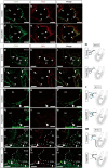
Figure 6. APJ-negative ECs sprout off from the SV and migrate into the embryonic atria and ventricles.
(af) Sagittal section through the SV of control (ac) and Ang1CKO embryos (df) at E10.5 immunostained for CD31 (green) and Tie2 (red). CD31 and Tie2 were uniformly expressed in the ECs of the SV (arrowheads). (gl) Sagittal section through the SV of control (gi) and Ang1CKO embryos (jl) at E10.5 immunostained for CD31 (green) and APJ (red). APJ-negative ECs were detected among the CD31-positive ECs in both control and Ang1CKO embryos (arrows). ECs in the SV expressing both CD31 and APJ were similarly observed in control and Ang1CKO embryos (arrowheads). (m) Schematic illustrations of the SV at E10.5 showing that the ECs were heterogeneous for APJ expression. (ns) Sagittal sections through the right atrium (RA) of control (np) and Ang1CKO embryos (qs) at E11.5 immunostained for CD31 (green) and APJ (red). (t) Schematic illustrations of the RA at E11.5 showing that all of the invading ECs were negative for APJ in both control and Ang1CKO embryos. (uz) Sagittal section through the RA and right ventricle (RV) of control (uw) and Ang1CKO embryos (xz) at E12.5 immunostained for CD31 (green) and APJ (red). The CD31-positive ECs invading the RV did not express APJ in either control or Ang1CKO embryos (arrow). In contrast, APJ was expressed in all of the CD31-positive ECs in the RA of control, but not Ang1CKO embryos (arrowheads). (aa) Schematic illustration of the RA and RV at E12.5 summarizing the expression of APJ. Blue line, APJ-positive ECs; black line, APJ-negative ECs. Scale bars, 50 μm. RA, right atrium; RV, right ventricle; SV, sinus venosus.
Figure 7
Figure 7. APJ-negative ECs precede APJ-positive ECs on the ventricle surface.
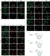
Sagittal sections through the RA and RV of wild-type embryos at E12.5 (ac), E13.5 (df), E14.5 (gi) immunostained for CD31 (green) and APJ (red). At E12.5 and E13.5, the APJ-negative (immature) ECs migrated at the forefront of the sprouting subepicardial coronary vessels (arrows in ac area 2, df area 4) and preceded the appearance of the APJ-positive (mature) ECs (arrowheads). At E14.5, all of the subepicardial ECs were double-positive for CD31 and APJ (arrowheads in gi). (jl) Schematic illustration of the SV, RA and RV at E12.5 (j), E13.5 (k) and E14.5 (l). Blue line, APJ-positive ECs; black line, APJ-negative ECs. Scale bars, 200 μm (upper panels in ai); 50 μm (insets). RA, right atrium; RV, right ventricle; SV, sinus venosus.
Figure 8
Figure 8. Ang1 enhances venous differentiation of Flk1+ immature endothelial progenitor cells synergistically with VEGF.
(ac) The venous marker protein COUP-TFII was upregulated in vascular progenitor Flk1+ cells by the addition of COMP-Ang1 to VEGF and cAMP. Flk1+ cells were immunostained with anti-CD31 antibody (red) and anti-COUP-TFII antibody (green). Nuclei were stained with DAPI (blue). (de) Quantitative expression analysis of the venous marker genes COUP-TFII and APJ in the Flk1+ cells (normalized to GAPDH mRNA; n=3). (d) The expression of COUP-TFII mRNA was increased exclusively by the combined treatment with VEGF, cAMP and COMP-Ang1. (e) The expression of APJ mRNA in the Flk1+ cells was significantly upregulated by stimulation with VEGF and Ang1 compared with treatment with VEGF alone. APJ mRNA was further upregulated by the addition of COMP-Ang1 to VEGF and cAMP. The results were expressed as relative intensity over cells treated with VEGF. Scale bars, 50 μm. Values are shown as means±s.e.m. for three separate experiments. One-way analysis of variance was used to compare differences. *P<0.05, **P<0.01, ***P<0.001 for the indicated groups.
Figure 9
Figure 9. Role of Ang1 on coronary vessel formation.
(a) Working model of coronary vein formation. The upper panel shows a schematic illustration of coronary vein formation in wild-type mice. The ECs in the SV at E10.5 consist of APJ-positive mature ECs (blue) and APJ-negative immature ECs (black). The APJ-negative ECs sprout off from the SV into the RA at E11.5. While the APJ-negative ECs migrate from the RA into the RV at E12.5, the APJ-negative ECs in the RA undergo venous differentiation in response to the action of Ang1 secreted from the myocardium, and differentiate into APJ-positive mature venous ECs. At E13.5, the APJ-negative ECs continue to migrate ahead of the APJ-positive ECs, which have emerged on the surface of the RV. At E14.5, all of the subepicardial CD31-positive cells have differentiated into mature venous APJ-positive ECs. In Ang1CKO mice, the migration, the proliferation and the venous specification of the APJ-negative ECs are impaired, resulting in defective coronary vein formation (lower panels). (b) Schematic diagram of coronary vessel formation in Ang1CKO embryo. Cardiomyocyte-specific deletion of Ang1 disturbs coronary vein formation, but does not impair coronary artery formation. Blue: APJ-positive ECs, Black: APJ-negative ECs. ep, epicardium; myo, myocardium; RA, right atrium; RV, right ventricle; SV, sinus venosus.

References

    1. Luttun A. & Carmeliet P. De novo vasculogenesis in the heart. Cardiovasc. Res. 58, 378–389 (2003). - PubMed
    1. Riley P. R. & Smart N. Vascularizing the heart. Cardiovasc. Res. 91, 260–268 (2011). - PubMed
    1. Del Monte G. & Harvey R. P. An endothelial contribution to coronary vessels. Cell 151, 932–934 (2012). - PubMed
    1. Guadix J. A., Carmona R., Munoz-Chapuli R. & Perez-Pomares J. M. In vivo and in vitro analysis of the vasculogenic potential of avian proepicardial and epicardial cells. Dev. Dyn. 235, 1014–1026 (2006). - PubMed
    1. Mikawa T. & Fischman D. A. Retroviral analysis of cardiac morphogenesis: discontinuous formation of coronary vessels. Proc. Natl Acad. Sci. USA 89, 9504–9508 (1992). - PMC - PubMed

Publication types

LinkOut - more resources