. 2014 Oct 23;426(21):3479-99.
doi: 10.1016/j.jmb.2014.07.017. Epub 2014 Jul 27.

Control of RecBCD enzyme activity by DNA binding- and Chi hotspot-dependent conformational changes

Affiliations

Control of RecBCD enzyme activity by DNA binding- and Chi hotspot-dependent conformational changes

Andrew F Taylor et al. J Mol Biol. .

Abstract

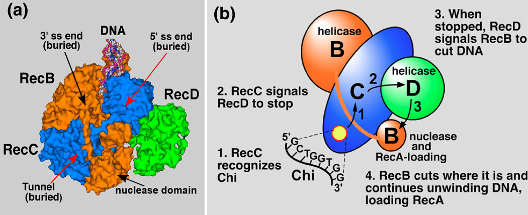
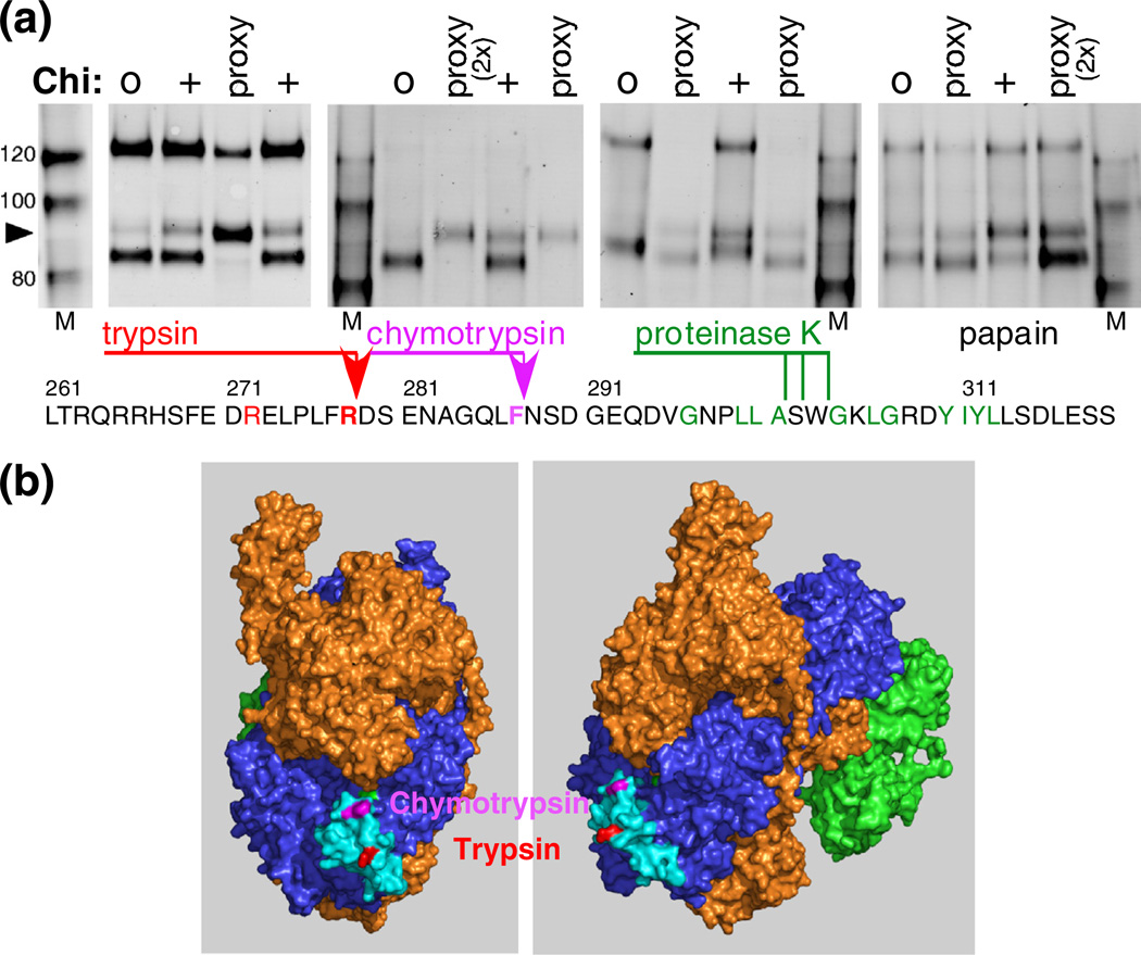
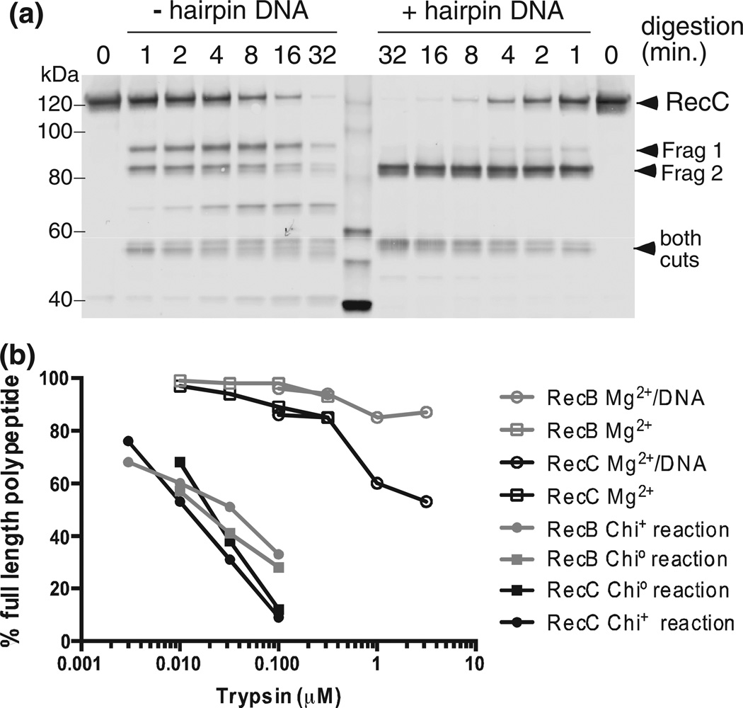
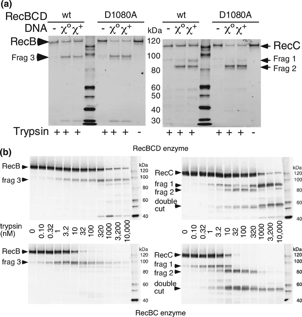
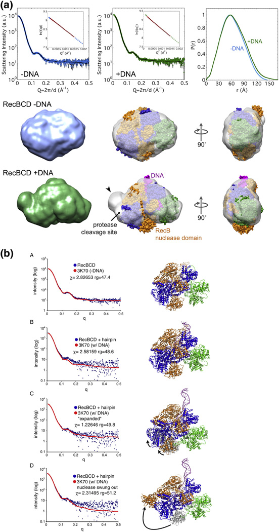
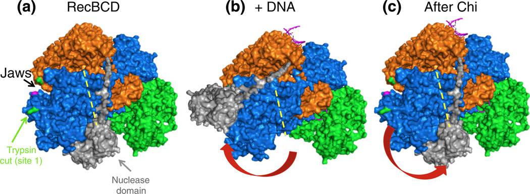
Faithful repair of DNA double-strand breaks by homologous recombination is crucial to maintain functional genomes. The major Escherichia coli pathway of DNA break repair requires RecBCD enzyme, a complex protein machine with multiple activities. Upon encountering a Chi recombination hotspot (5' GCTGGTGG 3') during DNA unwinding, RecBCD's unwinding, nuclease, and RecA-loading activities change dramatically, but the physical basis for these changes is unknown. Here, we identify, during RecBCD's DNA unwinding, two Chi-stimulated conformational changes involving RecC. One produced a marked, long-lasting, Chi-dependent increase in protease sensitivity of a small patch, near the Chi recognition domain, on the solvent-exposed RecC surface. The other change was identified by crosslinking of an artificial amino acid inserted in this RecC patch to RecB. Small-angle X-ray scattering analysis confirmed a major conformational change upon binding of DNA to the enzyme and is consistent with these two changes. We propose that, upon DNA binding, the RecB nuclease domain swings from one side of RecC to the other; when RecBCD encounters Chi, the nuclease domain returns to its initial position determined by crystallography, where it nicks DNA exiting from RecC and loads RecA onto the newly generated 3'-ended single-stranded DNA during continued unwinding; a crevice between RecB and RecC increasingly narrows during these steps. This model provides a physical basis for the intramolecular "signal transduction" from Chi to RecC to RecD to RecB inferred previously from genetic and enzymatic analyses, and it accounts for the enzymatic changes that accompany Chi's stimulation of recombination.

Keywords: SAXS; crosslinking; helicase–nuclease; limited proteolysis; recombination.


Conflict of interest statement

Conflict of Interest: The authors declare that they have no conflict of interest.

Figures

Fig. 1
Fig. 1
Structure of RecBCD enzyme bound to DNA and a “signal transduction” model for the Chi-dependent alteration of RecBCD enzyme. (a) The crystal structure of RecBCD bound to hairpin-shaped DNA (PDB entry 1W36) [18]. The RecB polypeptide is orange, RecC is blue, and RecD is green. In this structure, the 3′-ended strand would encounter the RecB nuclease domain upon exiting the RecC tunnel, in which Chi is likely recognized. (b) A “signal transduction” model for the Chi-dependent change of RecBCD [17]. When Chi is in the RecC tunnel (yellow disk), it prompts RecC to signal RecD to stop unwinding, which in turn signals RecB to nick the 3′-ended strand of DNA near Chi and to begin loading RecA.
Fig. 2
Fig. 2
Reaction with Chi+ DNA sensitizes RecC to cleavage by trypsin. (a) RecBCD was treated for 1 min with trypsin (3.2, 10, 32, 100, 320,or 1000 nM) during reaction on phage lambda DNA (48.5 kb) lacking (Chi0) orbearinga Chi site 3.5 kb from the right end (Chi+). RecC polypeptides and their fragments were detected by Western blots using polyclonal antibodies. (b) As in (a) but showing polypeptides detected with anti-RecB polyclonal antibodies. (c) Quantification of Chi0 data in (a), with fragments identified by their molecular mass in kilodaltons (kDa). 91 + 55, sum of the 91-kDa and 55-kDa fragments, the latter likely resulting from trypsin cleavages at both sites 1 and 2. (d) Quantification of Chi+ data in (a). Diagrams at the bottoms of (a) and (b) indicate the positions of trypsin cleavages and the designations of the tryptic fragments.
Fig. 3
Fig. 3
Further characterization of the Chi-dependent protease cleavage of RecC. (a) RecBCD enzyme was reacted with uncapped lambda Chi0 or Chi+ DNA, as in Fig. 2, or with lambda substrates capped at the left or right end of lambda, marked L and R, to block RecBCD entry at the capped end. Chi is active only if RecBCD enzyme enters from the right end [8]. Reactions were with 5 mM ATP and 2.5 mM Mg2+ (low Mg2+) or 8 mM Mg2+ (high Mg2+), as shown, and reacted with 10 nM trypsin for 1 min before Western blot analysis using RecC-specific polyclonal antibodies. (b) As in (a) but showing polypeptides detected with anti-RecB polyclonal antibodies. (c) Quantification of the Chi-dependent fragment in (a) and in parallel reactions that used 3.2 and 32 nM trypsin; x and o, Chi+ and Chi0 reactions; L and R, left- and right-end caps. (d) Persistence of RecC’s Chi-dependent sensitization to trypsin. RecBCD was reacted with Chi+ or Chi0 DNA for 1 min; the reactions were stopped by addition of EDTA to 23 mM, incubated for the further times indicated, and then treated for 1 min with 160 nM trypsin. RecC fragments were detected as in Fig. 2. Continuous and broken lines, ~91-kDa Chi-dependent fragment 1 and ~82-kDa Chi-independent fragment 2. (e) DNA substrate used in (a), (b), and (c). Phage lambda DNA (48.5 kb) with a Chi site (χ+D) 3.5 kb from the right end. Hairpin DNA (gray, data not drawn to scale) ligated in some cases to one strand at one or the other end of the lambda DNA substrate prevents RecBCD from unwinding from the capped end. Arrow, direction RecBCD must unwind DNA to be changed by χ+D [8].
Fig. 4
Fig. 4
Chi-dependent and proxy fragments of RecC comigrate and retain the native carboxy-terminus. (a) RecBCD enzyme was treated with trypsin for 1 min under the conditions noted, and the reaction terminated with trichloroacetic acid and concentrated. Traces of lanes from the separate and mixed samples were aligned by the flanking size markers. The similarity of the width of the peak, without evidence of splitting into two peaks, for the ~91-kDa fragments (Frag 1) in the separate and mixed samples indicates that the tryptic cleavage site is at the same position (±3 amino acids, by interpolation from size markers) in the proxy and Chi reactions. (b) As above, but with chymotrypsin. (c) The Chi-dependent tryptic fragment and the fragment produced under proxy conditions extend from the same or similar position in RecC to the C-terminus of RecC. RecBCD tagged with six histidines at the C-terminus of RecC (RecBCHis6D) was digested under a proxy condition with 2.5 mM Mg2+ and 10 µM trypsin or during reaction on Chi0 or Chi+ DNA (digested with 32 nM trypsin). The products were analyzed using rabbit anti-RecC and chicken anti-His tag polyclonal antibodies, which were detected with fluorescent species-specific secondary antibodies (RecC, red; His tag, green). “Fleck” indicates a small, local imperfection in the scanned membrane.
Fig. 5
Fig. 5
Location of Chi-dependent protease cleavages to a small exposed area of RecC. (a) RecBCD reacting with Chi0 or Chi+ DNA was treated with 32 nM trypsin, 100 nM chymotrypsin, 10 nM proteinase K, or 32 nM papain. RecBCD in buffer with DNA plus Mg2+ (proxy condition) was treated with the indicated protease (3 µM), and RecC was analyzed as in Fig. 2a. Sites of cleavage, determined by sequencing the trypsin- and chymotrypsin-derived Chi fragments (arrowheads), are shown on the RecC amino acid sequence. The location of the proteinase K cleavage was estimated by comigration with trypsin-cleaved substrate, using gels similar to those in Fig. 4. The substrate specificity of each protease includes the amino acids in color on the sequence. (b) The Chi-dependent sites of cleavage in the RecC polypeptide are in a disordered loop (cyan; residues 250–293) on the surface of the crystal structure [18]. The left view is rotated ~90° about the vertical axis relative to the right view.
Fig. 6
Fig. 6
Protease cleavage under proxy conditions is suppressed by DNA binding and requires much more trypsin. (a) RecBCD (35 nM) without or with 140 nM hairpin DNA similar to thatin the original crystal structure [18], with five additional T residues at the 5′ end, was treated for 1, 2, 4, 8, 16, or 32 min with 0.1 µM trypsin, and RecC was analyzed as in Fig. 2a. Similar results were obtained using K-PO4 buffer (Fig. S6). (b) Chi0, Chi+, and proxy reactions were performed and analyzed as in Fig. 2. The percentages of the visible bands that are full-length RecB or RecC are shown. Additional data are in Figs. S1 and S6.
Fig. 7
Fig. 7
Chi-dependent protease cleavage is reduced in a nuclease-deficient mutant RecBCD enzyme but is enhanced in the absence of RecD. (a) RecBCD enzyme or RecBD1080ACD enzyme was reacted in the absence of DNA or during reaction with Chi0 or Chi+ DNA and treated with 32 nM trypsin. Additional data are in Fig. S2. (b) RecBCD enzyme or RecBC enzyme (50 nM in ME buffer) was digested for 1 min with trypsin at the indicated concentrations. Reaction and sample preparation were as in Fig. 2.
Fig 8
Fig 8
Chi stimulates UV-dependent crosslinking of RecC to RecB. (a) RecBCF287BpaD enzyme was reacted with Chi0 or Chi+ DNA. Reactions were started by addition of ATP and were irradiated at 259 nm from 15 s to 75 s. Samples were prepared and analyzed as in Fig 2. (b) Reactions as in (a), using the buffers (20 mM) shown. [Mg2+] was 5 mM except for samples marked with an asterisk, which contained 8 mM Mg2+. Reactions, in sets of three, were without DNA, with Chi0 DNA, and with Chi+ DNA. Additional data are in Figs. S3 and S4. (c) A surface view of a portion of RecBCD enzyme (PDB file 3K70); the left panel is an expansion of part of the right panel, which shows the entire RecBCD enzyme (Fig. 1). The RecB helicase domain is orange, and its nuclease domain is gray. RecC is blue, and RecD is green. RecC amino acid F287 (magenta) shows the position of the Bpa in RecBCF287BpaD and the adjacent chymotrypsin cleavage site (Fig. 5) [19]. RecC amino acid F277 (red) shows the position of Bpa RecBCF277BpaD and the nearby trypsin cleavage site (between amino acids 278 and 279). RecB amino acids A108 and R119, the most likely locations of the Bpa crosslink in RecBCF277BpaD, are cyan and green, respectively. The measured distances between the C alpha of F277 and A108 or R119 are indicated by red arrows.
Fig. 9
Fig. 9
SAXS analysis of DNA-bound and unbound RecBCD enzymes. (a) Envelopes of RecBCD enzyme determined by SAXS (data in upper panels) of free enzyme and enzyme bound to hairpin DNA in K-PO4 buffer with glycerol (Figs. S7 and S8; Table S2). Ab initio shape reconstructions agree well with the data; Guinier analyses (insets) show linear fits. The crystal structure, with subunits colored as in Fig. 1a and oriented to be optimally docked within the SAXS envelope, is superimposed on the SAXS envelope. Note that swinging of the RecB nuclease domain (carat) improves the fit and accounts for the change of protease sensitivity (relatively sensitive −DNA and resistant + DNA). (b) SAXS all-atom modeling analysis of the above data. In (b–A), data from enzyme without DNA were tested against PDB structure 3K70 [19] with DNA computationally removed; in (b-B), data from enzyme with a bound DNA hairpin were tested against PDB structure 3K70; in (b–C) and (b–D), data from enzyme with a bound DNA hairpin were tested against two possible conformational changes in the enzyme, one (b–C) involving movement of the lower lobes of RecB and RecC moving toward the helicase domain of RecB, perhaps reflecting jaws closure, and the other (b–D) involving swinging of the nuclease domain to cover the protease-sensitive region. Additional data are in Figs. S7–S16.
Fig. 10
Fig. 10
Model for the Chi-dependent conformational changes of RecBCD enzyme, based on the crystal structure [18] and the results reported here. Subunits are colored as in Fig. 8c. (a) In the absence of DNA, the nuclease domain (light gray) is on the right side of RecC, as in the crystal structure (Fig. 1a), next to the RecC tunnel (yellow broken line). A surface patch of RecC is exposed to cleavage at R278 (green) by trypsin or at F278 (magenta) by chymotrypsin (Figs. 2, 3, and 5). (b) Upon binding to DNA, the nuclease domain (positioned manually) swings to the left and protects the RecC surface patch from proteolysis (Fig. 6). In addition, the “jaws” between the RecB helicase domain and RecC close partially (hidden by the nuclease domain). (c) Upon addition of ATP plus Mg2+, RecBCD unwinds the DNA, and the jaws close more. When Chi moves through the RecC tunnel, the nuclease domain swings back to the right, again exposing the RecC surface patch to proteolysis (Figs. 2 and 3), and the jaws close even more (data not shown). The nuclease domain is then in position to nick the 3′-ended strand a few nucleotides to the 3′ side of Chi [8] and, upon rotation, to load RecA protein onto the 3′-ended strand [12,42] and, at high [Mg2+], to occasionally nick the 5′-ended strand [10,11].

Similar articles

Cited by

References

    1. Cromie GA. Phylogenetic ubiquity and shuffling of the bacterial RecBCD and AddAB recombination complexes. J Bacteriol. 2009;191:5076–5084. - PMC - PubMed
    1. Smith GR. How RecBCD and Chi promote DNA break repair and recombination—a molecular biologist’s view. Microbiol Mol Biol Rev. 2012;76:217–228. - PMC - PubMed
    1. Ganesan S, Smith GR. Strand-specific binding to duplex DNA ends by the subunits of Escherichia coli RecBCD enzyme. J Mol Biol. 1993;229:67–78. - PubMed
    1. Taylor AF, Smith GR. RecBCD enzyme is a DNA helicase with fast and slow motors of opposite polarity. Nature. 2003;423:889–893. - PubMed
    1. Taylor A, Smith GR. Unwinding and rewinding of DNA by the RecBC enzyme. Cell. 1980;22:447–457. - PubMed

Publication types

LinkOut - more resources