. 2014 Jul 28;30(2):224-37.
doi: 10.1016/j.devcel.2014.06.008.

EGFR modulates DNA synthesis and repair through Tyr phosphorylation of histone H4

Affiliations

EGFR modulates DNA synthesis and repair through Tyr phosphorylation of histone H4

Ruey-Hwang Chou et al. Dev Cell. .

Abstract

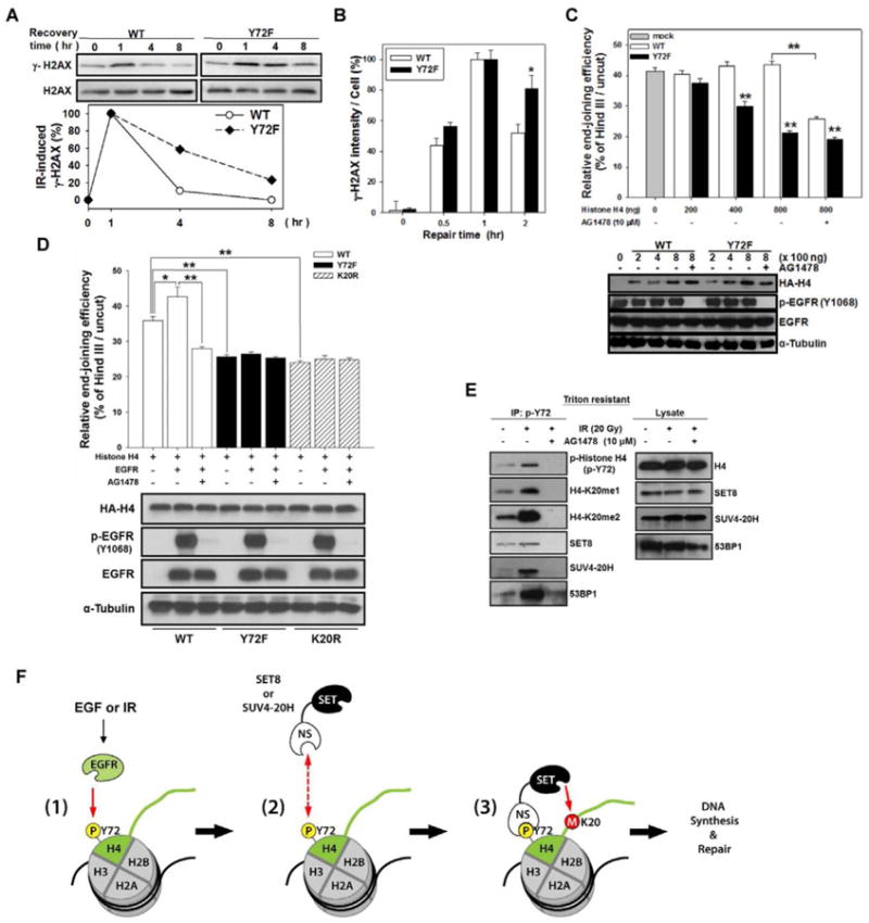
Posttranslational modifications of histones play fundamental roles in many biological functions. Specifically, histone H4-K20 methylation is critical for DNA synthesis and repair. However, little is known about how these functions are regulated by the upstream stimuli. Here, we identify a tyrosine phosphorylation site at Y72 of histone H4, which facilitates recruitment of histone methyltransferases (HMTases), SET8 and SUV4-20H, to enhance its K20 methylation, thereby promoting DNA synthesis and repair. Phosphorylation-defective histone H4 mutant is deficient in K20 methylation, leading to reduced DNA synthesis, delayed cell cycle progression, and decreased DNA repair ability. Disrupting the interaction between epidermal growth factor receptor (EGFR) and histone H4 by Y72 peptide significantly reduced tumor growth. Furthermore, EGFR expression clinically correlates with histone H4-Y72 phosphorylation, H4-K20 monomethylation, and the Ki-67 proliferation marker. These findings uncover a mechanism by which EGFR transduces signal to chromatin to regulate DNA synthesis and repair.


Conflict of interest statement

Conflict of interest: The authors have no conflicts of interest to declare.

Figures

Figure 1
Figure 1. Histone H4 interacts with EGFR
(A) Histone H4 and EGFR reciprocal immunoprecipitation (IP), followed by immunoblotting (IB) with the indicated antibody. (B) Histone H4 IP/IB of serum-starved MDA-MB-468 cells treated with or without EGF and/or AG1478 for 30 min. (C) Histone H4 IP/IB of serum-starved MDA-MB-468 cells treated with or without IR and/or AG1478 for 30 min. (D) The in situ subcellular interaction between EGFR and histone H4 determined by Duolink proximity ligation assay in serum-starved MDA-MB-468 cells treated with or without EGF (20ng/ml) or post-irradiation (20 Gy) for 30 min (arrows indicated red spots). Blue color: DAPIstained nucleus. (E) The in vitro translated myc-tagged FL-, ECD-, or ICD-EGFR (top) was individually incubated with purified recombinant histone H4 protein (middle) and immunoblotted to determine their interactions (bottom). (F) HEK-293 cells were co-transfected with HA-histone H4 plasmid with myc-ECD-EGFR ormyc-ICD-EGFR plasmid for 24 h. Cell lysate was examined by reciprocal IP/IB with antibodies against myc and HA, respectively. (G) Phosphotyrosine (p-Y) of histone H4 was examined by IP/IB with anti-p-Y or anti-histone H4 antibody, respectively, in 24 h-serum-starved cells treated with or without EGF and/orAG1478 for 30 min. See also Figure S1.
Figure 2
Figure 2. EGFR mediates tyrosine phosphorylation of H4-Y72
(A) Mass (MS/MS) spectrometric analysis of A431 nuclear extract immunoprecipitated with anti-histone H4 antibody. Top, the flanking regions of histone H4-Y72 among different species. Dotted circle, phosphotyrosine-containing peptide fragments. (B) Cells were serum-starved for 24 h and then treated with or without EGF and/or AG1478 for 30 min. Top, the level of histone H4-pY72 determined by IP/IB. Bottom, the endogenous levels of indicated proteins examined by IB. (C) HEK-293 cells were co-transfected wild- type (WT) or Y72F mutant (Y72F) histone H4 plasmid with or without myc-EGFR plasmid and/or AG1478 or gefitinib for 24 h. Exogenous histone H4-Y72 phosphorylation was detected by IP/IB. (D) MDA-MB-468 cells were infected with lentivirus containing shRNAs against luciferase (Luc) or EGFR (EGFR-1 or EGFR-2). Phosphorylated histone H4 at Y72 of each transfectant was examined. See also Figure S2.
Figure 3
Figure 3. Phosphorylation at Y72 of histone H4 enhances its methylation at K20
(A) The expressions of indicated proteins from 4-day-serum-starved MDA-MB-468 cells treated with or without EGF and/or AG1478 for or gefitinib 30 min. (B) HEK-293 cells were co-transfected with HA-histone H4 or -H4Y72F plasmid with or without myc-EGFR plasmid and/or AG1478. The exogenous HA-histone H4 or HA-H4Y72F was immunoprecipitated with anti-HA antibody, followed by IB with specific antibodies against methylated histone H4 at K20. (C) HEK-293 cells were co-transfected with HA-tagged histone H4 plasmid with or without ΔNLS- or wild-type-EGFR plasmid. Nuclear and cytosolic of the indicated proteins are shown below. (D) SET8 or SUV4-20H HMTase was immunoprecipitated from the nuclear extract of MDA-MB-468 cells with specific antibody. An increasing amount of immunoprecipitated SET8 or SUV4-20H nuclear extract (50, 100, or 200 μg) was incubated with recombinant histone H4 or H4Y72F protein in the HMTase reaction buffer. Histone H4 K20 methylation and input were detected by IB. (E) The recombinant wild-type and Y72F mutant histone H4 proteins were treated with or without EGFR kinase, followed by HMTase assay with immunoprecipitated SET8 or SUV4-20H from nuclear extract (50 μg). The histone H4-K20me1, H4-K20me2, and input of the histone were also examined. (F) The same sequential in vitro EGFR kinase and HMTase assay as in (E) was performed with or without EGF and/or AG1478. See also Figure S3.
Figure 4
Figure 4. Phosphorylation at Y72 of histone H4 enhances its interaction with SET8 and SUV4-20H
(A) Serum-starved MDA-MB-468 cells were treated with or without 20 ng/ml EGF and/or AG1478 (10 μM) for 30 min. Triton-resistant extract was isolated and immunoprecipitated with anti-histone H4 antibody to determine its association with SET8, SUV4-20H, or SUV39H1HMTases by IB. (B) Histone H4 in nuclear fractions from indicated stable transfectants with or without AG1478 (10 μM) was immunoprecipitated to determine its association with SET8 or SUV4-20H(top). Endogenous levels of indicated proteins in the nuclear fraction (bottom). (C) Purified GST-SET8 and -SUV4-20H2 proteins were stained with Coomassie blue (top) orimmunoblotted with specific antibodies (bottom). (D) Different amounts of histone H4-Y72 peptides (IRDAVT-Y-TEHAKR) containing non-phospho- (NP) or phospho-residue at Y72 (p-Y72) were dotted onto the PVDF membrane, stained with Ponceau S solution (bottom), and then incubated with purified GST-SET8 or GST-SUV4-20H2 protein. Binding of SET8 or SUV4-20H to the coated H4 peptides was determined by IB (top). (E) Histone H4-pY72 peptide was dotted onto membrane, incubated with GST-SET8 or GST-SUV4-20H2 protein with or without H4-Y72 peptide (NP or p-Y72) followed by IB. (F) Histone H4-pY72 peptide was conjugated on agarose beads and then applied to pull down against GST-SET8, GST-SUV4-20H2, and GST-SUV39H1 recombinant proteins. (G) HEK-293 cells were transfected with full- length (FL), SET-containing (S), or non-SET-containing (NS) region of SET8 or SUV4-20H2 for 24 h. Expression of each fragment was examined by IB against V5. (H) Cell lysate from each transfectant was incubated with the H4-pY72 peptide-coated membrane followed by IB against V5. See also Figure S4.
Figure 5
Figure 5. Histone H4-Y72 phosphorylation is involved in DNA synthesis
(A) Stable transfectants were serum starvated for 24 h, then were treated with or without EGF and/or AG1478, and pulse-labeled with 100 μM BrdU for 12 h. Histone H4-associated newly synthesized DNA was determined. Top right, expression of HA-histone H4 in each stable transfectant. (B) Stable transfectants were pulse-labeled with 100 μM BrdU for 4 h. Subsequently, the DNA contents and the incorporated BrdU in each transfectant were determined by flow cytometry. The percentage of cells distributed in each phase of cell cycle is shown. (C) Stable transfectants were treated with nocodazole and/or AG1478 for 24 h. The distributions of cells in G1, S, and G2/M phases of cell cycle were analyzed by flow cytometer (Figure S5A). The bar plot is the G1 (%) in each treatment. (D) Stable transfectants were synchronized at G1 phase and then released to normal culture medium for indicated time intervals. The distributions of cells in each stage of cell cycle were analyzed by flow cytometry (Figure S5B). Bar graph shows quantitative results. (E) Relative number of cells of each stable transfectant was determined at indicated time intervals. Error bars, mean ± SD. See also Figure S5.
Figure 6
Figure 6. Histone H4-Y72 phosphorylation is involved in DNA DSB repair
(A) Top, each stable transfectant was irradiated with 20 Gy and incubated at 37 °C to recover for the indicated time. Expression of γ-H2AX was examined by IB. Bottom, relative quantity of γ-H2AX was normalized to H2AX 1 h after irradiation. (B) The quantitation of γ-H2AX in Figure S6A. (C) HEK-293 cells were co-transfected with circular (cir-pGL3) or linearized (lin-pGL3) luciferase plasmid with myc-EGFR and increased amounts of wild-type (WT) or Y72F mutant histone H4 plasmid for 48 h with or without AG1478. The relative end-joining efficiency was determined by the percentage of luciferase activity from cells with lin-pGL3 over that with cir-pGL3. Error bars, mean ± SD. Bottom, the expression of transfected plasmids as indicated. (D) A similar experiment as described in (C) was also performed using another histone H4K20R mutant plasmid (800 ng) with or without EGFR and/or AG1478. (E) Serum-starved MDA-MB-468 cells were treated with or without irradiation and/or AG1478. After 1 h, Triton-resistant fraction was immunoprecipitated against histone H4-p-Y72 followed by IB with the indicated antibodies. Right, endogenous level of indicated proteins. (F) A proposed model of nEGFR-mediated H4-Y72 phosphorylation in the regulation of H4-K20 methylation. See also Figure S6.
Figure 7
Figure 7. Y72 peptide demonstrates tumor suppressive activity in vivo, and histone H4-pY72 and K20me1 correlate with EGFR expression in clinical samples of human breast cancer
(A) MDA-MB-468 cell lysates were subjected to IP with anti-EGFR antibody in the presence or absence of Y72 or scrambled peptide (200 μM) followed by IB against histone H4 and EGFR. (B) The tumor volume of mice treated with PBS or the indicated peptides. Error bars, mean ± SD. (C) Bar graph shows the quantative tumor weight in each group. Error bars, mean ± SD. (D) Protein lysates from tumors in (C) were subjected to Western blot analysis with the indicated antibodies. To detect histone H4-pY72, lysates were subjected to IP first with anti-histone H4-pY72 antibody followed by IB with anti-histone H4 antibody. (E) Correlations between expression of EGFR and histone H4-pY72, histone H4-K20me1, or Ki-67 in surgical specimens of breast cancer were analyzed by Pearson Chi-Square test. M=membrane, N= nucleus, C= cytoplasm. (F) IHC staining of human breast tumor tissue sections for EGFR and histone H4-K20me1. The plot represents the quantitative results of histone H4-K20me1 in the tissue sections with different EGFR status. EGFR N+, nuclear EGFR positive; EGFR C+, cytosolic EGFR positive; EGFR-, EGFR negative. Error bars, mean ± SD. See also Figure S7.

Similar articles

Cited by

References

    1. Beck DB, Burton A, Oda H, Ziegler-Birling C, Torres-Padilla ME, Reinberg D. The role of PR-Set7 in replication licensing depends on Suv4-20h. Genes Dev. 2012;26:2580–2589. - PMC - PubMed
    1. Blagoev B, Kratchmarova I, Ong SE, Nielsen M, Foster LJ, Mann M. A proteomics strategy to elucidate functional protein-protein interactions applied to EGF signaling. Nat Biotechnol. 2003;21:315–318. - PubMed
    1. Botuyan MV, Lee J, Ward IM, Kim JE, Thompson JR, Chen J, Mer G. Structural basis for the methylation state-specific recognition of histone H4-K20 by 53BP1 and Crb2 in DNA repair. Cell. 2006;127:1361–1373. - PMC - PubMed
    1. Chen HC, Appeddu PA, Isoda H, Guan JL. Phosphorylation of tyrosine 397 in focal adhesion kinase is required for binding phosphatidylinositol 3-kinase. J Biol Chem. 1996;271:26329–26334. - PubMed
    1. Chen YJ, Huang WC, Wei YL, Hsu SC, Yuan P, Lin HY, Wistuba II, Lee JJ, Yen CJ, Su WC, et al. Elevated BCRP/ABCG2 expression confers acquired resistance to gefitinib in wild-type EGFR-expressing cells. PLoS One. 2011;6:e21428. - PMC - PubMed

Publication types

LinkOut - more resources