. 2014 Oct;57(10):2155-64.
doi: 10.1007/s00125-014-3337-2. Epub 2014 Jul 30.

Exosomes participate in the alteration of muscle homeostasis during lipid-induced insulin resistance in mice

Affiliations

Exosomes participate in the alteration of muscle homeostasis during lipid-induced insulin resistance in mice

Hala Aswad et al. Diabetologia. 2014 Oct.

Abstract

Aims/hypothesis: Exosomes released from cells can transfer both functional proteins and RNAs between cells. In this study we tested the hypothesis that muscle cells might transmit specific signals during lipid-induced insulin resistance through the exosomal route.

Methods: Exosomes were collected from quadriceps muscles of C57Bl/6 mice fed for 16 weeks with either a standard chow diet (SD) or an SD enriched with 20% palm oil (HP) and from C2C12 cells exposed to 0.5 mmol/l palmitate (EXO-Post Palm), oleate (EXO-Post Oleate) or BSA (EXO-Post BSA).

Results: HP-fed mice were obese and insulin resistant and had altered insulin-induced Akt phosphorylation in skeletal muscle (SkM). They also had reduced expression of Myod1 and Myog and increased levels of Ccnd1 mRNA, indicating that palm oil had a deep impact on SkM homeostasis in addition to insulin resistance. HP-fed mouse SkM secreted more exosomes than SD-fed mouse SkM. This was reproduced in-vitro using C2C12 cells pre-treated with palmitate, the most abundant saturated fatty acid of palm oil. Exosomes from HP-fed mice, EXO-Post Palm and EXO-Post Oleate induced myoblast proliferation and modified the expressions of genes involved in the cell cycle and muscle differentiation but did not alter insulin-induced Akt phosphorylation. Lipidomic analyses showed that exosomes from palmitate-treated cells were enriched in palmitate, indicating that exosomes likely transfer the deleterious effect of palm oil between muscle cells by transferring lipids. Muscle exosomes were incorporated into various tissues in vivo, including the pancreas and liver, suggesting that SkM could transfer specific signals through the exosomal route to key metabolic tissues.

Conclusions/interpretation: Exosomes act as 'paracrine-like' signals and modify muscle homeostasis during high-fat diets.


Figures
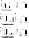
Fig. 1
Fig. 1
(a, c, e) Phospho-Akt and Akt quantified by western blot in gastrocnemius muscle of SD- and HP-fed mice incubated ex vivo with 10−7 mol/l insulin for 15 min (a), in C2C12 cells treated with 0.5 mmol/l palmitate (Palm) for 18 h (c) and in C2C12 cells 9 h post palmitate treatment (e). Data are unitless (phospho-Akt/total Akt intensities). *p < 0.05; **p < 0.01, for indicated comparisons. (b, d, f) Quantities of exosomes released from quadriceps of SD- and HP-fed mice (b), from C2C12 cells treated with 0.5 mmol/l palmitate for 18 h (d) or from C2C12 cells 9 h post palmitate treatment (f). White bars and black bars represent incubation without and with insulin, respectively. *p < 0.05
Fig. 2
Fig. 2
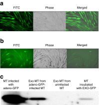
(a) C2C12 myotubes (MT) infected with adeno-GFP (magnification × 10). (b) MT incubated for 48 h with EXO-GFP released from infected MTs described in (a) (magnification × 20). (c) Western blot detection of GFP in infected MTs (1 μg) or in their released EXO-GFP (1 μg) or in MTs incubated with EXO-GFP (50 μg); Exo-MT, exosomes from myotubes
Fig. 3
Fig. 3
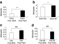
Akt quantified by western blot. Actin was used as loading control. Data are unitless (total Akt/actin intensities). (a, b) C2C12 myotubes incubated with C2C12-released exosomes collected from cells pre-treated with EXO-Post Palm vs EXO-Post BSA (a) or with quadriceps-released exosomes from HP-fed mice vs SD-fed mice (b). (c, d) C2C12 myotubes incubated with CM of cells pre-treated with palmitate (CM-Post Palm) (c) or with CM-Post Palm depleted of exosomes (CM-DE) (d)
Fig. 4
Fig. 4
Gene ontology (GO) pathways (levels from 3 to 9) significantly enriched in genes differentially regulated in C2C12 cells treated with EXO-Post Palm vs EXO-Post BSA (p < 0.05). White bars, upregulated genes; grey bars, downregulated genes
Fig. 5
Fig. 5
Expression levels of Ccnd1, Il-6, Slc2a4, Myod1 and Myog quantified by qRT-PCR. (a) C2C12 cells pre-treated with EXO-Post BSA (white bars), EXO-Post Palmitate (black bars) or EXO-Post Oleate (grey bars). (b) C2C12 cells pre-treated with palmitate (black bars) or BSA (white bars). (c) mRNA expressions in gastrocnemius muscle of SD (white bars) or HP (black bars) mice. All data are unitless (mRNA/Tbp mRNA level). n = 4 biological replicates; *p < 0.05, **p < 0.01 vs EXO-Post BSA, BSA or SD
Fig. 6
Fig. 6
C2C12 myoblasts (n = 8) were incubated with serum-free DMEM supplemented with EXO-Post Palm or EXO-Post Oleate. (a, b) Impedance measurement with the xCELLigence System. (a) The presence of the cells on top of the electrodes will affect the local ionic environment at the electrode/solution interface, leading to an increase in the electrode impedance. The more cells attached to the electrodes, the larger the increase in electrode impedance. (b) Data is graphed as ‘Delta Cell Index’, which is the change in impedance from the time point immediately after cells were added (*p < 0.05 vs EXO-Post BSA). (ce) Myoblast sizes determined by the Scepter 2.0 handheld automated cell counter after 48 h proliferation. (c) Serum-free DMEM (grey line) or EXO-Post BSA (black line). (d) EXO-Post BSA (grey line) or EXO-Post Palm (black line). (e) EXO-Post BSA (grey line) or EXO-Post Oleate (grey line)
Fig. 7
Fig. 7
Quantification of fatty acids in exosomes. EXO-Palm (black bars) and EXO-BSA (white bars) from C2C12 treated for 18 h with BSA or palmitate
Fig. 8
Fig. 8
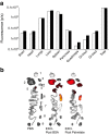
Biodistribution of C2C12 exosomes. EXO-Post BSA (6.02 × 1010 particles/ml; white bars) and EXO-Post palmitate (3.44 × 1010 particles/ml; black bars) were used for tail injection in C57Bl/6 mice. (a) Quantification of fluorescent signals for each organ, normalised to background. (b) Organs targeted by C2C12 DiR exosomes; 1 = brain, 2 = liver, 3 = heart, 4 = lungs, 5 = gastrointestinal tract, 6 = spleen, 7 = pancreas, 8 = quadriceps, 9 = kidney

References

    1. Pedersen BK, Febbraio MA. Muscles, exercise and obesity: skeletal muscle as a secretory organ. Nat Rev. 2012;8:457–465. - PubMed
    1. Thery C, Boussac M, Veron P, et al. Proteomic analysis of dendritic cell-derived exosomes: a secreted subcellular compartment distinct from apoptotic vesicles. J Immunol. 2001;166:7309–7318. doi: 10.4049/jimmunol.166.12.7309. - DOI - PubMed
    1. Mathivanan S, Ji H, Simpson RJ. Exosomes: extracellular organelles important in intercellular communication. J Proteome. 2010;73:1907–1920. doi: 10.1016/j.jprot.2010.06.006. - DOI - PubMed
    1. Lopez-Verrilli MA, Court FA. Exosomes: mediators of communication in eukaryotes. Biol Res. 2013;46:5–11. doi: 10.4067/S0716-97602013000100001. - DOI - PubMed
    1. Guescini M, Guidolin D, Vallorani L, et al. C2C12 myoblasts release micro-vesicles containing mtDNA and proteins involved in signal transduction. Exp Cell Res. 2010;316:1977–1984. doi: 10.1016/j.yexcr.2010.04.006. - DOI - PubMed

Publication types

LinkOut - more resources